Thursday, February 2, 2023

Iron Deficiency Anemia.

                                                             Iron Deficiency Anemia.

                                                       PKGhatak, MD


 Anemia is defined by the WHO as having less than 12 gm /dL of hemoglobin in women and less than 13gm /dL in men. By this criterion, 33 % of the world's population has anemia and half of the cases are due to iron deficiency. Children below 5 years old and women of childbearing age are particularly susceptible to iron deficiency anemia in sub-Saharan Africa and South Asia. Iron deficiency anemia is the number one nutritional disorder in the world.

Iron deficiency anemia develops when the iron store is depleted and the daily loss of iron exceeds the daily iron intake. The consequences of iron deficiency anemia are many, chief among them are dysfunctions of several systems of the body, and notably among them are limitation of physical work, reduced energy production, depressed immune function, recurrent infection, poor digestive function, and various neurocognitive functions. The diagram below is an outline of iron utilization. 


Iron containing proteins in humans.

Four general categories of proteins contain iron: (1) heme proteins (e.g., hemoglobin. (2) mononuclear iron proteins (e.g., superoxide dismutase), (3) diiron-carboxylate proteins (e.g., ribonucleotide reductase, ferritin), (4) iron-sulfur proteins (e.g., aconitase), Hemoglobin is the most abundant iron-containing protein in humans.  More than one-half of total-body iron is contained within hemoglobin.

Basic physiology of Iron in humans.

Iron is a very reactive metal and combines readily with oxygen, when it reacts with H2O2 (hydrogen peroxide) it generates oxygen radicals. Oxygen radical sickens living cells and in the end, kills cells. To protect tissue from its harmful effects,  a protein, ferritin, combines with iron. Moving in and out of cells requires conversion of ferric to ferrous state and must pass special areas of the cell membrane called gates. Once inside the cells, iron is converted back to ferric state and in the blood and inside the cells iron remains combined with ferritin.

The generation of RBC is called erythropoiesis. Demand for erythropoiesis comes from: (a)Tissue oxygenation, (b)Erythrocyte turnover, (c) Blood loss from hemorrhage.

(a)Tissue oxygenation remains more or less stable in health.

(b) Approximately 20 mL of old erythrocytes die daily, and 20 mg of iron is recovered from the dead RBC.  The immediate source of iron for erythroblasts is mono or diferric transferrin, found in high concentrations in plasma. The sources of diferric transferrin are the gut (diet), macrophages (recycled iron), and the liver (stored ferritin iron). the diet. 

2.5mL of whole blood contains 1.0 mg of iron. 1.0 mg of iron is absorbed from the diet daily and 20 mg of iron from recycled erythrocytes is available to support erythropoiesis. Once iron stores are depleted, dietary and recycled erythrocyte iron is not usually sufficient to compensate for acute blood loss. In a normal person, less than 2 mL of blood is lost daily in the stool.  But this tiny amount of blood in stool does not give a positive occult blood test. A minimum of 60 mL/d is needed for a positive occult blood test to detect the presence of blood in the stool. Women in childbearing age lose additional blood due to menstruation. Repeated nosebleeds and regular blood donation lower body stores of iron.

Iron in pregnancy:

It is estimated that about 1.2 gm of iron is required from conception through delivery. The breakdown is as follows: (i)Mother's erythrocyte mass should increase from 350 to 450 mL. And that needs 450mg of iron (ii) Cessation of menstruation saves 600 mg of iron (iii) A full tern newborn has 280 mg (iv) Placental loss 90 mg.

At birth, the fetal red cell mass is 50 mL/kg. (Compared with 25–30 mL/kg in adults). Even in anemic mothers, the fetal ferritin levels remain 10 times higher than the maternal ferritin, indicating nature's preferential treatment of a growing child over the mother.

Common causes of iron deficiency in poor countries.

1. Poor diet. Red meat, organ meat and egg are good sources of iron. Poor people can hardly provide meat or eggs on a regular basis. Bush meat in Africa, at one time adequately supplied the local population, but increased demand by the growing urban population has dried up this source. Vitamin C improves iron absorption, but tea and coffee interfere with absorption. Inorganic iron absorption requires multiple mechanisms,  but infections and inflammation depress iron absorption by hepcidin inhibitory action. 

2. Malaria. Malaria is a special circumstance. Intravascular hemolysis from the effects of parasites and Blackwater fever produces severe anemia. In addition, malnutrition depresses iron absorption.

 3. Hookworm. It is an intestinal parasite, that is a significant source of GI blood loss in millions of people in South Asia and Africa. Defecation in the open, and walking in bare feet in flooded fields make it possible for hookworms to enter the body. Each worm takes 0.3 to 0.5 mL of blood from the upper small intestine and produces anemia. [ see footnote ]

4. Gastrointestinal factors. Suppressed gastric acid secretion from taking a proton pump inhibitor or gastric mucosal atrophy from gastritis, prevents the release of iron from organic iron in food. Tropical sprue and other duodenal pathologies hinder iron absorption from the gut. Liver diseases may decrease ferritin production and in chronic infection, excess hepcidin decreases GI iron absorption. And finally, Helicobacter infection of the stomach produces gastritis and causes decreased iron absorption.

A look at the peripheral blood in iron deficiency anemia.


Blood of iron deficient anemia.


                                       Blood of a normal person.

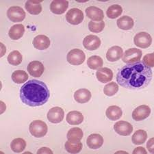
The characteristic features of iron deficiency anemia are

The RBCs are microcytosis (small sized cells) and hypochromic (pale). In addition, a combination of increased red cell distribution width (RDW), decreased red blood cell (RBC) count, decreased MCH (mean corpuscular hemoglobin), and decreased mean cell volume are manifested. Red cells are pale in the center, smaller in size and the shape is variable and easily identifiable on blood smears. Because of non-steady state hemopoiesis, the RDW shows wider variation and is usually over 15%.  This is marked in contrast with the Sickle cell trait. The red cells in the sickle cell trait are also small and pale in the center and the hemogram appears similar to iron deficiency but RDW remains in the normal range due to the fact that hemopoiesis is steady because the iron stores in the body are full and so also the of serum iron and ferritin saturation index.  In iron deficiency, the serum ferritin level is 15 μg/L or below.  In research centers, the iron concentration of the reticulocytes, erythroblasts, the bone marrow and the liver is determined, but in clinical practice, those tests are not needed for diagnosis.  Iron deficiency causes increased release of soluble transferrin (transport protein) from erythroblasts. Therefore, ratios of soluble transferrin receptor and ferritin are used to detect iron-deficient erythropoiesis. 

Treatment of iron deficiency anemia,

Replacement of the iron stores is the first priority. Oral iron preparations are of two kinds. Inorganic and inorganic iron. Inorganic iron preparations are affordable but difficult for patients to tolerate because they produce constipation, nausea and anorexia. Organic iron preparations are easier to tolerate but expensive. In previous generations, intravenous iron therapy was problematic because of frequent hypersensitive reactions. The improved formulation has made rapid correction of anemia possible.

Advances in Iron knowledge.

 The most significant advance is the discovery of hepcidin. Hepcidin expression is highly variable and influenced by a circadian rhythm. That knowledge should improve dosing regimens.  When iron therapy does not improve iron deficiency anemia, TMPRESS6 gene mutation is suspected. In normal conditions, the TMPRESS6 gene provides instructions for the Matriptase-2 protein molecule, which is a part of the controlling mechanism of hepcidin production in the liver.

________________________________________________________________________________________

Footnote:


1. Larva entering by piercing skin of feet---> enter venous blood and carried to the right heat chambers, then to pulmonary capillaries----> larva moving out from lung alveoli to airways and crawls up the trachea----> coughed up and swallowed----> enters the stomach and settles in upper small intestine---- sucks blood, matures, mates, releases eggs----> egg containing feces deposited in open fields and the cycles continue.

********************************************************************


Sunday, January 29, 2023

Cardiomyopathy

                                                Cardiomyopathy.

                                         PKGhatak, MD

Cardiomyopathy is a weakened condition of the Heart Muscles and the heart chambers increase in size and subsequently the heart fails. It is suspected that 1 in 5,00 adult people may have cardiomyopathy without being aware of it. People seek medical attention only when cardiomyopathy becomes symptomatic. The initial symptoms are shortness of breath and a marked decrease in energy level to carry on the usual work.

There are wide varieties of conditions that lead to cardiomyopathy and based on pathophysiology the disease is classified as Dilated, Hypertrophic, Restricted, and Arrhythmogenic Cardiomyopathy.

Cardiomyopathy also happens due to inherited defects of genes, producing structural changes in the heart muscle or metabolic defects, which cause accumulation of iron and other material in the heart muscles.

 I. Dilated Cardiomyopathy:

The heart is enlarged and the muscles are stretched and become thin. Thinned out heart muscles fall behind in pumping blood out of ventricles into the circulation. Accumulated blood causes congestion of the lungs, and produces shortness of breath and in other organs. Dilated cardiomyopathy is diagnosed by clinical examination and confirmed by a chest x-ray and echocardiogram. 

 X-ray criterion is the transverse diameter of the heart exceeds 50% of the chest diameter in PA view.

A few common causes of dilated myopathy.

1. Virus infection.

Viral myocarditis of a mild nature happens frequently. In most cases, myocarditis resolves spontaneously without any health consequences. The common viruses causing these conditions are Parvovirus B-19, Coxsackie A & B, HIV, Covid-19, Cytomegalovirus, Epstein-Barr virus and Adenoviruses.

Damage to heart muscles in viral infection happens in two stages. In the initial phase, the inflammatory cells infiltrate the heart muscle. Immunocytes release acute phase inflammatory cytokines namely IL-1 beta, TNF alpha, and Interferon alpha and these agents should be sufficient to avert a full blown inflammation, but in some cases, they fail. In the second phase, about 7 to 14 days after the initial symptoms, T-cells accumulate. Vasoconstriction and release of Perforin molecule create multiple pores like apertures in heart muscles. Immunocytes release excess amounts of  IL-1, IL-6, CCL5, and TNF beta. Interferon decreases B-Cell activities and facilitates T-Cell activation. Cytokines continued to appear in the heart in response to the presence of viral particles in the myocardium. Because of the close resemblance of the viral antigen to muscle protein, the cytokines make mistakes one for the other. and damages the heart muscle.

A diagnosis of viral cardiomyopathy requires endocardial biopsy and identifying viral antigens. Heart failure and other organ insufficiencies are treated with medications. A cardiac transplant is a definitive treatment.

2. Chagas Disease:

Chagas disease is due to a protozoan parasite, Trypanosoma cruzi. It is an important cause of dilated cardiomyopathy in the Americas. In Central and South American countries, the Chagas disease is endemic. It is estimated that 5 million people are suffering from Chagas disease with an annual rate of infection of 200,000 and annual deaths of 10,000.

About 40,000 people were identified with Chagas disease in the USA among the South American migrant communities in 2019.

The parasite is carried by a bug, Triatoma infestans, commonly known as the kissing bug. The bug bites humans during sleep and leaves a fecal deposit at the site. The contaminated puncture wound, usually on the face or forehead, is the entry point of the parasite. Local inflammation and adenopathy may or may not produce symptoms. About 5 % of victims develop myocarditis. In the chronic phase, 40% develop dilated cardiomyopathy from the continued presence of parasites at the heart and resultant immune inflammation, tissue necrosis and scars replacing heart muscles. Cardiac conduction abnormalities are common and later heart failure predominates.

The diagram shows the life cycle of T. cruzi.

3. Alcoholic Myopathy.

People are well aware of liver disease due to alcohol; however, many will be surprised to learn about 25 % of all cardiomyopathy. Alcohol acts as an important factor in causing cardiomyopathy. About 2% of heavy alcohol users risk cardiac complications. Colored people have more risks of cardiomyopathy than light colored people. Families with Alcoholdehydrogenase deficiency are more likely to have cardiomyopathy. Alcohol is a Mitochondrial poison. As mitochondrial functions falter, acetaldehyde accumulates in the cells and oxygen radicals form as a consequence and the combined effects of these produce cell deaths. Cardiac muscles are replaced by weak scar tissue, and cardiomyopathy develops in the same way as described earlier. There are no distinctive histological characteristics of alcoholic myopathy, however enlarged and disorganized sarcoplasmic reticulum, fat and glycogen deposition and dilated intercalating discs are seen under electron microscopy in addition to various stages of diseased and disappearing mitochondria.

4. Chronic coronary arterial disease and Hypertension.

These two diseases are silent killers of heart muscles. Acute coronary events produce damage to heart muscles, even when emergency Angioplasty is successfully performed and helps to salvage most of the ventricular muscles, some loss is unavoidable. Chronic insufficiency produces almost identical damage but the pace is slow and takes place over a much longer time. Scar formation eventually leads to dilated myopathy and heart failure.

Hypertension produces ventricular muscle hypertrophy initially. If left untreated, the hypertrophic muscles eventually flatten out. A flabby enlarged heart fails. Hypertension is usually associated with coronary artery disease and induces cardiomyopathy but in the absence of coronary artery disease, hypertension alone is an independent cause of cardiomyopathy.

5. Diabetes mellitus.

Diabetes mellitus produces microvascular changes. The exact statistic is variable based on the nature of the inquiry but consistently higher than the controlled groups.

6. Thyroid disorder.

Both hypothyroidism and hyperthyroidism produce Cardiomyopathy but the mechanism is different. Hypofunction of the thyroid decreases metabolism in general and specially in the heart, delaying cardiac muscle repair and replacement. That leads to an enlarged flabby heart and often a viscous pericardial effusion. In hyperthyroid conditions, the cardiac muscles are overworked due to sinus tachycardia, increased demand for more cardiac output and associated hypertension. As the heart fails, the cardiac chambers dilate.

II. Hypertrophic Cardiomyopathy.

Hypertrophic cardiomyopathy (HCM) is an autosomal dominant inherited disease. One copy of the mutated gene is all that is needed for HCM to manifest. Of the several genes responsible for HCM. These gene mutations are common- MYH7 and MYPPC3 gene; other mutated genes are MYPBC3, TNNT2, TNN13, TPN1, MLC2, and MLC3. 1 in 700 people in the Americas are carriers of these genes and about 75,000 people have HCM at a given time. But most are unaware of the presence of mutant genes, and unfortunately, a sudden cardiac arrest may be the first sign of it. 

                                  Normal heart                    HCM heart

In the right-hand picture please note the thickened partition between the ventricular cavities. That part blocks the path of blood going out into the aorta during the ventricular ejection phase. In addition, the Mital valve is displaced and often deformed producing mitral insufficiency and reducing left ventricular ejection fraction further.
Calcium iron enters the myocardium in excess amounts and binds with actin-myosin. The cardiac impulse conduction path and coronary arterial system are altered. These are additional causes of abnormal heartbeats, the most serious one is ventricular tachycardia and it is often fatal. The initial symptoms are delayed till the teenage years, and a sudden collapse during a sporting event is usual. Diagnosis is made on clinical examination and confirmed by an echocardiogram. Treatment of HCM is multipronged.  Beta blocks and calcium channel blockers are effective as initial therapy. A recently approved drug, Mevacamten, is a new class of drug and is a reversible cardiac myosin ATPase inhibitor. It reduces the formation of actin-myosin cross-bridges. This action counteracts the inherited defect of this enzyme. The drug promotes energy savings in the myocardium and reduces outflow tract obstruction.
The definitive therapy is surgery. It involves removal of the defective parts and repairs. Technically known as Septal myomectomy, an alternative surgery is Alcohol septal ablation. In failed cases, a cardiac transplant is the only option. Genetic counseling is an essential part of therapy and all close relatives of newly diagnosed HCM patients should be examined for the presence of HCM by examination and echocardiogram.

 III. Restrictive Cardiomyopathy.

Restrictive cardiomyopathy is much less prevalent and accounts for only 5 % of all cases of diagnosed cardiomyopathies. This entity also consists of varied clinical conditions. The common pathophysiology is that abnormal proteins or abnormal cells accumulate between the muscle fibers of the ventricular wall, making the ventricular muscles less pliable and muscles fail to stretch fully. Returning blood accumulates in the atrium and causing both atria to dilate. The cardiac output falls and biventricular failure develops. In addition, the stagnant blood in heart chambers may clot and produce systemic and pulmonary embolism, which are additional features of restrictive cardiomyopathy.

Restrictive cardiomyopathy is a disease of the older generation. Shortness of breath and cardiac arrhythmia are presenting symptoms, additional symptoms are marked loss of weight, fatigue and the effects of arterial embolism like strokes, renal infarction, or limb ischemia.

1. In the Western world, the common causes of restrictive cardiomyopathies are Amyloidosis, Systemic sclerosis, Sarcoidosis and Hemochromatosis. Incidence is increasing in cases of post radiation therapy for malignancy and cancer treatment with Adriamycin.

 2, Primary Endomyocardial Fibrosis and Restrictive Cardiomyopathy.

Taking the world as a whole, Primary Endomyocardial Fibrosis (EMF) is the most prevalent in this group and affects 12 million people. Most cases are seen in equatorial Africa and less frequently in tropical and subtropical Asia and in South America. EMF is somewhat similar to Loeffler eosinophilic endocarditis fibrosis seen in Non-tropical countries. The role of the eosinophil in EMF is still debated, some believe that eosinophils infiltrate the heart muscles due to dead and dying cardiac muscles. Others believe cytokines released by eosinophils produce myocardial necrosis and fibrosis.

The primary EMF is inherited as an autosomal dominant trait with variable penetration. Mutation of genes encoding Sarcomeric proteins - Troponin I, Troponin T alpha & beta, cardiac actin-myosin heavy chain are responsible for this disease.

[  For more information see footnote  ]

IV. Arrhythmogenic Cardiomyopathy.

Arrhythmogenic cardiomyopathy is suspected to be present in 1 in 5,000 people, many of whom are asymptomatic. It is an inherited autosomal dominant condition. Mutation of at least 13 genes is identified. These genes are called Desmosomal genes and also called PKP2 genes. They provide instructions for making components of cell structures called Desmosomes. Under normal conditions, desmosomes keep the muscle fibers of the heart bound together so that all the heart muscles receive cardiac impulses from the sinus node in an orderly fashion and the ventricles can contract in unison.

In this disorder, the cells of the myocardium detach from each other and die. The damaged heart muscles are replaced by fibrous tissue and disrupts cardiac impulse transmission, leads to arrhythmia. The right ventricle is the prime site of this pathology, but since 2008, the same pathology was identified in the left ventricular wall.

PKP2 gene mutated people develop symptoms between 20 and 30 years of age. Sudden syncopal attacks are a usual presentation. Cardiomegaly and heart failure follow the same patterns as other forms of cardiomyopathy. PKP2 gene mutation was detected in 60% of cases, the other genetic abnormality is under investigation.

____________________________________

Footnote:

For further reading, please look at blogs dated -

 1. Amyloidosis …. dated August19, 2000.

2. Hemochromatosis.......Oct 18, 2020.

 3. Radiation pneumonitis...... January 10, 2021.

 4. Connective tissue & MCTD...... Dec 14, 2022.

 5. Sarcoidosis. This is a multisystem disease of unknown cause. Lung lesions resemble pulmonary tuberculosis, and extensive lymph node engagement is a distinctive feature. Many vital organs like the heart, eyes, and liver are commonly affected. Diagnosis requires tissue biopsy demonstration of non-caseating granuloma like TB but no acid-fast organisms are present. Other important lab findings are elevated serum YKL-40, ACE and IL-2R.

 6. Adriamycin. Adriamycin is a potent cardiotoxic drug. It permanently damages heart muscles and produces fibrosis.

*********************************************

Saturday, January 21, 2023

Immune Cells of the Brain & Spinal Cord

                                           Immune cells of the CNS 

                                         PKGhatak, MD


Two kinds of cells are present in the Central Nervous System (CNS). Nerve cells, and Supporting cells. The general term for the supporting cells is Glial cells. The glial cells consist of Astrocytes, Microglia, Schwann cells, Oligodendrocytes and Ependymal cells. Outside the CNS, the supporting cells are called Satellite cells, and these cells are present in the nerve ganglia.


A neuron upon receiving information generates nerve stimuli and a stimulus is either transmitted to other neurons via synaptic connections or to an effector organ for action. The Glial cells surround the neurons, supply nutrition, produce insulation, Myelin, remove waste products, maintain immune surveillance, initiate and maintain inflammatory reactions till the infection or disease is controlled. These cells also help in the repair and regeneration of supporting cells inside the CNS.

The immune cells of the nerve ganglia are called Satellite cells. Satellite cells perform similar functions as microglia. Nerve ganglia are found in the posterior root ganglia and autonomic nerve ganglia.

                                     Peripheral ganglion and Satellite cells


                                                   Microglia.


Microglial cells are immune cells of the CNS and perform functions similar to Dendritic cells and macrophages of the other tissues.

Unlike any immune cells of the rest of the body, microglia can function as proinflammatory and anti-inflammatory cells depending on the circumstances.

Microglia cells are widely distributed throughout the brain and spinal cord. These cells are very sensitive to pathological changes in the CNS and changes in the CSF potassium concentration. They are capable of quickly detecting invading pathogens in the CNS. The blood-brain barrier prevents antibodies and most other protein molecules from entering the brain. Microglia manufacture antibodies and other important products that are unable to enter the CNS.

Origin of Microglial.

Microglia develop from Yolk Sac cells which migrate in very early fetal development to the neuroectoderm and as the CNS begins to take shape, the microglia differentiate as immune cells by acquiring properties like both dendritic cells and macrophages of hemopoietic cell origin.

Change in microglia morphology.

A.  Microglia change shape according to location and require them to perform specific functions.

 1. Resting state.

The cell body of microglia, at a resting state, is small and has multiple arms like tentacles. The cell body does not move, but the tentacles move in all directions to detect any foreign agents. The resting cells do not release cytokines.

2. Reactive stage.

In the reactive state, most of the tentacles are withdrawn. Cells undergo rapid proliferation and the cell population grows to a large number. The cells release proinflammatory cytokines. These changes are triggered in response to increased potassium concentration (from dead neurons), TNF (tissue necrosis factor) and membrane lipopolysaccharide.

3. Amoeboid state.

The microglia move all around and phagocytize dead cells, foreign agents and cellular debris and clear the field for regeneration to take place.

 4. Phagocytic state.

The cell bodies are engorged with phagocytic material and the cells secrete cytokines to attract other glial cells and transform themselves into T-cell like functional states and begin to generate antibodies.

 5. Glitter cells.

Microglia with a heavy load of ingested substance become inactive, stationary and clump together. The presence of glitter cells indicates a post-inflammatory state.

 6. Juxtavascular cells.

The cell bodies lie in contact with the basal lamina of blood vessels and secrete MHC II (major histocompatibility complex molecules) I & II, etc.

Pro-inflammatory and post infection repair and replacement of cell population are two arms of microglia. For repair to begin, the microglia release cytokines designated for such functions. Other glial cells migrate to the site and the repair takes place.

In overwhelming infection of the CNS, the junction between endothelial cells of CNS capillaries weakens and the blood-brain barrier is broken. The immune cells of blood and antibodies, if present, take over the immune function. And after the completion of infection, microglia again take over the immune function.

Microglial immune activities have taken on a new importance because researchers speculate it may hold a clue to prevent or slow down neurogenerative diseases. A number of genes have already been identified and the interest at present is whether these genes could be modified to achieve the desired effects.


**************************************************


.


Tuesday, January 17, 2023

Cornea and Cornea Transplant.

                                           Cornea and Cornea Transplant

                                         PKGhatak, MD


Eye to eye contact is an essential characteristic of humans. It is said one can see the soul of a person looking through the eyes of others. Medical science has not advanced to that level to know where the soul resides, but what a person sees looking directly into the eyes of the other is a colored iris within which is a central beautiful transparent zone, the dept of that remains elusive, probably the source of the idea of the location of the human soul. That central transparent zone is the pupil of the eyes, a part of the Cornea.

                                              Parts of the cornea.


Eyes are extensions of the central nervous system and develop at the same time as the brain is taking shape. The outermost layer of the eye is made up of tough collagen tissue, the white of the eyes is called the sclera and the transparent part is the cornea. The main function of the sclera is to protect the inner delicate eye structures and prevent infection. The function of the cornea is to bring objects in focus on the retina for vision. Cornea is one of a few immune privileged tissues, which are out of reach of immune surveillance cells and immune directed inflammation. Others such as tissue/organs are the placenta, fetus, sperm and articular cartilage.


Anatomy and the property of the cornea.

The cornea is 11 to 12 mm in diameter, the thickness varies from 0.5 mm in the center to 0.8 mm at the periphery. The cornea is devoid of blood vessels and lymphatic channels.

Histology of cornea.

                    Cornea from outside to inside.

Epithelium.

This layer of non-keratinized stratified epithelium consists of 6 layers of cells. The epithelium of the cornea is continuous with the conjunctiva of the eye. The cells of the basal layer of epithelium have fast regenerative capacity and quickly replace any damaged cells.

Bowman's membrane.

It is a tough tissue made up of collagen I fibrils, and the fibrils are tightly interwoven and adhere to each other. Bowman's capsule is about 14 micrometers in thickness. and devoid of any cells.

Stroma.

It is also called the substantia propria. This layer is made of regularly arranged collagen fibrils of type I collagen, arranged in thin sheets like pages of a book and has 200 layers. In this stoma, a few scattered but interconnected keratinocyte cells are present, these cells are responsible for the daily maintenance and repair of the stroma.

Descemet's membrane.

This layer is also called the posterior limiting membrane. It is 10 micrometers thick and made up of collagen IV fibrils. The thickness of Descemet's membrane increases with age and can be 20 micrometers in thickness. A tough layer of the innermost part of Descemet's membrane is known as the Dua layer, which is about 15 micrometers in thickness but can withstand 2 bars of pressure.

Endothelium.

Anatomically, the term endothelium is appropriate but these cells do not come in contact with blood. The endothelium is only one layer of cells, 5 micrometers in thickness, and the cells contain a large number of mitochondria. These cells are bathed in aqueous humor and regulate a proper fluid balance of the cornea. The endothelium cannot regenerate. When one cell dies, the adjoining cells stretch to cover the empty space. When a significant loss of cells happens, the cornea swells and becomes opaque and vision falls.

Nerve supply.

The cornea is innervated by sensory fibers of the ophthalmic division of the trigeminal nerve. Unmedullated fibers are very sensitive and carry pain sensation. The pain receptors of the cornea are 500 times greater than the skin and any corneal injury is excruciatingly painful.

The nerve terminals enter the cornea through three sites – at the episclera, sclera and conjunctiva. The nerve fibers form a network in three levels -midstromal, subbasal and epithelial and from these networks, all the structures of the eye are innervated. 

Optical property of cornea.

1. Refractive index. The cornea is highly transparent and allows 95% of daylight waves to penetrate inside. A radial colored diaphragm, the Iris of the eye, by varying its size of the pupil, regulates the amount of light to enter the eye. The size of the pupil can vary between 1.5 mm to 8 mm in diameter. As stated earlier, the cornea is composed of layers of cells of different types, the refractive index of the epithelium, the stromal anterior and posterior wall is 1.401. 1.38, and 1.373 respectively.

The refractive index of the cornea, as a whole, is n = 1.3765 +/- 0.0005. (see footnote)

Diopter of eye.

The property of refracting light in ophthalmology is expressed as Diopter (D). The eye contains two focusing lenses, the cornea and the crystalline lens. The cornea has 45 diopters and the lens 15 diopters for a combined 60 diopters of focusing power. The accommodation apparatus of the eye can add additional diopters required for near vision.

Cornea Transplant.

In December 1905, Edward Zirm, an Austrian ophthalmologist, was the first person to successfully perform a cornea transplant on a human. Cornea transplants are relatively problem free because the cornea is a privileged tissue and does not attack the transplanted cornea. Various modifications and advances in corneal transplant, like selected corneal layers rather than full-thickness cornea grafts, refined sutures and the use of the surgical microscope and eye banks, have resulted in high demand worldwide for cornea transplants. However, for every 1 successful transplant,  70 others are waiting because of the limited availability of donated cornea.

Indication of cornea transplant.

The opacity of the cornea from any conditions and even with associated other eye conditions amenable to medical treatment like glaucoma and cataract is considered for a cornea transplant.

Statistics.

About 10 million people in the world are blind due to corneal diseases.

In 2012, some 46,000 people in the USA had corneal transplants; 185,000 corneal transplants were performed in 116 countries of the world in the same year.

Eye diseases are treated by a cornea transplant.

1. Penetrating injury to the cornea.

2. Opacity from corneal ulcers or wounds

3. Keratoconus. In this condition, the cornea bulges forward due to structural weakness.

4. Fuchs dystrophy. It is an inherited condition acquired by dominant inheritance. The endothelial cells begin to die out slowly, as one cell dies, the adjoining cells stretch to cover the void. When many cells are gone, the remaining cells form little clumps. Fluid in the stromal layer accumulates and the opacity of the cornea becomes evident.

5. Thinning and tearing of cornea.


Types of transplants. The medical term for a corneal transplant is Keratoplasty.

 1. Full-thickness Keratoplasty.

In penetrating wounds of the cornea. a full-thickness graft is best suited. A circular portion of the damaged cornea is cut out and a graft, exactly matching the removed portion, is transplanted and kept in place by placing 16 sutures. In general, it takes 12 weeks for this type of graft to be fully functional and may still be dislodged in blunt trauma to the eye.

2. Endothelial transplant.

Descemet's stripping automated endothelial keratoplasty is the choice of operation for Fuchs endothelial dystrophy and bullous keratopathy.

3. Deep Anterior Lamellar Keratoplasty.

Conditions like keratoconus and corneal stromal scar are suitable for this operation.

4. Posterior Lamellar Keratoplasty.

Corneal opacity due to damage to the inner layers of the cornea is treated by this method. Only the damaged layers are replaced by the same layer of tissue of the donated cornea. Technically, this operation has an advantage over a full-thickness graft, only 2 sutures can hold the graft in place and in 2 weeks, the graft is fully functional and the refractive power matches with preoperative evaluation. The graft is not displaced in blunt injury to the eye.

5. Artificial Cornea Transplant.

The Boston K-Pro company made an artificial cornea by using medical-grade poly-methyl-meth-acrylate (PMMA). A cornea graft tissue is encased in two layers of PMMA. Dr. Francis Price performed the first artificial cornea transplant in Indiana, USA in 2004.

Indication for artificial cornea graft.

1. Artificial cornea grafts have been used successfully in cases of failed grafting on multiple previous attempts.

2. Steven Johnson syndrome. 

3.Ocular cicatricial pemphigoid.

 4. Systemic autoimmune disease produces corneal opacity.

5. Ocular burns.

6. Aniridia (absence of the iris) and other conditions.

_________________________________________________________________.


Footnote:

Refractive Index of Cornea.

The refractive index is the bending of light rays when entering another medium. The refractive index varies with the wavelength of light; the rainbow is a result of this phenomenon. The diagram below shows the angle of the entering rays at the contact surface and the angle the refracted light makes inside the other medium. The ratio of these two angles is known as the refractive index.

Focal Point of a lens.

The point is where parallel light rays converge into a point in the case of a convex lens.

Focal Length of a lens.

Focal length is the distance in mm from the distance from the lens to the focal point.

For a concave lens, these two definitions are the same, except in a concave lens the light rays diverge out from the parallel axis and the focal length is given in a negative value.

Diopter.


In medicine, the focal length of the cornea or the crystalline lens is expressed as Diopter (D). It is the reciprocal of the focal length of the lens expressed in meters. 1D is equal = 1m – 1. or, 1000/ focal length of the lens in mm.  If additional eyeglasses are required for clear vision, the prescription is written + D for correction of far vision, and - D for correction of near vision. The patient is said to have near vision when the image falls in front of the fovea centralis (used for reading) of the retina and a negative D lens will make the image on the fovea centralis.  For patients with far vision, the reverse is true and for correction, a positive value D lens is needed.

 The diagram shows the difference between convex and concave lenses.


      Converging lens (+D)  
                                                            Diverging lens (-D)

Additional graph representing a concave lens forming an image.


***************************************************************                                                                


Thursday, January 12, 2023

Nasal Septum


                                                         Nasal Septum

                                                PKGhatak, MD

Humans have one nose but two nasal openings called nasal passages. The two passages are separated by a central partition, made up of bony plates and one piece of cartilage. The diagram below shows pieces of bone and one cartilage forming a vertical nasal septum.


1. perpendicular plate of ethmoid bone (pink)

2. vomer bone. (green)

3. cartilage of the septum (khaki).

4. crest of the maxillary bone. (brown)

5. crest of the palatine bone. (blue)


A CT scan view of the nasal cavity and nasal septum.


How the nasal septum developed:

In the early embryonic stage of development, two nose buds (nasal placode) develop from the neural crest. Evolutionary embryology documented how the human nose transformed from an olfactory organ to dual functions of respiration and olfaction through various stages of transformations. In the end, several facial and skull bones participate to form the nose and the nasal septum. The cartilages fill in places where injuries and deformities are likely to take place. 

Nostril in other animals.

Sperm whales, dolphins, beluga whales and orcas have one nostril, also called the blowhole, the second nostril is modified as an echolocation organ.

Fish use gills for respiration and have 2 to 4 nostrils. One set is used to take in water to a chamber for detection of odor and the other two to discharge water.

Octopuses, crabs and butterflies have no nostrils. Snakes have no nostrils, an opening of the airway called the gullet is located just behind the tongue and is kept closed until the snake swallows air, then directs air to its lungs. Snakes can protrude the gullet to one side of the mouth while swallowing a large prey and at the same time breathe. The snake collects odor molecules from the air by flicking out its tongue repeatedly and when the tongue is retracted into the mouth, a special organ located on the palate generates the sense of smell.

Variation of size and shape of the nasal septum.

It is said that people in hot and humid tropical areas have flat, wide noses and a flatter nasal septum because for a given amount of air, the oxygen content is lower than what is present in a cold climate. As a result, a larger amount of air is necessary and a wide short nose is more suitable for such a purpose. In a cold climate, the air has to be warmed up to body temperature quickly. A higher nasal bridge and a larger nasal septum increase the surface area and warm up the air in a shorter time.

Embryology:

Nasal septum development is a part of the development of the face. It is difficult to summarize its embryology in a few short sentences. The link below can be used to access a more detailed description.

https://www.ncbi.nlm.nih.gov/pmc/articles/PMC7965203/figure/F4/

[Also see the footnote].

Head ectoderm, neural crest and prechordal plate by repeated fusion and separation from a common oral-nasal cavity. The midbrain neural crest migrates below the nasal placode (a plate like thickened epithelial layer) and develops as the lateral nasal process and the forebrain neural crest forms the medial nasal process. As these cavities begin to deepen, a middle ridge appears. This medial ridge develops as the vertical plate of the maxillary bone. Other bones and cartilage follow the same pattern and ultimately a nasal septum comes into shape.

Histology of Nasal Septum.


The nasal septum is about 2 mm thick. Both surfaces of the nasal septum are covered by mucous membrane and the submucous tissue contains blood vessels and nerve tissue. The surface of the mucus membrane is covered by epithelial cells, which have 3 different forms. See the diagram below. 


At the front end of the septum, the surface is covered by keratinized stratified squamous epithelium. The major portion of the septum is covered by respiratory epithelium which is the pseudo-stratified columnar ciliated epithelium. The uppermost part, below the cribriform plate, is covered by olfactory epithelium. These cells are tall columnar cells and support the olfactory cells.

Olfactory epithelium.

The olfactory epithelium contains Odor sensing neuronal cells and supporting cells, basal cells and brush cells.

Olfactory cells. These are bipolar cells, the top part facing the nasal cavity is supplied by hair cells (modified cilia). Its function is to trap odor molecules, helped by watery secretion from glands in the submucosa. From the base of the bipolar cells, the nerve fibrils emerge and the nerve fibers pass through the cribriform plate and make synaptic connections with the Mitral cells of the olfactory bulb.

Supporting cells. These are pseudo-stratified ciliated columnar cells. Functionally, cells are of two types. The sustentacular cells and microvillar cells. The sustentacular cell metabolically supports the respiratory epithelium. Microvillar cells are endowed with immune functions.

Basal cells divide actively and transform into other cell types, including bipolar nerve cells.

Brush cells. They are columnar cells and have microvilli. Brush cells are general somatic sensory receptor cells of the trigeminal nerve branch of the nose.

Cavernous tissue. Erectile tissue covers the turbinate (overhanging structures of the lateral wall of the nasal cavity), it is also present at the junction of bone with the cartilage. The blood in the cavernous tissues quickly warms air when the artery is dilated by parasympathetic signals.

Blood supply to Nasal Septum.

The nasal septum has a rich blood supply and several arterial branches of both the internal and external carotid arteries, which join together into an arterial plexus. From this plexus, the septum receives its arterial supply. The anterior inferior part of the vestibule (the place people put their fingertips) is known as Little's area and is the main area of the source of nose bleeds. The four arterial branches that join together to form the Kieselbach plexus are - the Sphenopalatine artery, a branch of the internal maxillary artery, which is a branch of the external carotid. The anterior ethmoidal artery, which is a branch of the ophthalmic artery, originates at the circle of Willis, comes from branches of the internal carotid artery. Septal branches of the superior labial artery, a branch of the facial artery. Posterior ethmoidal artery, a branch of the maxillary artery.

Venous drainage.

A submucosal venous plexus drains blood to the Sphenopalatine vein and ophthalmic vein. These veins make a connection with the cavernous sinus of the brain (a potential source of brain infection).

Nerve supply to Nasal Septum. The origin of the Olfactory nerve is stated earlier. The general somatic sensation (pain, pressure, heat & cold) is supplied by the sensory division of the 5th cranial nerve. The ophthalmology branch of the 5th cranial nerve supplies the upper anterior of the septum, the maxillary division of the 5th supplies the posterior and inferior part of the septum.

Parasympathetic secretory innervation of nasal glands originates in the parasympathetic nucleus of the 7th nerve, the preganglionic fibers synapse in the pterygopalatine ganglion and postganglionic fibers innervate the nasal glands. Stimulation of this nerve dilates blood vessels and increases nasal secretion.

The sympathetic nucleus is present in the Thoracic 1st segment of the spinal cord, preganglionic fibers travel up and make a synaptic connection in the Caudal sympathetic ganglion. Postganglionic fibers travel along blood vessels to reach the nasal glands, Stimulation of this division produces vasoconstriction and relieves congestion.

Lymph drainage of the nasal septum to the posterior deep cervical and submandibular lymph nodes.

Structural abnormalities of the nasal septum.

A common deformity comes from a direct injury to the nose resulting in a fracture of the bones of the septum but the cartilage withstands many less severe blows because of its flexibility and side to side mobility. Both congenital and acquired diseases can alter the shape and character of the septum.

Genetic abnormality.

Cleidocranial dystocia is an autosomal dominant trait. It is characterized by short stature, sloping shoulders, absent collar bones and deformed nasal septum.

Down syndrome babies have saddle shaped nose, a short neck, a protruding tongue, and exhibit mental developmental delays.

Congenital.

Congenital syphilis produces destruction of nasal cartilage and a saddle nose, frontal bossing, saber shins and many other abnormalities.

Acquired condition.

Relapsing Polychondritis.

It is an autoimmune disease that develops around midlife. Inflammatory destruction of respiratory cartilages, a saddle nose is the common finding.

Cocaine abuse. Cocaine snorting produces ischemia and as a result perforation of the nasal septum develops. Cocaine use causes frequent sinus infections.

Leprosy. Untreated leprosy can destroy cartilage and bones of the face and nose, and many other systemic symptoms.

Leishmaniasis. Mucocutaneous sores may erode nasal bones and soft tissue and other areas of the mouth and face, leading to many secondary infections.

-------------------------------------------------------------------------------------------

Footnote: -

Root. The uppermost portion of the nose is just below the eyebrows.

Bridge. The part that connects the root to the rest of the nose.

Apex. The tip of the nose.

Nostril. The opening of the nose is also called naris (pleural – nares).

Ala. The side wall of the nostril (pleural – alae).

Philtrum. The part connecting the tip of the nose to the upper lip. It is concave in shape.

**************************************

Sunday, January 8, 2023

Eardrum

                                                         Eardrum

                                            PKGhatak, MD


The eardrum is known as the Tympanic membrane (TM). The word Tympanum is a Greek word, it means to beat or strike. The Latin of the eardrum is Myringa and inflammation of the tympanic membrane is known as Myringitis.

The eardrum is about 1 cm in diameter, located at the end of the ear canal, positioned in a slant fashion, with the external surface facing downwards and forward, looking towards the face.

Structure. It is made of three layers. The outer surface. The middle fibrous portion and the inner surface.

A thin layer of skin tissue covers the outer surface, these cells are stratified squamous keratinized epithelial cells. The middle layer is a tough connective tissue made up mostly of type II and type III collagen and this layer contains blood vessels and nerve fibers. The inner surface is made of cuboidal epithelial cells, which are continuous with the lying cells of the middle ear.

Embryology.

In a developing embryo, an invagination of the first pharyngeal groove joins the first pharyngeal pouch. These two layers form the TM. The outer layer is Ectodermal in origin and the inner layer is derived from the Enteroderm. The middle layer is derived from the neural crest, a mesenchyme tissue.

Blood Supply. The TM has two different blood supplies. The blood supply of the outer surface is provided by the deep auricular branch of the Maxillary artery. The inner surface is supplied by the anterior auricular artery, which is a branch of the maxillary artery. The posterior part of the inner surface is supplied by the posterior tympanic artery, a branch of the posterior auricular artery.

Nerve supply. The inner and outer surface of the TM is supplied by separate nerves.

The sensory supply of the outer surface. The cranial nerves – the 3rd (Trigeminal), the 7th (Facial), the 10th (Vagus) and the 11th (Glossopharyngeal) supply different areas of the outer surface.  The inner surface is supplied by the cranial nerve 11th.

Attachment of TM to the bones.

The outer rim of the TM is thick and cartilaginous. That fits snugly with a groove of the mastoid bone; however, the ring is not complete, a segment of the top is devoid of the ring, as a result, this part is less taught and called pars flaccida.

Bone attachment to TM:

In the middle of the inner surface of TM, the manubrium of the Malleus bone is attached. It pulls the TM inwards and gives it a conical shape; the inner side takes a convex shape and the outer side assumes a concave appearance.

When the TM is observed under direct vision with a scope, the different areas are identified with specific names; a diagram is easier to point this out.


                                              The Tympanic membrane

Protective muscle of the middle ear.

Two small muscles, the tensor tympani, and stapedius muscles protect delicate hearing organs by reflex action. The motor fiber for the Tensor tympani comes from the Trigeminal nerve (motor division) and the Stapedius muscle is supplied by a branch of the facial nerve. Tensor tympani by contracting increases the tension of TM and reduces the amplitude of vibration. Stapedius can disengage the stapedius bone from the oval window of the inner ear in order to protect the delicate sound receptors and the hair cells.

                                       Tensor tympani & Stapedius muscles.

Function of TM.

TM is a physical barrier between the middle ear and the ear canal. It prevents water, dirt, dust, small insects,  etc. from getting inside the middle ear.

TM transmits sound waves to the 3 small bones directly, the bones in turn transmit the sound waves to an opening of the bony Cochlea, the Oval window and the sound is transferred to the endolymph of the membranous cochlea and thereby to the hair cells.

The surface area of TM is 64.3 mm square. The surface area of the Oval window is 1/120 of the surface area of TM. The sound waves are magnified 27 times by the time it reaches the endolymph of the cochlea.

Diseases of TM.

In adults, diseases of TM are not common. Injuries usually result from blast injuries in certain professions using dynamite and in warfare. Inflammation or infection from the ear canal can spread to the TM. Such infections usually develop in swimmers, due to moisture-loving bacteria like Pseudomonas or Atypical tubercular bacteria.

In children, TM injury or infection is common. Injuries result from improper use of Q-tips. Throat infections rather easily spread to the middle ear and then to the TM because the Eustachian tube in children is short and does not drain so easily as in adults.

A few special diseases of TM.

Bullous Myringitis.

Bacteria, Mycoplasma and Virus infections occasionally produce not only acute infection of the TM but also produce a fluid filled blister on the TM. Blisters may be hemorrhagic. This was found to be common in Mycoplasma infection, subsequently, bullous lesions are also observed in bacterial and viral infections. Blisters may be hemorrhagic. Myringitis is a painful febrile illness, that often results in perforation of the TM and a temporary decrease in hearing.

Swimmer's ear itches.

Cotton from the Q-tip gets dislodged in the ear canal, which blocks the drainage path. Stagnant water favors Pseudomonas aeruginosa and Staphylococcus aureus growth. Acute infection later becomes a chronic spreading infection from the TM to the entire external ear.

Swimming pool granuloma. Mycobacterium marinum can cause a chronic ear canal and TM infection, usually from minor wound infection. It produces granulation tissues and damages the tissue if not properly treated.

Ramsey Hans Syndrome. It is an uncommon illness characterized by paralysis of one side of the face and the appearance of a bunch of blisters,  including on the TM due to reactivation of the chicken pox virus, often called shingles. Pain in the ear, loss of hearing on the side of facial paralysis and difficulty in speech and eating are some of the main symptoms.

Cholesteatoma of the ear.

Retracted TM or perforated TM left untreated for a long time causes accumulation of dead skin in the inner side of TM and in the middle ear. Occasionally skin grows into a lump or cysts develop. The accumulated wax starts to eat away the bone and recurrent infections lead to the formation of osteomyelitis in the mastoid bone. Fowl smelling discharge, loss of hearing, sensation of fullness of ear and dizziness develop. 

Rupture and perforation of TM.

Ruptures are mostly accidental due to a sudden blast of air hitting the eardrum in milliseconds before the protective reflex action of the stapedius muscle can disengage from the Oval window.

Barotrauma. Air pressure of both sides of the TM is equal due to a reflex action of swallowing, yawning or clearing the throat. In a sudden change of air pressure, like an airplane taking a nose dive or loss of cabin pressure and many other situations, the decreased ear canal pressure causes TM to bulge out so suddenly that it actually ruptures.

Perforation of TM. Acute otitis media with large effusion or pus formation in the middle ear pushes the TM outward, and treatment is delayed, the TM ruptures and drains pus outside. Other causes of TM perforations are instrumentation, attempts to clean wax using unorthodox methods. Severe head trauma. Deep water diving or Caisson's disease.

Deliberate act. Bajau people of the Philippines, Indonesia, Malaysia,  engaged in deep water hunting, used to deliberately puncture their ear drums so to prevent accidental rupture during deep dive, and miss to earn a living wage for a few weeks.

Retracted ear drums. Retraction of TM is a common occurrence, in most cases pars flaccida is retracted and nearly all resolve spontaneously. Only a few instances, the whole TM is retracted and due primarily to pharyngotympanic tube blockage. It is one of the causes of cholesteatoma.

 Cancer of TM:

Cancer of the ear drum is very rare, because the outer surface is skin, the following cancers are possible: Basal cell carcinoma, Squamous cell carcinoma, and Melanoma.

The inner surface is the same as the lining membrane of the middle ear, if cancer develops, it would be adenocystic carcinoma and adenocarcinoma. 


************************************************


Archive

  Archive. P.K.Ghatak, MD 2025  March. Laser Endoscpoe 2025 February. Use of Ultrasound in Medicine MRI 2025 January. Bacteriology and Rober...