Friday, December 1, 2023

Lung Flukes


Lung Fluke- Paragonimus westermani

                   PKGhatak, MD


Humans are infected by Paragonimus westermani from eating uncooked or poorly cooked crab, crayfish and pork. Metacercariae stage of the parasite travel from the intestine to reach the lungs through the diaphragm. And within a month of reaching the lungs, the worms attend maturity and a pair of adults cross fertilize each other and begin laying eggs in the bronchus. The eggs are coughed up and spat out or swallowed. Eggs hatch in water and newly released miracidia seek out snails. Within the muscles of the snails, the parasites multiply and develop into a motile form of cercariae.  Cercariae leave the snail. Circariae moves along the bottom of rivers or lakes and finds the 2nd intermediate hosts – crabs, shrimps and crayfish. Again the parasites multiply and develop into infective from metacercariae and are ready to infect humans or any mammals of the cat family.


In East Asia, lung fluke infections are mostly seen, however, the lung fluke infection is prevalent in a wide area of the world, wherever the people eat raw crab meat, or pickled carbs and shrimps and raw crayfish. It is estimated that 200 million people are at risk of lung flue infection.


Symptoms and diseases produced by paragonimus parasites:

Following ingestion, the eggs hatch in the intestine and migrate to the lungs. For 2 days to 2 weeks, during migration of the parasites, the patients have symptoms of abdominal pain and diarrhea. These symptoms are followed by fever, chest pain, cough, rusty sputum production, blood eosinophilia and various forms of infiltrates in the lungs detected by chest x-rays. Occasionally pneumothorax and a small pleural effusion develop.

Weeks later, the main symptoms become chronic cough, with a low grade fever, fatigue and sputum containing traces of blood, resembling pulmonary tuberculosis and or, chronic bronchitis with bacterial infection.

In heavy parasite infection, in 25% of hospitalized patients, the parasites move into the cranial cavity and produce symptoms of meningitis and meningoencephalitis, and seizures. Occipital and temporal lobes are commonly infected. Diplopia to blindness develops. Characteristic soap bubble like pictures are seen within the lateral and occipital ventricles of the brain on CT or MRI of the brain. The CSF examination shows high eosinophil counts.

 Diagnosis.

Sputum examination detected P. westermani eggs, each one measures 80 -120 x 4 – 6 micro M., and are diagnostic. Specific IgM and IgG ELISA tests are also used, specially in CNS infections.

 Treatment:

Praziqyental orally for 3 days is quite effective.

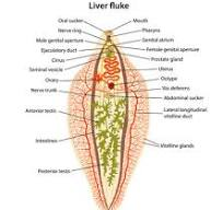
 Anatomy of P.westermani:


The four stages of the parasite are as follows- Eggs, Cercaria, Metacercaria and adult worm.

Eggs – An egg is 80 -120 x 40-60 micro M, brown in color, and resembles a coffee bean. 

Cercaria and Metacercaria are similar in size and appearance to any other Trematode worm,

Adult – the adult worm is 7 -12 mm long and 4 -6 mm wide. The outer wall is covered with scales like spines. It has two suckers like any other Trematodes and each one has both the male and female sex organs. But unlike Liver flukes Lung flukes meet as a pair and cross fertilize each other. Adult worms produce fibrous cavities in the bronchi filled with their excreta and altered blood


Life Cycle of P. westermani.

In general P. westermani needs a snail as an intermediate host but unlike the liver fluke P.westermani requires another host – crabs and crayfish for the multiplication and development to an infective form. P.westermani use any snail species that are locally available, unlike liver flukes.

************************************************************

.


Tuesday, November 28, 2023

Fasciola Hepatica


Fasciola hepatica

Liver fluke

PKGhatak, MD


Fasciloa heptica is a flatworm belongs to Trematodes. It infects humans and other ruminant mammals.

Three other species of liver flukes, namely Chonorchis sinensis, Opisthorchis vivirini and Opisthorchis felinecus have similar life cycles and modes of human infection.

In endemic areas, the liver fluke infects the hepatobiliary system and is an important risk factor for Cholangiocarcinoma (CCL). According to the WHO, people in areas of the Mekong River basin, the liver fluke infection is between 30 to 70 % of the population and the death rate from Cholangiocarcinoma is 3 %. CCL is the second most common cause of death in East Asian countries. Thailand has the highest rate of CCL, about 40 in 100,000 people, followed by China, Japan, and other East Asian countries.

The anatomy of Fasciola hepatica.


The adult worm is 3 to 8 cm in length and looks like a leaf. It has a tough outer membrane. The mouth functions as sucker and in addition has an anchoring sucker on its ventral side. It has no inner body cavity, it lives on blood of its victim. Both male and female reproductive systems are present inside the body and a worm usually lives for 9 years inside the bile duct of humans and mammals.

Life cycle:

Each fasciola releases 20,000 to 25,000 eggs daily in the bile. The eggs are plentiful in the stool. The eggs when come in contact with water, transform into embryonated eggs and seek out snails. Inside the snail, the embryos develop into elongated larvae with a long tail. This stage of the parasite is known as Miracidia. Miracidia leave snails and swim by undulating their tails to the nearby vegetation or fish, crabs and crayfish. In this second hosts the larvae transform into cysts are called Metacircarisae. Grazing animals or humans eat contaminated water crest, other vegetation and crustacea and become infected.

In human victims.

The outer wall of the cysts prevents digestion in the stomach and the cysts are propelled into the duodenum. Here the eggs are released. The larvae burrow through the intestinal wall and enter the peritoneal cavity and move to the liver. The worm grows rapidly in the liver and then enters the bile duct system and takes its final residence in the common bile duct or its branches. Within 90 days of entering the human body, the liver flukes become adult worms and begin releasing eggs in the bile.

Diseases produced by Fasciola.

Many people remain symptoms in the early stage of infection. Others develop abdominal pain, nausea and mild malaise within 4 to 7 days when the larvae are migrating from the intestinal wall to the liver. An enlarged, tender liver and abnormal liver enzymes and eosinophilia are usually detected.

This period may last about 7 days and may last for months.

In the chronic stage. Months may pass without any symptoms. Then symptoms of biliary disease begin to occur.  The common symptoms are related to gallstones, Cholangio hepatitis, hepatitis, allergic skin lesions, and the blood eosinophil count is high. are usual symptoms. In heavy infestation of the bile duct obstructive jaundice may occur but usually, a secondary infection is the usual cause of jaundice.

The most serious illness from the liver fluke infection is Cholangiocarcinoma. The bile wall mucosa is damaged by the worm, endothelial metaplasia and fibrosis become recurrent and chronic. This leads to cancer development. Patients seek medical help in a late stage when surgery is not feasible or if performed the results are not good.

Diagnosis:

Examination of stool is a simple test but diagnostic. In very early cases of suspected, asymptomatic infection the saliva, urine and blood tests reveal the presence of Fasciola antigen. Antibody test in 3 weeks post infection becomes positive.

In advanced countries like Japan, Taiwan and others, endoscopy, ERCP and MRI are available to detect parasites in the hepatic and biliary system.

Medical treatment.

Triclabendazole is very effective and requires only two oral doses.

People at risk:

A large population from Malaysia to Japan, China and Russia are at risk of infection of liver fluke infective cysts. Humans are accidental victims due of their life styles or lack of knowledge. The liver of sheep, goat and water buffalos when eaten raw or barely cooked is another source of infection besides drinking water contaminated and other food items already mentioned.


Whipple's disease

  Whipple's disease: Whipple's disease is a bacterial infection of jejunum manifest as malabsorption of fat, fat soluble vitamins...