Tuesday, November 14, 2023

Dracunculiasis / Guinea Worm Disease


Dracunculiasis

Guinea worm Disease

PKGhatak, MD


Guinea worm disease and the name of President Jimmy Crater are intertwined, to recall one, the other name pops up immediately. President Jimmy Carter single highhandedly brought the misery of African people suffering from Guinea worm disease to our consciousness.

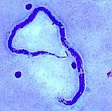
Dracunculus medinences, the parasitic nematode (roundworm) has been a curse of humanity known since 1000 BCE. The worm lives a symbiotic life with the water fleas called Copepods which are abundant in the small pools of water in Sub-Saharan Africa. Farmers and others drink water from these pools during hot summer months and ingest Copepods and become infected with the Guineas worm larva.

Life cycle of Drancunculus medinences:

Humans, domesticated dogs, and cattle are victims of the roundworm. Dogs and cattle are infected because of closeness with their masters. People develop incessant burning pain during the release of larvae by the gravid female. Farmers dip their feet in the pool in order to get some relief of the burning sensation. The worm releases thousands of larvae which are food for the Copepods.

Thirsty men drink water from the pool during summer days working in their fields under the hot sun. The outer cuticle of the copepods dissolves by the gastric juices. The released larvae enter the small intestine and begin to move along the tissue planes to the abdominal and thoracic muscles. In about 3 months the worms reach sexual maturity. There, the male and female worms mate and soon the male worm dies. The female starts her migration towards inferior extremities and finally settles underneath the skin of one leg. It takes 12 to 14 months to complete this migration. Once the right moment arises the worm breaks through the skin of the foot or lower leg, forming an ulcer, and continues to deliver larvae for 10 weeks.

The victim suffers burning pain during the entire period. There are no medications to kill the worm or any vaccine to prevent infection. The only solution, as President Carter saw, was to provide people with simple water filters and educate, educate and educate people on how to protect themselves. And he nearly achieved his goal.

Guinea worm diseases:

Nausea, vomiting, and diarrhea following drinking contaminated water are common.

Painful blisters on the legs, leg ulcers, secondary bacterial infection, draining wounds, abscesses, and gangrene of limbs are similar symptoms in all the villagers.

About 1 % fatality from septicemia.

Control of Guinea worm:

This task is a WHO project.

People at risk of Guinea worm infestation:

People living in the countries located in sub-Saharan countries from Angola to South Sudan are at risk.

The success story:

In 1986, 3.5 million people had dracunculiasis and in 2022 only 13 people were found with guinea worm disease. 17 counties out of 21 were free of guinea worm. That is a 99.99 % success rate.

Treatment :

No improvement has taken place over the age old custom of grabbing the worm with a tweezer as breaks the skin. Tie the worm to a small stick and periodically twist the stick with the worm. Slowly and bit by bit the entire I meter long and 1 to 2 mm wide Drancanculia will be out of the leg. But multiple worm infestations is the norm and so the misery of the sufferers continues.

The name Drancunculus is a misnomer, the worm does not drink blood like Dracula or another worm – Hookworm.

***********************************************************************


Monday, November 13, 2023

Elephantiasis and Tropical Pulmonary Eosipholia


Elephantiasis and Tropical Pulmonary Eosinophila.

PKGhatak,MD


Round worm infestation of people living along the coast of the Bay of Bengal causes  Filariasis. The common nematodes are Wuchereria bancrofti, Brugia malayi,and Brugia tremor. Elephantiasis is the result of Lymphatic channels obstruction by the adult nematodes producing gross deformation of the legs of the victims, resembling elephants' legs. Tropical Pulmonary Eosinophilia is produced by the type I hypersensitive reaction to microfilaria antigen which is released intermittently from the trapped microfilaria in the lung parenchyma.

Life story of the filaria worm:

All the nematodes have a similar life cycle. It consists of 5 stages, part of them in the humans and rest in mosquitoes. A wider variety of mosquitoes - Culex, Anopheles, and Aedes are vectors of human filariasis. The female mosquitoes are infected at the time of feeding on the blood of the infected patients. In the gut and thoracic muscles of the mosquitoes, the microfilaria molt twice and the 3rd stage larvae are infective microfilariae which move to the salivary apparatus of the mosquitoes and wait for the opportunity to infect humans and carry on to complete two more moltings and take up permanent residence in in the lymphatic channels, lymph nodes, and spleen of victims as adults worms. The male and female worms unite and a female gives birth to thousands of larvae every day. These microfilaria come out at night and circulate in the systemic blood, hoping to be ingested by a mosquito and to continue the life cycle.

Elephantiasis:

120 million people in a wide area of the world, spanning from India, South Asian countries, Western Pacific islands, Tropical Africa, Brazil, Haiti, Dominican Republic and Guyana are at risk of filariasis.

The adult filaria worms preferentially reside in lymph nodes of the groin and neck. The female worms remain fertile for 5 years out of 9 years of their lives. Lymphatic obstruction produces repeated Staphylococcus and fungal infections and scarring. The lymph flow disruption causes the thickening of the skin, and the skin turns hard and lumpy, and the legs become enormous in size. In W. Bancrofti infection the skin of the perineum thickens and causes disfigurement and deformities of the genitalia. The lymph edema that develops from Brugia infection spares the perineum and external genitalia.

Obstruction of the thoracic duct produces bilateral pleural effusion, the fluid is turbid due to the presence of high fat content, specially after a fatty meal. Abdominal pain and Chylous ascites result from abdominal lymphatic obstruction.

Complications: Ulceration and abscess formation, sinus formation from chronic ulcers develop in patients who are not properly cared for. Depression and loss of employment are generally common.

Tropical Pulmonary Eosinophilia (TPE):

Tropical Pulmonary Eosinophilia is much more common in India and in the adjoining countries than Elephantiasis.

TPE is a hypersensitivity eosinophilic inflammation of the respiratory organs. Nocturnal cough, wheezing, fever, loss of weight, blood stained sputum and eosinophilia, at one time thought to be Psudopulmonary eosinophilic tuberculosis. Dr. Weingarten was the first to use the term Tropical Pulmonary Eosinophilia in 1943. The eosinophil count is generally over 3,000/ml. Serum IgE over 1000 mg/dl.

Chest x-ray shows interstitial infiltrates to reticular interstitial pulmonary fibrosis.

Pathology of TPE.

An eosinophils release basic and acidic proteins, Peroxide and neurotoxic chemical in the tissues around the larvae. This weakens the microfilaria and restricts their activities. Complement activation increases opsonization and destruction of microfilaria. The Thymic Lymphocytes type II activation produces IL-4 and IL-5, filaria specific IgM, IgG and IgE and eosinophils. IL -4 potentiates inflammation and Interferon-gamma suppresses inflammation.

Diagnosis of filariasis:

Old standard diagnostic test of direct visualization of microfilaria in the nocturnal blood samples are difficult to exercise and often negative, specially in Elephantiasis. Various methods of concentration of blood for easier detection of microfilaria are practically replaced by the PCR test to detect filarial antigen and indirect ELISA antibodies are more in use at present.

Aspiration of lymph nodes and detecting of microfilaria in the fluid occasionally provide positive results. Also in some cases, microfilaria are detected in ascites and pleural fluids.

Treatment of Elephantiasis:

Adult worms are difficult to remove even by surgery. Ulcerated skin and gross deformed skin segments are removed by surgery.

Treatment of TPE:

In India, where more TPE is seen than Elephantiasis, is customary to use steroids initially for a few days then Diethylcarbamazine is used for 21 days. The results are excellent. Recurrence of TPE is due to reinfection rather than failure of treatment.

Albendazole and Ivermectin are also used but on a limited scale and case by case basis.

****************************************

Sunday, November 12, 2023

Onchocerciasis


Onchocerciasis


PKGhatak,MD.


Onchocerciasis is known as River Blindness. 31 nations in Africa, Yemen and several countries in South and Central America where Onchocerciasis is endemic.

A black fly of Simulium group, is the vector, the worm is Onchocerca volulus and humans are victims and harbor this Nematode worm. In 1915, Dr.Rodolfo Robles found the worm and linked it to eye diseases.

The life cycle of Onchocerca is almost identical to that of Loia loia worm. The areas of exceptions are the vector is black fly, the habitat of black fly is the fast running rivers and the nematode is Onchocerca volulus.

The important difference in the pathogenicity of human illness is that the microfilaria are allergenic to humans, while the adult worms are not. The microfilaria wonders around the body underneath the skin in the subcutaneous tissue and produces several different types of skin lesions. The eye diseases produced by Onchocerca are conjunctivitis, corneal scar, uveitis, glaucoma, macular edema and optic atrophy and blindness. Chronic sclerosing keratitis is the main cause of blindness. Onchocerca is the second most common cause of mass blindness. 17 million people are at risk and 800, 000 have already lost their eyesight.

Onchocerca microfilaria is in symbiotic relation with the bacteria Walachia group. The dying microfilaria releases bacterial antigen that produces sensitization and allergic reaction, and when Ivermectin produces mass killing of microfilaria the overabundance of antigen produces anaphylactic shock and deaths.

WHO has elimination programs for this illness. WHO distributes Ivermectin tablets to the participating nations. And has already eliminated it from several countries in South America, Columbia being the first. Ivermectin kills the microfilaria but not the adult worm, as a result, Ivermectin had to be repeated every 6 to 12 months intervals.

Serological tests and PCR tests are available for diagnosis but visualization of microfilaria in the blood is the mainstay locally.

******************************************************

Loiasis


Loiasis

PKGhatak,MD.


Loiasis is a human parasitic disease produced by a nematode - Loa loa. It belongs to the Filaria group of round worm. Loiasis is endemic in 11 counties of Central and West Africa. In the rainy season, the disease activity is maximum which coincides with the breeding season of Chrysops fly. The fly is a deer fly, locally known as Mango fly or Mangrove fly.

20 million people are at risk of Loiasis and annual incidence is 3 to 10 million. The illness was thought to be benign, now, that is questioned by the finding that the mortality reaches 14 % in local areas where parasitemia is unusually high – over 30,000 microfilaria /ml of blood. Another worm disease, Onchocerciasis, is also endemic in several countries in the very area; and the treatment of Onchocercia by Ivermectin leads to the development of encephalitis and deaths of unsuspected patients having both these two diseases simultaneously.

The first case of Loiasis was reported from San Domingo in 1770, by a French surgeon Mongin who saw the Loa loa worm in the eye of a woman but was unsuccessful in removing it.


The Chrysops fly, is an unusually aggressive and determined fly. It lacerates the skin of its victim with its sharp saw like proboscis and then licks the blood from the wound. The bites are quite painful and attempts to drive the fly away, lead to more bites by the same determined fly who must have a bloody meal for her egg development.

The life cycle of Loa loa:

In the gut of the fly blood containing microfilaria undergoes development to a 3rd stage of infective microfilaria and in 10 days the microfilaria moves to the proboscis of the fly and is ready to begin its life in humans.

The skin wound and the draining lymph nodes swell and become tender. In 6 months to a year, the worm becomes an adult. The adult worm moves around in the subcutaneous tissue and the sexually mature worms unite and the female worm gives birth to about 20,000 microfilaria every day. The microfilaria move into the pulmonary circulation, and from the lungs, they enter the systemic circulation every day during 10 AM and 3 PM. There they wait for the fly bite and begin their lives inside the fly. Then the cycle repeats. An adult worn can live unto 15 years.

Symptoms produced by the parasite:

Both the adult worm and microfilaria are allergenic to humans.

Most victims, however, are asymptomatic. Generalized itching, urticaria, recurrent muscle and joint pain and tender lumps on the skin over the underlying worm develop. These lumps are common around knees, ankles and other joints and are called Calabar swellings. The migrating adult worm in the subconjunctiva of the eye and eyelids is a characteristic feature of Loiasis and is an Africa Eye Worm Disease. Adult worm in the eyes occasionally enters the vitreous humor of the eye and secondary infection may lead to blindness. The risk of encephalitis when Invective is given is not to be underestimated.

Diagnosis requires visualization of Microfilaria in the blood, collected during daytime and blood smears are stained with Giemsa stain. Serological tests and PCR antigen recognition tests are neither locally available or standardized.


Treatment: Three medications namely, Diethylcarbamazine, Albendazole, Ivermectin are used in the elimination of both the adult worms and microfilaria. The selection of a particular drug based on the the microfilaria load, patient's symptoms and allergic history.

**************************************

Whipple's disease

  Whipple's disease: Whipple's disease is a bacterial infection of jejunum manifest as malabsorption of fat, fat soluble vitamins...