Friday, October 21, 2022

What Cerebellum Does


What Cerebellum Does

PKGhatak, MD


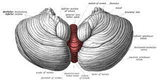
The human cerebellum occupies a small space at the back of the head, just above the neck, and it is a part of the hindbrain. Sensory information from joints, tendons and soft tissues from the limbs and truck travel to the cerebellum. The motor stimuli coming from the cerebrum motor cortex, make connections with nuclei of the cerebellum before reaching the lower motor nuclei of the spinal nerves and cranial nerves. This interconnection of the motor cortex with the cerebellum results in a smooth sustained movement of the body, able to maintain the head position in space and in relation to the rest of the body and keep balance without falling. And that much knowledge people had till 1990.


Now the cerebellum is a subject of intense research for a variety of reasons.

Research in developmental biology documented three separate stages of growth of the cerebellum in humans. As the forebrain is added cerebral cortex, which has increased the mental faculties, knowledge gained about self, mind, higher state of consciousness, language and development of culture and made humans as humans. But the cerebellum did not lag behind.

The growth of the cerebellum is more spectacular than the cerebral cortex. Cerebellum weighs 150 grams and contains 70 billion neurons, whereas the cerebral cortex weighs 1300 grams but contains only 16 billion neurons. The surface area of the cerebellar cortex is 1500 square centimes, which is about 80 % of the surface area of the cerebral cortex. The cerebellar cortical folds are arranged like pages of a book - lots of surface area in a small space.


Compared with the cerebral cortex the layout of neurons and their connections in the cerebellum is much simpler and more uniform. Cerebellum receives input and sends out output to the same side of the body that is also a contrast with the cerebrum which controls the opposite side of the body. The central part of the cerebellum is old, but the lateral parts, the cerebellar hemispheres, kept pace with the growth of the cerebrum.

Cerebellum is the site of the formation and storage of working memory and the integration of automated sequences of thoughts and actions. This results in developing behavioral patterns, speech, dexterity of complex movements, a skill needed for technological advances and language development.

In association with the cerebrum, it forms kinetic memory, plans coordinated muscle movement patterns, retains reflex memory for self preservation and executes normal speech.

Cerebellum with two way communication with the thalamus and basal ganglia and receive direct input from the spinocerebellar tract and maintains balance and a stable head position both in states of body movements and at rest.

Vestibular sensory information is closely monitored by the cerebellum, that information is used to control conjugate movements of eyes, prevent double vision and reflex eye movements.

Rapid eye movement (REM) sleep includes Implicit memory involving perception and body movements. Implicit memory uses cerebellar retained memories.

*********************************


Tuesday, October 18, 2022

Science of Skin Color


Science of Human skin color

PKGhatak, MD


The skin is one of the body's large organs. The color of the skin prevents or minimizes the harmful effects of Ultra Violet (UV) light of the sun.

The darker the shade of color the lesser amount of UV light penetrates the skin and decreases Vitamin D synthesis, the reverse is true for the lighter shades of color. It is said that women have lighter skin color because more vitamin D is needed for childbearing.

Not every part of the skin of an individual has the same shade of color and variability depends on the degree of exposure to sunlight. The sole of the feet and palms of the hands have different thicknesses of skin and a much lighter color, and that lighter shade is most noticeable in very dark colored people.

Theory of color variation in humans.

Scientific communities believe that the human race originated in equatorial Africa. Having dark skin gave humans an advantage over the other predators to hunt for a longer time each day. Because the black skin color developed as the coarse hairs were replaced by fine body hair. This combination allowed the body from overheating. That advantage led to a population boom and subsequent migration of people to other lands. When people settled in the subtropical area, their dark skin turned to a disadvantage; the lack of calcium produced weaker siblings, and most did not survive beyond their early childhood. A genetic mutation made the color of the skin lighter shade which allowed more UV light to penetrate the skin and generate more vitamin D. and the population bounced back.

The so called white colored skin is the color of the pale white color of the subcutaneous tissue shining through the light-colored skin.

What are the color pigments of human skin:

Melanin is the primary color and minor color contributions come from hemoglobin carotenoids and oxyhemoglobin.

Perception of color:

The combination of colors of melanin, oxyhemoglobin (proportional to blood flow) and carcinoids produce the final color perceived by the observer's retina.

Melanin:

Melanin is a group of chemicals very closely resemble each other in molecular structure. The skin Melanin in humans is Eumelanin and Pheomelanin. Neuromelanin is present in the brain.

Eumelanin and Pheomelanin are the same chemical but differ only in the number of polymer bonds in them.

Two colors of melanin:

Eumelanin is a dark pigment, has a range from brown to black, and Pheomelanin has a shade between yellow to pink color. The pink color of the lips is due to pheomelanin.


Eumelanin synthesis.

Amino acid Tyrosine is converted to Dihydrophlylalamine (DOPA) by the enzyme Tyrosinehydrolase and in the presence of cofactor Tetrahydrobiopterin. DOPA is converted to Dopaquinone by Tyrosinase and then undergoes several steps of polymerization to form eumelanin or pheomelanin.

Site of melanin production:

Melanin is produced by the Melanocytes present in the basal layer of the skin. The pigment granules are nicely packed in an envelope and are called melanosomes. Melanosomes are carried along the long arm of the melanocyte and the pigment packets are deposited within the Granular cells of the keratinocyte layer. One melanocyte and 36 granular cells and one Langerhans cell make one keratinocyte unit. It is estimated one melanocyte can supply 500 granular cells with melanosomes in its lifetime.

Stimuli for melanin formation:

UV radiation stimulates the synthesis of melanin. A hormone, produced by the Pars Intermedia of the Pituitary gland, Pro-Opiomelanocortin, is turned into Alpha Menaocytotropic hormone (alpha MSH). UV light also increases ACTH (adrenocorticotropic hormone) secretion. MSH and ACTH increase melanin production. Androgen also influences melanin deposit of pigment in the form of male patterns.

Genes controlling Melanin.

In humans chromosome 11 is the primary chromosome and three additional chromosomes, 17, 6 and 13 have also influence melanin. The location of the genes on these chromosomes are 11q24.2, 17q23.2, 6q25.1 and 13q33.2.

Defective genes and abnormal skin color:

1. Albinism.  It is a Hereditary disease due to an inheritance of defective genes. Skin, hair, retina of the eyes are devoid of melanin and is known as Oculocutaneous albinism (OCA). It is inherited as an autosomal recessive trait. At present 7 varieties of OCA have been identified.

2. Hermansky-Pudlak Syndrome.  Hermansky-Pudlak patients lack skin pigment and in addition, have a tendency to bruise easily and bleed spontaneously. Patients have additional symptoms which may involve GI, Pulmonary and CNS systems. So far 11 gene mutations have been identified. This abnormality is also inherited as an autosomal recessive pattern.

3. Chediak-Higashi syndrome.  It is an autosomal recessive hereditary disorder consists of partial OCA, platelet disorder, immune deficiency due to abnormal Lysosomes and in addition to hematophagy lymphohistiocytosis syndrome

4. There are many other syndromes due to inherited defective genes. Symptoms involve various organ systems and skin color. Just to name a few – Acanthosis nigricans, Xeroderma pigmentosa, Treitz syndrome, Histiocytosis lymphadenopathy plus syndrome, Lengius syndrome, Cruzion syndrome, Incontigentia pigmenti, etc.

5. Loss of melanocytes.  Skin abrasions may peel off the basal layer of skin containing the Melanocytes, when skin lesion heals the area is devoid of skin color.

5. Autoimmune diseases. Some acquired autoimmune diseases may attack melanocytes producing pigment voided spaces.

6. Vitiligo. It is suspected to be an auto-immune disease. The color free areas are patchy and can be any spot on the skin. No genetic link has so far been documented.

7. Localized dark pigmentation on skin:

Freckles, moles, Mongolian spots on buttocks, Caffe au lait spots, and Birthmarks are examples of uneven distribution of melanin in keratinocytes. Some of these spots are transient, and others disappear and reappear later in life. Caffe au lait spots, if multiple, are associated with neurofibromatosis.

All these are benign conditions unless one starts to grow in size and becomes darker or bleeds.

 8. Melasma. Symmetrical dark patches on the face due to estrogen – progesterone concentration variation, as seen in pregnancy and menopause. Melasma has no health consequences and is important from the cosmetic point of view.

Carotenoids:

Carotenoids are Terpenoids having formula C40H56. Carotenoids are plant pigments and humans must depend on plant sources for carotenoid supply. Yellow, orange, or red vegetables and fruits contain carotenoids. They are fat soluble chemicals; animal fat is also a source like Salmon. Cooking shredded vegetables in oil increases the bioavailability of carotenoids. In humans these molecules are antioxidants, boost immunity and provitamin A. Vitamin A is an essential vitamin for vision.

An essential organ:

The skin is an essential organ. The skin protects the body from the physical mechanical and thermal injury, prevents harmful agents from getting inside, maintains body temperature, synthesizes vitamin D, performs immune functions and the sensory receptors of pain, touch, temperature and pressure are located in the skin. Melanin in the skin protects tissues from the radiation damage of the sun.

All 8 billion people:

The color of the skin comes from one pigment - Melanin. All 8 billion people on earth have the same melanin. Color variation of the skin is due to the variation of melanin content in the skin cells.

People seeking advantage and try to divide human families into many color groups, like India did through the caste system. Businesses interested in selling their products claim about 110 tones of skin colors and have marketed matching colored products for sale.

Science has steadfastly maintained the scientific data which anyone can verify and as it stands today, all humans carry the same skin pigment melanin.

The difference in skin color is only in the view of the observer.

******************************
Sunday, October 16, 2022

Speech Disorder


Speech disorder

PKGhatak, MD


Speech is defined as the ability to express thoughts and feelings by articulated sounds. Language is in the same domain as speech but not the same. Language is the medium of communication with said words and symbols. 

1. Speech disorder in children is primarily developmental and congenital, after that comes others.

2. Developmental delay presents as uncoordinated brain function manifest as Stuttering. 

3. The next group comprises Autism, dyslexia and cerebral palsy.

4. Birth defects such as tongue-tie, defects of the floor of the mouth, hare-lip and cleft palate and occiput occipito-facial abnormality. Many of them are surgically correctable.

 5. Apraxia and dysarthria are apparently similar defects but in apraxia, the brain fails to formulate a motor plan to supply nerve impulses to muscles of the larynx and vocal cords; in Dysarthria the muscles of the mouth and tongue fail to move properly due to mechanical and /or coordination abnormalities and fails to produce a normal speech.

 6. Congenital deafness or onset of early childhood deafness, leads to absent speech known as muteness. However, the mute babies did cry at the time of birth proving that absent speech in these babies comes from deafness.

Speech disorder in adults.

Speech problems in adults are always acquired after having normal speech for years.

There are three categories of this disorder - I. Transient and reversible. II. Secondary. III. Primary.

 I. Transient:

Alcohol intoxication is the most common cause of stuttering, fumbling for words and inappropriate answers. Opioid addiction and use of recreational drugs, tranquilizers, sleeping pills are additional causes.

II. Secondary:

Multiple sclerosis (MS) produces various neurological deficits including speech. One special feature of MS is the exacerbation and remission of symptoms. ALS, Ascending paralysis, Myasthenia gravis, and myopathy of various types. and diseases involving the larynx, vocal cords and paralysis of the recurrent laryngeal nerve are further examples of speech defects.

III. Primary:

The brain is the center for speech formation (ideation), generation of motor signals and transmission of information to muscles involved in the execution and articulation of spoken words. Any disruption of the flow of information to the effector organ/tissues will adversely affect speech. Some of the pathological causes are interruption of blood supply, pressure from space occupying lesions, inflammation and infection of the brain, and traumatic injury to speech centers.

Ideation:   One can easily recall teachers telling “engage your brain before you blurred out words”. That is the ideation – the speaker has instant knowledge of what he is going to say in a conversation or at the podium before a lecture. Ideation is the result of coordinated activities of many brain centers, including visual, auditory, and memory centers which are located in the different areas of the cerebral cortex.

Motor function:   Cerebral motor center is located in the Pyramidal cortex of the brain. The axons from pyramidal neurons make synaptic connections to the lower motor neuron of the motor nuclei of cranial nerves and anterior horn cells of the spinal cord. Nerve impulses are carried to the muscles of the larynx, vocal cords, throat, lips, mouth, tongue, diaphragm and respiratory muscles of the chest wall transmitted by the axons of the lower motor neurons.

Monitoring and modification of speech are active processes and require communication via synaptic relays between various brain centers.

Localization of lesion:

At an earlier time, every speech abnormality was documented by neurological examination and then verified by autopsy. The medical practice of that time required memorizing named syndromes to identify each speech defect. Now neuroimaging by functional MRI and CT, with and without contrast, have made the diagnosis more refined and accurate at the same time names attached to lesions are not required to be remembered.

Types of speech abnormalities in adults.

 1. Broca's Aphasia.

Broca's aphasia is both a language and word disorder due to damage to Broca's area of the brain. Broca's area is a special speech center located in the left frontal lobe of the cerebral cortex. It is situated in the depth of the main horizontal fissure dividing the cortex into frontal and middle lobes and partially covered by the tip of the left temporal lobe. Anatomically, it is located in the inferior frontal gyrus. Blood supply of the Broca's area comes from the branch of the middle cerebral artery. Interruption of the blood supply produces a language and speech disorder known as Broca's Aphasia.

In Broca's Aphasia, the patient retains the capacity of Ideation but loses languages due to partial loss of capacity to construct either the spoken or written language. Broca's aphasia has two components – Expressive aphasia and Receptive aphasia. In expressive aphasia, the patient is able to say important words but leaves out prepositions, pronouns and functional words. Speech becomes piecemeal, comes out in sprats and stops, says wrong words, missing the word orders. This type of speech is known as Telegraphic speech (at one time the telegram was the fastest mode of communication but expensive and the charges were based on the number of words to be transmitted. That leads to cutting out words). In Broca's aphasia, the patient is cognizant of his disability and becomes visibly frustrated in his attempt to find his speech back.

In Receptive aphasia, the patient is able to speak in grammatical sentences but lacks semantic significance and exhibits trouble in comprehension.

Thrombosis or emboli obstruction of the Left middle cerebral artery produces Right sided paralysis including the right lower half of the face and loss of sensation from the right side of the body and loss of right visual field and Broca's aphasia.

 2. Wernicke's area. Werneck's area is located in the parietal cortex next to the lateral sulcus at the junction of the parietal and occipital lobes. In this area, the visual and auditory words are comprehended and an area for selecting words and phrases to construct a meaningful sentence. Patients with damaged Wernicke's area, from alcoholism and other diseases, produce articulated sentences but many lack any meaning. And some patients may speak fluently and are not aware they are uttering meaningless words. Alcoholism may lead to Korsakoff psychosis. During an episode of Korsakoff psychosis, the speaks freely but confabulates and tells amazing stories.


3. Aphonia:

A lack of ability to produce any sound is known as aphonia. Direct injury to vocal cords, tracheostomy and bilateral recurrent laryngeal nerve damage can produce aphonia. And in addition, there are instances when a person is about to give an important talk and gets up to the podium and then is unable to make any sound. Some psychological or emotional neuronal activities overwhelm the natural order of speech making process.

4. Apraxia and Dysarthria are common in stroke and other lesions of the brain involving speech forming areas.

*************************

Leprosy

                                                  Leprosy                                              P.K.Ghatak, MD It is the perception ...