Wednesday, December 6, 2023

Ascaris lumbricoides


Ascaris lumbricoides infestation

PKGhatak, MD.


Ascaris lumbricoides is a foot long intestinal parasite. The human infection has been known since the ancient time. The first written record of human infection was given by Linnaeus in 1758.

Ascaris lumbricoides is a roundworm, the sexes are separate and the female worm is larger in size. Adult worms live in the small intestine of the human. Ascaris also infects pigs, monkeys and other animals. A female ascaris lives one to two years and can lay 200,000 eggs a day. The eggs are mostly fertile but unfertilized eggs are also present in the stool of the victims. If a female worm does not find a male worm, it moves around, and crawls into the throat. If enters the larynx it causes respiratory distress and a bout of violent cough till it is dislodged. It may also come out of the nostrils.


People at risk:

The WHO says about 1 billion people are at risk of infection. The infective state of the worm is the fertilized 3rd stage eggs or embryonated eggs. Unlike any other parasite, ascaris eggs are well protected by their thick outer shell and can live for 3 years in the soil. People unknowingly swallow eggs carried on their fingers due to their unhygienic habits or ingestion of contaminated water and food. Humans and farm animals act as reservoirs of ascaris.


Life history of Ascaris:

The fertilized eggs are deposited in the soil, either by spraying the human waste on the agricultural land as fertilizer or, by people defecate out in the open. The eggs undergo further development in the soil and become embryonated eggs in 15 -18 days.

Children playing in the darts carry soil and eggs on their fingers or, under their nails. The eggs enter the mouth when children eat with their fingers. The same way adults are infected or, contaminated food and drinks by the food handlers.

Inside the duodenum, the larvae emerge. The larvae move to the lungs for further development. The larvae reach the lungs by way of the Portal vein and finally in the pulmonary circulation. In the lungs, the larvae become juvenile worms in about 15 days. The juvenile worms journey back to the small intestine by way of trachea and are coughed up and swallowed by the victim. Back in the intestine, for the second time, they reach maturity and mate with the opposite sex and begin laying eggs in 3 months from the time of infection.

Researchers are not sure why ascaries larvae must travel to the lungs. Several reasons are put forward but still remain unknown.


 Human disease.

During initial infection. Most of the victims are unaware of infection and remain symptom free. Some develop abdominal cramps and diarrhea.

During migration to the lungs. This period is generally asymptomatic.

The period of stay in the lungs. Symptoms are mostly due to the physical presence of foreign bodies in the airways and all related pulmonary complications, secondary infections and allergic asthma with eosinophilia.

During migration from the lungs to the small intestine. Cough and the horror of live worms being coughed up.

Small intestinal stay. Though the worms do not suck blood like hookworm never the less,  they steal nutrients from the children and children become stunted and malnourished. Motile worms in the intestine can enter the bile duct, gall bladder, and appendix causing acute obstruction and infection. A heavy parasitic load in the small intestine causes bowel obstruction.

During adult female searching for a male worm. When the worm reaches the pharynx and sometimes enters the larynx cough and respiratory distress develop.


Diagnosis:


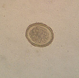
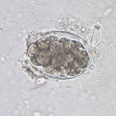
Fertilized ascaris eggs are diagnostic. Unfertilized eggs sink while fertilized eggs float in water. Each egg is oval to round in shape, measuring 75 x 50 micrometers, and has a thick mamillated outer shell and is usually stained brown by bile.

Adult worms are also diagnostic. The worms are long, slender, cylindrical, unsegmented and colored light yellow. The mouth end is surrounded by 3 lips. The male worm is 30 cm long and 4 mm in diameter, and has a curved tail end. A female measures 40 cm ( over a foot long) X 6 mm. The genital aperture is in the upper third of the body and 2/3 of the body contains sacs containing 25 million eggs.


Treatment. Albendazole and Mebendazole are effective in killing the adult worms. A daily dose for 3 days is recommended for cure. Reinfections are common in endemic areas.


Monday, December 4, 2023

Hookworm


Hookworm.

PKGhatak, MD.

The name Hookworm is given to this roundworm because the worm has anchoring teeth or, hard plated in the mouth to latch onto the interstitial wall of its victims. An adult worm is 8 mm long and the female is 11 mm. The worms are round in shape, have a long thin unsegmented body with tapering ends, and are pale creamy color. Each worm carries both sex organs. The outer wall is made of a tough cuticle

Human diseases produced by hookworm:

  1. Iron deficiency anemia and protein malnutrition

  2. Allergic skin lesions and eosinophilia. Cutaneous larva migrants

  3. Eosinophilic enteritis.

The life cycle of hookworm.

Human excreta contaminates the soil where hookworm infection is common. Children playing bare feet on the fields or farmers working on their fields, come in contact with the infective stage of hookworm larva. It takes only 5 minutes for the larvae to find the tiny hair openings and or penetrate the skin. In the dermis, the larvae begin migrating to locate the capillaries or venules and enter. The blood carries the larvae to the right side of the heart and then into the pulmonary alveolar capillaries. This part of the migration takes 10 days, The larvae penetrate into the alveoli and enter the airways. Ultimately, the larvae enter into the major airways and are coughed up and swallowed. In the stomach, they are protected by the outer tough cuticle. Once inside the small intestine, the larva molts twice and develops a buccal capsule with teeth and a muscular esophagus. The larvae attach to the mucous membrane of the intestine from the lower duodenum and upper ilium. The larvae secrete tissue resolving enzymes which dissolve tissue and expose the blood vessels. The larvae bite and remain attached to the blood vessels of the intestine with their teeth or buccal plates. In humans, the parasites do not multiply.

The larvae quickly grow and attain maturity in a month and after mating, begin to release eggs. A female can release 30,000 eggs daily. Adult Nicator worm lives up to 5 years and Ancylostoma species for one year only.

In the soil.

The eggs release the immature Rhabdotiform larvae in a day or two, The larvae feed on the excreta and molt twice. In 7 days rhabdotiform larvae become the infective Filariform larvae. The filariform larva has a short life outside and dies if unable to find a victim within 3 weeks.


;


Diagnosis:


The stool test for eggs is commonly done for the diagnosis of hookworm infection. The eggs with multiple larvae are diagnostic. Each egg measures 60 x 40 micrometers. The usual practice is to release the eggs from the fecal matter by placing a portion of the stool sample in a glass jar diluted with water and agitated it thoroughly. The eggs float on the top of the water and are collected by merely touching the top of the water with a glass slide.

PCR tests are available but are hardly utilized.

When the larvae are in migration and until the adult worm starts producing eggs, the stool tests will be negative. The PCR tests are needed for confirmation,

Treatment.

Albendazole is effective and requires a daily dose for 3 days or just one heavy dose.

Iron and vitamin supplements are also required.

Infection of hookworm does not provide protection from future infection. Vaccines are developed but not commercially available.

Epidemiology.

According to a 2010 WHO report, about 120 million people had hookworm infection. The countries where most cases are seen are poor - spanning from Sub Sahara Africa to South Asia, East Asia and the Americas.

Species infective to humans.

Necator americanus infestation is the most common in humans, taking the world as a whole, followed by Ancylostoma duodenale.

Other hookworm species infective to humans are Ancylostoma ceylanicum,

Ancylostoma caninum

Ancylostoma braziliense.

A. barziliense commonly produce eosinophila with creeping skin lesions called Cutaneous larva migrants.

Besides the common mode of infection, some species can infect humans via contaminated drinking water and breast milk in children. Ancylostoma craninum usually remains dormant in the skeletal muscles and produces eosinophilic dermatitis, bronchitis and asthma. Then move to the intestine to propagate.

Amount of blood loss and anemia.

It is reported that each adult Necator worm consumes 0.03 ml of blood and 0.2 ml of blood by Ancylostoma species. In addition, blood is also lost in the stool. On average, in a heavy infestation, humans loose 1 ml of blood per day per adult worm. In general, many dozens of worms live in the intestine at the same time. A heavy infestation is determined by the presence of over 4,000 eggs /gram of feces.

It does not take much time for the children to become severely anemic and the hemoglobin levels fall to 4- 5 grams/dl.

An adult worm lives 1 to 5 years based on the species. An infestation of hookworm does not produce immunity against future infections.

Vaccines are known to protect against fresh infections in people but are not utilized and not produced commercially.

**************************************

Whipple's disease

  Whipple's disease: Whipple's disease is a bacterial infection of jejunum manifest as malabsorption of fat, fat soluble vitamins...