Immune cells of the CNS
PKGhatak, MD
Two kinds of cells are present in the Central Nervous System (CNS). Nerve cells, and Supporting cells. The general term for the supporting cells is Glial cells. The glial cells consist of Astrocytes, Microglia, Schwann cells, Oligodendrocytes and Ependymal cells. Outside the CNS, the supporting cells are called Satellite cells, and these cells are present in the nerve ganglia.

A neuron upon receiving information generates nerve stimuli and a stimulus is either transmitted to other neurons via synaptic connections or to an effector organ for action. The Glial cells surround the neurons, supply nutrition, produce insulation, Myelin, remove waste products, maintain immune surveillance, initiate and maintain inflammatory reactions till the infection or disease is controlled. These cells also help in the repair and regeneration of supporting cells inside the CNS.
The immune cells of the nerve ganglia are called Satellite cells. Satellite cells perform similar functions as microglia. Nerve ganglia are found in the posterior root ganglia and autonomic nerve ganglia.
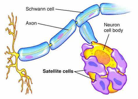
Peripheral ganglion and Satellite cells
Microglia.
Microglial cells are immune cells of the CNS and perform functions similar to Dendritic cells and macrophages of the other tissues.
Unlike any immune cells of the rest of the body, microglia can function as proinflammatory and anti-inflammatory cells depending on the circumstances.
Microglia cells are widely distributed throughout the brain and spinal cord. These cells are very sensitive to pathological changes in the CNS and changes in the CSF potassium concentration. They are capable of quickly detecting invading pathogens in the CNS. The blood-brain barrier prevents antibodies and most other protein molecules from entering the brain. Microglia manufacture antibodies and other important products that are unable to enter the CNS.
Origin of Microglial.
Microglia develop from Yolk Sac cells which migrate in very early fetal development to the neuroectoderm and as the CNS begins to take shape, the microglia differentiate as immune cells by acquiring properties like both dendritic cells and macrophages of hemopoietic cell origin.
Change in microglia morphology.
A. Microglia change shape according to location and require them to perform specific functions.
1. Resting state.
The cell body of microglia, at a resting state, is small and has multiple arms like tentacles. The cell body does not move, but the tentacles move in all directions to detect any foreign agents. The resting cells do not release cytokines.
2. Reactive stage.
In the reactive state, most of the tentacles are withdrawn. Cells undergo rapid proliferation and the cell population grows to a large number. The cells release proinflammatory cytokines. These changes are triggered in response to increased potassium concentration (from dead neurons), TNF (tissue necrosis factor) and membrane lipopolysaccharide.
3. Amoeboid state.
The microglia move all around and phagocytize dead cells, foreign agents and cellular debris and clear the field for regeneration to take place.
4. Phagocytic state.
The cell bodies are engorged with phagocytic material and the cells secrete cytokines to attract other glial cells and transform themselves into T-cell like functional states and begin to generate antibodies.
5. Glitter cells.
Microglia with a heavy load of ingested substance become inactive, stationary and clump together. The presence of glitter cells indicates a post-inflammatory state.
6. Juxtavascular cells.
The cell bodies lie in contact with the basal lamina of blood vessels and secrete MHC II (major histocompatibility complex molecules) I & II, etc.
Pro-inflammatory and post infection repair and replacement of cell population are two arms of microglia. For repair to begin, the microglia release cytokines designated for such functions. Other glial cells migrate to the site and the repair takes place.
In overwhelming infection of the CNS, the junction between endothelial cells of CNS capillaries weakens and the blood-brain barrier is broken. The immune cells of blood and antibodies, if present, take over the immune function. And after the completion of infection, microglia again take over the immune function.
Microglial immune activities have taken on a new importance because researchers speculate it may hold a clue to prevent or slow down neurogenerative diseases. A number of genes have already been identified and the interest at present is whether these genes could be modified to achieve the desired effects.
**************************************************
.

No comments:
Post a Comment