Saturday, November 11, 2023

Myiasis


Myiasis

PKGhatak,MD.


Several species of flies seek living animals, including humans, for lying eggs, so that the newly hatched maggots will have instant source of food to feed on and grow rapidly. Maggot infestation of humans was first described by Frederick William Hope of Jamaica in 1840 when he described a man with maggots eating away his flesh.

The term Myiasis is reversed for invasion by maggots of otherwise normal individuals. Maggots deliberately applied for cleaning and debridement of chronic wounds are not included under this term.

An outline of the life of maggots: Two species of flies - Blowfly and Housefly, seek out humans to lay eggs on any exposed part of the body, from the scalp to the sole of feet. Each female fly lays about 100 to 300 eggs. Depending on the outside temperature and the type of fly, these eggs hatch in 8 to 12 hours and immediately borrow under the skin and begin feeding and growing. In about 50 to 60 hours they are fully grown and all the maggots are similar in size and maturity, They stop feeding and fall off the body and pupate. They later emerge as adult flies.

These three species of fly are responsible for most Myiasis – Botfly, Tumbufly and Screwworm fly.

Myiasis is described under several categories based on the location of the maggots and the symptoms they produce. These categories are Cutaneous, Creeping, Wound, Body cavities, and Accidental Myiasis.

Types of Myiasis.

Cutaneous Myiasis: The back of the head and skin of the back are the favorite place of this fly to lay eggs. Growing maggots produce bumps on the skin. These lesions are painful and itchy. On close examination, a hole is visible on the top of a bump, through which the maggot gets its air to breathe. These wholes are used to pull maggots out with a pair of forceps.

Creeping Myiasis: Humans are an accidental host for this parasitic maggot. Maggots can not develop in humans, so the maggots move around underneath the skin and give the victims a creepy sensation. Surgical removal of maggots is necessary.

Body cavity Myiasis: The fly deliberately targets ear canals, nose, mouth and eyes. Growing maggots produce secondary infection and usually lead to serious respiratory, gastrointestinal and neurological complications. From the roof of the nose or eye sockets the maggots penetrate the base of the brain. Meningitis, encephalitis and brain abscesses are usual complications. Surgical removal of maggots is often required.

Wound Myiasis: Open wounds are within easy reach of flies. Maggots eat away dead and dying tissue and, in general, do not invade normal living tissues.

Accidental Myiasis: Farmers in Africa and South American countries, at times have to drink water from the nearby streams, which are usually contaminated with fly eggs. These eggs hatch in the stomach of the victims. Growing maggots produce nausea, vomiting and diarrhea. Maggots die due to low oxygen in the G-I tract or are removed by administration of medication and purgation.

In the USA Myiasis is not seen in the local population.

South American countries and tropical Africa are endemic to Myiasis.


**********************************


Friday, November 10, 2023

Rickettsiapox


Rickettsiapox


PKGhatak,MD


Rickettsiapox is a milder form of systemic disease, the predominant lesion is the skin infection produced  by Rickettsia akari. This bacterium is a parasite of hose mouse mite. In overcrowded squalid apartment building infested with mice harboring R. akari as parasite.

A cluster of cases in New Your City in 1946 led to the discovery of the mite and bacterium by an amateur entomologist Charles Pomerantz. Earlier investigators considered rickettsiapox as modified chicken pox.

The mite bite produces a red papule, which turns into a vesicle and heals leaving a black eschar. A week later patient develops a sudden onset of chills, fever, headaches, diffuse body aches and pain and photophobia. After 2 to 4 days the entire body is covered with red maculopapular eruptions, soon they turn into vesicles. The skin lesions heal in 10 days and the scabs are shed.

Skin biopsy when treated with conjugated antirickettsia globulin can detect rickettsia antigen. PCR tests are also developed. The 4-fold rise in antibody titer is a standard initial diagnostic test. Antibiotic Doxycycline is given for 7 days.

The disease is milder in comparison with other forms of rickettsiosis and is usually self -limited.

Rickettsiapox is endemic in Balkan States, Korea, Ukraine, South Africa and major US cities. The average incidence of rickettsiapox is about 30 /year in the USA.

***************************************************************

Epidemic Typhus


Rickettsia prowazekii.

PKGhatak,MD


Rickettsia genera have many pathogenic species and out of them, Rickettsia prowazekii is the deadliest for humans. During WWI, a score of soldiers fought and died in the trenches and another score died in the field hospitals because the soldiers were infested by a parasite, body lice loaded with R. prowazekii.

US army developed a vaccine, using inactivated R. prowazekii, that saved many solders on the US side but the vaccine was abandoned because of toxicity. No new or effective vaccine is produced since then.


The disease is known as Epidemic Typhus. Typhus means hazy – the term aptly describes the mental conditions of the soldiers. There are two other forms of the same illness are present, and fortunately, both of them produce milder symptoms and fewer fatalities, The initial infection is followed by several months or years of normal health then R. prowazekii, which had remained dormant in the lymphatic tissue, reemerge and produce illness. The disease is called Brill Zinsser Disease. In the southern states of the USA, flying squirrels are harbors of R. prowazekii and humans are accidental victims.

Body lice are infected by sucking patients blood. R. prowazekii multiplies in the gut of the louse and then bursts open. The bacteria remain alive in the dead lice and in the feces of lice. As patients itch, the bite sites get smeared with the bacteria. Dried feces along with the bacteria can float in the air and infect people as they inhale the contaminated air. This characteristic of R. prowazekii leads to certain countries use this bacteria as a terrorist weapon. Consequently, the US government prohibited the culture of R. prowazekii in laboratories. Only in government facilities, under strict conditions, culture are permitted.

The incubation period is 10 to 14 days. The skin bite sites and the local lymph nodes may become tender. Sudden fever, conjunctivitis, headaches and mental confusion are usual initial symptoms. Deafness due to the 8th cranial nerve lesion, macular skin rashes which spread centrifugally from the axilla but spare the palms and soles and later become confluent and hemorrhagic, and enlarged spleen are characteristic features.

The pathology of the Epidemic typhus is vasculitis. This results in multiorgan infection. Renal failure, pneumonia, myocarditis, gangrene of extremities, encephalitis, and death.

In epidemics the diagnosis is based on clinical grounds and treatment is started immediately without waiting for any laboratory test results. The choice of antibiotic is Doxycline but Chloramphenicol or Riphampine can be used as a substitute in special circumstances.

In Brill Zinsser disease the presence of serum IgG antibodies are common. In Endemic typhus, IgM antibodies appear in the blood in 5 to 12 days. The PCR test is rarely used, because as of now, 7 different genotypes are in circulation.

The mortality is 10%. Morbidity is significant, with most having an amputation or organ impairment.

Recent endemics:

Isolated Epidemic Typhus occurs in Siberia, But Russia experienced an local outbreak of Epidemic Typhus. Peru and Burundi had also Epidemic Typhus recently. The endemic areas of Epidemic Typhus are Central and Northeast Africa, central and South America

****************************************************


Rocky Mountain Spotted Fever

  Rocky Mountain Spotted Fever

  P K Ghatak, MD


Rocky Mountain spotty fever is a catchy name but unfortunately, this disease is neither that common in the Rocky Mountain area, nor, the only spotty fever. This illness is caused by an unusual bacterium, Rickettsia rickettsii; and transmitted to humans by a dog tick.


Skin rashes of various types are common findings in diseases transmitted by tick bite, flea bite, and bites from lice and mites.

Rickettsia family of organisms has many characteristics similar to viruses and other features like bacteria. Rickettsia lives as a parasite in the arthropods without harming them. Humans and other animals are susceptible to illness and if the treatment is delayed, deaths generally follows. In 1896, a US Army major Dr. Marshall Wood described Rocky Mountain Spotted Fever (RMSF). In 1899, the first description of a RMSF was published in a medical journal. The case came from the Snake River valley of Idaho. In 1906, Dr. Howard Ricketts identified the pathogen in the blood of a patient; also recovered the same organism from a guinea pig, after inoculating it with the eggs of infected ticks.


At risk people:

The majority of RMSF occur in US States east of the Mississippi River, most frequently from Carolinas and Virginias. Arkansas, Oklahoma, and Tennessee. In Arizona, the brown dog tick is the vector. In recent years the incidence of RMSF is on the rise in Arizona.

The annual incidence of RMSF in the USA is 2.2 per million people.

Clinical feature:

The incubation period is 2 to 14 days. The initial symptoms are like any other viral illness. The skin rash generally appears on 2 nd day onwards and by 5 days the majority will develop red petechia which gave the disease its name. The petechia start on the wrist the appear successively on forearms, ankles, legs and taros. These rashes are tiny flat pink colored macule and nonpruritic. Rashes also appear on palms and soles. The rashes change color to brawn and towards the end of the illness turn as black eschars and finally fall off the body.

If the patient remains untreated just for a few days, the bacteria spread rapidly through the entire body. The patient becomes deadly sick and develops multisystem failure.

Pathology:

Rickettsia rickettsii invade directly the endothelial cells of blood vessels. The organism rapidly multiply and spread. Just in a day or two, all the major organs of the body are inflamed.

Diagnosis:

Blood cultures are difficult to grow in laboratory. Cultures medium must contain nucleated living cells.

Rise of antibody titer 4 times over the base value, is too late for the patients to wait for treatment. The treatment must begin with the suspicion of RMSF. Skin biopsy is very valuable. Identifying the Rickettsia with immuno- histologic staining is relied upon but skin biopsy must be obtained before starting antibiotics. Antibody level does not rise till the 2nd week of the illness, so it is not helpful in clinical situations, PCR test is the other diagnostic test.

Treatment:

Doxycycline is the preferred antibiotic. The treatment must be continued till the patient is febrile. In pregnancy chloramphenicol or, Rifampin can be used as an alternative.

A report says

RMSF has become increasingly more common in certain areas of Arizona. Between 2003 and 2018, approximately 430 cases were reported with an associated case-fatality rate of approximately 5%The mortality rate in untreated cases of RMSF is 20-25%. Mortality rates can be as low as 5% with proper antibiotic therapy and as high as 70% in untreated elderly individuals. Death in 5 days can be expected in fulminant casesThe classic clinical triad of fever, headache, and rash may be present in less than 5% of patients in the first 3 days of illness but increases to 60-70% by the second week after tick exposure. The absence or delayed appearance of a rash increases the difficulty of diagnosis.”


************************************************************

Thursday, November 9, 2023

Lyme Disease

  Lyme Disease by P.K.Ghatak,MD.


Lyme disease was unknown in the USA but in 1970 an outbreak of a tick borne disease in Lyme, Connecticut established its name and proved to be the most common vector borne disease in the USA. About 30,000 cases of Lyme disease is reported each year. The bacterium belongs to Spirochete family and Lyme disease is due to Borrelia burgdorferi. Other important member of this family is syphilis.

Spirochetes are gram negative, spiral in shape measuring 3 to 500 micrometer long and 0.03 to 3 micrometer in diameter and is motile due to presence of variable numbers of flagella arranged along the axis

The vector is a tick – Ixodes scapulars, Rarely another tick - Amblyomma americium (Lone star tick) transmit the spirochetes in the southern states.

Population from Maine to Maryland and New Jersey, Pennsylvania, Wisconsin are prone to increase tick bites due  to increasing number of deer in the woods.

The adult tick must feed 48 - 72 hours during that time, B. burgdorferi enters the wound form the tick's saliva contamination.

The incubation varies from a few days to several weeks following the tick bite.

Based on the time of presentation and symptoms the disease is conveniently separated into 3 groups.

  1. Initial. Skin rash and slight fever. The skin rash is the hallmark of Lyme disease at this stage. The rash is red to violet in color, target like lesions called Erythema marginatum.

  2. Early disseminated disease usually develop in 2 -3 weeks but may be late, as late as 10 weeks. The presenting symptoms is painful selling of one knees or ankle, musculoskeletal pain, conjunctivitis, heart block, and Bell's palsy.

  3. Late or chronic. After months of quiet period, the asymmetrical arthritis of hands and spine, headaches, peripheral neuropathy, muscle weakness, cranial nerve palsy develop and persists for a long time inspire of treatment.

The skin rashes are due to an inflammatory reactions from the presence of spirochetes in the skin. The spirochete may enter fibroblasts and live in them near permanently. In early state 50% patients are seropositive, it becomes 100 % as weeks and months progress. Arthritis develop from cross reaction of the spirochetal membrane protein with the hosts' connective tissue and neural tissues. The newly formed complex is antigenic. The antibodies react with the complex and organ damages take place from released pro-inflammatory cytokines. People  having HLA-DR$ and HLA-DR2 are likely suffer most.

Chronic neurological manifestations are the results from the presence of the remnant DNA of the spirochetes in the nervous system of the victims which triggers an excess production of Interferon alpha.

CDC recommends the following procedures.

Initial stage: ELISA immunoassay of IgM and IgG.

Confirmatory stage: Western blot testing.

a). Symptomatic for less than 30 days. Perform both IgM and IgG Western blot tests.

b). Symptomatic for more than 30 days,  only IgG Western blot test is advised.

c).New newer test- C6 peptide test, which was prevalent in Europe, has been approved as an alternative to Western blot test. C6 peptide test is less expensive and equally sensitive as Western blot test.

Initial stage: The preferred antibiotics is Doxycycline by mouth for 10 to 21 days. Children, pregnant and lactating women should have Amoxicillin or Cefuroxime axetil PO for 10 to 21 days.

With cardiac or, neurological or, musculoskeletal involvement the treatment should be continued for 28 days.

In late stage: Parenteral Ceftriaxone, or Cefuroxime or, Penicillin G in high doses given for 14 to 28 days.

Post-treatment Lyme Disease:

About 20% patients who completed recommended treatment continue to be moderately symptomatic for 6 months or longer. The common symptoms are fatigue, musculoskeletal pain, hearing loss, headaches, ambulatory and balance problems, parenthesis, depression and sleeping difficulties, and other symptoms.

************************************************************

St.Louis Encephalitis

  St. Louis Encephalitis

  P K Ghatak, MD


St. Louis encephalitis virus is an indigenous Americus virus. Songbirds, finches, house sparrows and other species of birds harbor this virus and are immune. Birds are reservoirs of the virus and are the source of human infection. The virus is active over a wide territory from Northern Canada to Argentina. But the St. Louis virus (STLV) infection is limited to the warm months of August to early October in the North-East, and Mississippi River water-shed areas. The annual incidence of SLEV infection varies between 3 to 70 cases per 100, 000 population.

Fever starts abruptly 1 to 3 weeks after the mosquito bite. The majority of patients suffer only a few days of sore throat, aches, and fever. Elderly people with underlying diseases like diabetes, cancer, heart disease are susceptible to more serious illnesses. More seriously ill patients continue to be sick and have headaches, confusion, neck pain, and transient cranial nerve palsy. In children, convulsions are common. Severely affected patients lose consciousness and develop coma. Fatality rates among elderly men may run as high as 20 % in some years.

In 1933 SLEV was identified in St. Louis, Missouri. In that year 1,000 cases of encephalitis were recorded. During 2014 -2015 a limited outbreak in Arizona followed by 2016-2017 in California occurred. Argentina in 2005, had an outbreak in Cordoba.

SLEV is transmitted by several species of Culex mosquitoes

Detecting viral antigen by PCR in the CSF and or serum is diagnostic. ELISA antibody IgM and IgG test is also an accepted diagnostic method.

The gray matter is mostly affected. And the white matter is spared. The lesions dominated in basal ganglion and other midbrain ganglia and cerebellum, and lesions are also present in the cerebral cortex and spinal cord. About 30 % of encephalitis cases develop hyponatremia from Inappropriate antidiuretic hormone secretion (SIADH); and its complications – cerebral edema and Central pontine myelinolysis

Children when recovered from encephalitis may suffer from poor intellectual development later in life.

*****************************************************************


Babesosis

  Babesiosis 

 P K Ghatak,MD


Babesiosis is a parasitic infection transmitted to humans by a tick.

In 1888 Viktor Babes in Romania detected the parasite from the blood of the infected cattle. Humans are accidental victims. In the USA the main organism is Babesia microti, however, B. divergens, B. cressa, B. ventorum, and B. duncan species can occasionally cause illness.

The vector is a tick, Ixodes scapulars. It usually feeds on white footed mouse and other small animals.

At risk of Babesiosis :

The people in the coastal areas of the Northeastern states and the Great Lake states are mostly at risk of contacting Babesia and in the recent years an increased in Babesiosis cases have been reported.

The parasite:

Babesia microti is an unicellular organism, lives in the RBCs of vertebrates. Humans are accidental victims. At a given time several forms of the parasites are seen inside the RBCs, Each one measures 2 x1.5 micrometers, usually ring-shaped but other forms are rods, pyriform, motile amoeba like and a characteristic Maltese form are also present.

The reservoir of parasites:

The usual animal is the white-footed mice, but other mammals are also act as reservoirs.

The vector:

The deer tick - Ixodes scapulars.

How human infection occurs:

In outdoor work or recreational activities, when people are not properly protected can get deer licks on legs, head and other expose areas of the body. As the tick feeds on the blood, the wound gets contaminated with the saliva of the tick containing the parasites.

In unusual circumstances, contaminated blood transfusion can infect recipients. Still rarely babies are infected in utero via the placenta. The incubation period is 1 to 4 weeks. 

Diagnosis:

The diagnosis of Babesiosis is difficult. Unless the healthcare provider has a high degree of suspicion of this illness, the disease can go undetected for a prolonged time.

Detecting ring form of the parasite is not easy from the artifacts and malaria parasites even with careful examination of Giemsa stained blood smears. A Maltese Cross shaped form of the parasite inside the RBC is diagnostic. The PCR test for parasite antigen in blood is diagnostic.

Treatment:

A combination of Atovaquone and Azithromycin for 7 to 10 days. Severely ill patients with higher parasitemia are treated with Clindamycin IV and Quinine by mouth for 7 to 10 days. Exchange transfusion may be required. Longer period of therapy may be required.

Many patients may develop significant adverse effects; in others, one or both combinations proved to be adequate to eliminate the parasite.

A brief description of the life cycle of Babesia microti:

The parasite has two multiplication cycles- (a)budding and (b) sexual reproduction: and needs two hosts – a tick and a mammal.

The deer ticks lay eggs on the grasses and vegetation. In the spring these eggs hatch. The young ticks are promptly infected by Babesia. Inside the adult ticks, the parasites multiply by asexual methods. These young forms are transmitted to small animals or humans by the adult ticks during their bloody meals. In the RBCs of mammals, the parasites multiply and transform to other shapes and some parasites change themselves into male and female units. And waits for an opportunity to be transferred to ticks at the next feeding time of another tick. Inside the ticks, the sexual units mate and reproduce many young parasites. And the cycle repeats.

**************************************************

Powassan Encephalitis

 Powassan virus encephalitis

  P.K.Ghatak, MD


Powassan is a town in Ontario, Canada. This virus causes febrile illness and in some cases encephalitis, meningitis, and Polio like illness. The POW virus was first isolated in Powassan in 1932 from the brain tissue of a 8year old boy. The virus is named POW virus (POWassan). POW virus is an arbovirus and belongs to the family Flaviviridae and genera Flavivirus. It is transmitted by a deer tick (Ixodes ) from the small ground animals to human. In the USA the two species of deer ticks are responsible for transmission of POW virus - Ixodes scapulars and Ixodes cookie. The deer tick in addition to POW virus, transmit Lyme disease (a spirochete) and Anaplasma phagocytotrophism previously known as Ehrlichiosis (a bacterium) and also Babesiosis ( an intracellular parasite). 

 People living in the northern hemisphere that includes Canada, Russia and the USA are at risk of infection while working outdoors, camping, fishing or other outdoor activities. In the USA land around the Great Lakes have a large deer population and so the POW virus, and people in New England Sates have seen in the increase incidence of POW infection. 

Incidence in the USA:

On the average 20 to 40 people  are infected with POW virus each year.

The incidence of encephalitis among the infected group is not known. Fatality rate of those who develop neurological complication is 10%. And 50% of those recovered show neurological impairment. Incubation period is 1 to 5 weeks. The bite of tick is usually short and in 15 minutes viral entry to body is complete.

Clinical features:

The initial symptoms are similar to Flu. The majority of the patients recover within 10 days. A handful of recovered patients develop the second phase of illness marked by severe headaches, high fever, meningeal irritation, various degree of confusion, coma, polio like illness. Diagnosis and management are no different than other viral encephalitis mentioned before. Humans are the end host and human blood does not carry an enough load of virus to infect a tick.

*********************

West Nile Fever

 Western Nile Fever

  P.K.Ghatak,MD


In 1999 the first case of West Nile fever was identified in New York City. Out of 183 cases were detected, 84 had meningoencephalitis and 40 died. The disease quickly spread to other states and Louisianan had the worst outcome. In 2002 outbreak 4,156 cases were confirmed in the USA and 2,354 cases of meningoencephalitis and 288 had died. That was the worst outbreak in anywhere in the western world. It was due to virus becoming more neurotrophic. The neurotropism was probably acquired in the Romanian outbreak during 1996.

The West Nile virus initially was isolated in 1937, from the blood of a febrile patient who came from West Nile district of Uganda. During 1951 to 1954, the disease was prevalent in the Mediterranean countries. Several large outbreaks occurred in the Nile Delta of Egypt. 60 % of population of Egypt became seropositive.

The virus:

The West Nile virus belongs to Flaviviridae family of viruses. This virus is closely related to St. Louis encephalitis virus, and Japanese B encephalitis virus There is serological similarity among these group viruses.

Vector: Culex mosquitoes.

Reservoir of West Nile virus: Crows, Ravens, Blue Jays and many other bird species, and dogs and horses.

Present risk of West Nile Fever in the USA:

The people in the Great Plains and western states are at risk of contacting West Nile fever. In 2023, 13 confirmed cases were recorded and 8 of them had neurological complication.

Incubation period : 2 to 14 days, usually 4 to 6 days.

Initial symptoms: Flu like.

Susceptible population : Over 60 years of age and in diabetics and immunocompromised patients.

Clinical types:

  1. Minimal. From asymptomatic to a few days of flu like symptoms followed by full recovery. 70 to 80 % of patients belong to this group.

  2. Encephalitis, meningitis and other neurological features, use to occur 1 in 200 infections: now incidence of encephalitis had gone considerably up.

Viremia: Generally the virus can be grown from blood of the patients within 24hrs. and viremia may persists for 6 to 12 days.

Diagnosis, treatment, surveillance and sequala are in the same line as other arboviruses.

Man to man transmission does not happen and no vaccine is available.


Japanese Virus Encephalitis


Japanese Virus Encephalitis

By

P.K.Ghatak, MD.


Japanese virus encephalitis (JVE) was originally known as Japanese B virus encephalitis when in 1934, the scientist Hayashi isolated the virus from the monkey brain and named it Japanese B virus to distinguish this summer virus from other viruses. In 1871, an endemic of encephalitis broke out in Japan, and most of the clinical and pathological features were documented during that time but the pathogen remained elusive. Many large and small epidemics happened since then in Japan and the neighboring countries. Japan claimed the virus originated in Malaysia peninsula, Malaysia thought the virus came from Indonesia. According to WHO, Japanese virus encephalitis is the most common viral encephalitis in Asia.

The virus:

JVE is an arbovirus, transmitted to humans by Culex mosquitoes. The wild water- birds, like heron and egrets, are natural harbor; the domesticated water -birds are important source and household pigs are amplifying factor for the virus.

The viral particles are 50 nanometer in diameter and 11kb in length, the genome of the virus is composed of several copes of capsid C-protein and the host supply the lipid covering membrane.

The vector: Culex mosquito.


Clinical features: Incubation period varies from 4 to 14 days, usually 6 to 8 days.

Initial symptoms: Most cases remain asymptomatic or only develop mild symptoms. High fever, headaches, variable mental status, convulsions in children. Mutism and speech impairment, abnormal movements like dystonia, chrioathetoid movements and Parkinson disease like features point to more severe inflammation in the basal ganglion of the midbrain and cerebellum.

Complications: Children are usually have worse outcome. In some endemics the mortality is as high as 25 % .. Those who recover, have significant leaning difficulties and ambulatory disorders,

Diagnosis: The virus is difficult to isolate or successfully grown in labs. EISA IgM antibody test is mostly relied upon for confirmation where there clinical and laboratory findings are indicate of viral encephalo-miningitis.

Treatment: Symptomatic.

Prevention: Individual protection from mosquito bites. Vaccination prior to visiting endemics counties, specially visiting agricultural area with intention of staying 30 days or more. The vaccine usually protects 5 years. WHO says the vaccine is under utilized.

Vaccine: The JVE vaccine is made using mice brain and expensive. Japan, Korea and Taiwan made compulsory childhood vaccine to its population and the illness is controlled in those counties.

People at risk:

India is top of list of nations keeping records, the outbreaks follows monsoon rain and flooded rice fields where Culex mosquito breeds. All south Asian countries, pacific island countries and Australia have recorded JVE encephalitis.

In USA:

Only passengers returning home from a visit of the endemic countries brought the disease with them. The total number was 13, giving an incidence of 1 in 1 million returnees.

************************************************************

Zika

  Zika

  P.K.Ghatak, MD


Zika.


Zika is another Aedes mosquito borne viral illness in humans. Zika resembles like Dengue fever and Chikungunya in clinical features and also has its own distinguishing features.

Incubation period is longer, 2 weeks; Zika is also transmitted by sexual contact and the virus may remain viable in the sperm of recovered males for 90 days. The febrile illness, last only 2 to 3 days and have similar skin rashes and muscles and bone pain, but neurological complications are more prevalent; and most of all Zika contacted by pregnant women  results in the development of deformed fetal brain and often a small brain called Microcephaly develop. Congenital cataract, macular scars and retinal pigment mottling are also seen. In adults Guillian-Barre syndrome, meningoencephalitis, transverse myelitis and other neurological complication may develop.

History:

Zika virus was first detected in Macaque monkey in Zika forest of Uganda in 1947. In 1952, human Zika cases were reported from Uganda. By 1980, Zika was widespread in Africa and South Asia. In 2015 an endemic in Brazil was wide spread and cause panic because of rising incidence of abnormal brain formation in newborns and neurological complications in adults. Puerto Rico ad US Virgin island followed by Miami, FL and Texas reported cases of Zika in 2016 and 2017. Now the disease is on the wane.

Diagnosis and treatment of Zika are in the same line as Dengue and Chikungunya.

No vaccine or any antiviral agents have been developed,

**********************************

Chikungunya

  Chikungunya

  P.K.GhataK, MD.

People in Tanzania who speak a derivative of Bantu language describe an febrile illness as “Bend over in pain” due to crushing bone pain. Chikungunya and Dengue have many similarities - in the clinical presentation, mode of transmission by infected Aedes mosquito bite, about the virus( though belong to a separate category,) has a similar single stranded RNA strand, no effective antiviral agent is available, and no vaccine is made so far. An important difference between the two is that the majority of Chikungunya cases are benign and last only 3 to 5 days. Skin rashes are common, appear as red bumps all over the body including palms and soles. Only a very few patients suffer from chronic ill joint and muscle pain in post acute phase. Post infection immunity lasts for life.

Chikungunya began in Tanzania, Africa in 1952, The virus mutated into 3 forms in Africa. Later, in 1963 the virus spread to Calcutta, India and subsequently spread to other Asian countries. In 2013 the Asian variety appeared in the USA. Now in South American countries have a different mutated form. A large section of the population of the world are at a risk of contacting Chikungunya wherever Aedes mosquitos are present.

Diagnosis of Chikungunya is by detecting the viral antigen and IgM and IgG antibodies.

A new vaccine, Ixchia is approved on 11/11/23 in the USA for people travelling to endemic areas of the world.

************************************************

Wednesday, November 8, 2023

Dengue

 Dengue

 P.K.Ghatak, MD.

Dengue Fever.


Dengue fever is a viral illness transmitted by mosquito bite. The virus belongs to Flaviviridi, genus Flavivirus. The Dengue virus is spherical in shape, the center of the virus is made up of viral genome and C-protein. The virus acquire a lipid envelope form its victim. The virus was identified in 1943, by Ren Kimra and Susumu Hotts from the blood samples collected during Dengue epidemic in Nagasaki, Japan.

The virus is a single-stranded positive-sense RNA virus and has 10,700 base pairs. Dengue virus mutates often in endemics and are known as DENV -1, DENV -2, DENV-3 and DENV-4. At a given time all 4 variants may be circulating in a given location.

Where Dengue is most prevent:

Febrile illness transmitted by mosquito is recorded since 1600, and were called Dengue but no serological tests were known at that time. Today, it appears that the so called Dengue is actually a collection of vector borne viral illnesses and one of them is dengue.

Dengue presently is endemic in India, and other South Indian Nations, South China, Taiwan, Pacific Islands, Caribbean, Mexico, Africa, and South and Central America. Dengue virus infects 390 million people and causes 25,000 deaths each year.

How Dengue spreads:


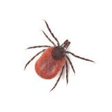
Two closely related mosquito species – Ades aegypti and Aedes albopictus acquire the virus by biting Dengue patients and then spread the virus to new victims. Other uncommon sources are - the pregnant mother can transmit dengue to her unborn child, infected organ transplantation and blood transfusions. The Dengue virus does not spread person to person.

What other virus illnesses are spread by Ades mosquito:

  1. Zika virus.

  2. Yellow fever.

  3. West Nile fever

  4. Chikungunya.

Previous endemics:

Many epidemics and endemics can be traced by looking into the CDC reports but confirmed endemics are as flows-

  1. In 2023, major urban centers of India have seen wide spread dengue fever following monsoon floods.

  2. In 2014, Brownsville Texas and Key West, Florida, and Hawaii have experience Dengue fever.

  3. CDC reports 2,000 cases of dengue in USA and its territories in 2022..

  4. In 1942, Nagasaki, Japan.

  5. During 2014 to 2016 Singapore recorded 33,000 cases of dengue. A biological method was used to control the endemic and the incidence dropped to 1/3 in 2017 by releasing male Ades aegypti mosquito infected with bacteria Wolbachia. This bacteria made male mosquito sterile and as a result the mosquito population

dropped and so the incidence of dengue.

Clinical feature:

Incubation period:

3 to 10 days, from the time of the mosquito bites to the onset of fever.

Symptoms:

 Abrupt on set of fever, headaches, retro orbital pain, body pain, bone pain, petechia, hematuria, hemorrhage in gastrointestinal tract and brain, prostration and some incidence of deaths.

Clinical types:

  1. Minor fever and bone pain. Most of the patients belong to this group. Fever lasts 2 to 7 days and the fever may be biphasic. In minority of patients the symptoms are - high temperature to 104F, severe headaches, retro-orbital pain, muscular and bone pain, ecchymosis, bleeding from gums, epistaxis, hematuria, petechiae and abdominal pain.

  2. Weakness, prostration and severe bone pain and severe hemorrhagic episodes called Dengue Hemorrhagic Fever.

  3. Dengue shock syndrome, consists of hypotension, shock and bleeding.

Special aspect of decreased Platelet count:

Low platelet count, usually around 150,000/mL is seen in most patients and develop on 3rd and 4th day. On about 7 to 10 days the platelet numbers returns to normal. In 10 to 20 % cases the platelet number falls below 50,000 and may reach a low 20,000/mL. Only about 5% of patients require platelet transfusions. One unit of platelet requires 4 units of blood.

What causes low Platelets:

Multiple processes of low platelet count are identified.

  1. Dengue virus infects various cell types including immune cells - monocytes, macrophages, dendritic cells, and Langerhans cells. Immune cells fail to swallow the virus, generate antibodies, fail to generate killer cells and limit spread of virus spread and fails to activate anti- inflammatory processes.

  2. Depression of megakaryocytes in the bone marrow.

  3. Destruction of platelets associated with activation of complement factor C3, and C5 binding to platelets. The DENV -2 alters platelets and activates macrophages to phagocytose platelets.

  4. Dengue antigen when attached to platelet IgM and IgG, becomes antigenic and starts producing antibodies that causes destruction of platelets.


Diagnostic tests:

Blood test detects Dengue NS1 antigen and also IgM and IgG antibodies. Test kits are commercially available and marketed by SD Bioline Dengue Duo test. NS1 antigen can be detected early in the stage of the illness. This test is moderately high sensitivity and very high specific. After 10 days following infection, the serology becomes positive. IgG antibody remains positive 10 months following infection.


Treatment:

More mild to moderate cases proper hydration are maintained. For fever and pain in bones and muscles Acetaminophen is used. Aspirin, NSAID (non steroidal anti inflammatory drugs) are contraindicated because those drugs can aggravate bleeding.

Those who develop low platelet count require close monitoring platelet count and boost platelets by platelet transfusions which may have to be repeated.

 The Janssen Pharmaceutical Companies of Johnson & Johnson (Janssen) announced, on October 2023, promising data from a Phase 2a human challenge study evaluating JNJ-1802, a first-in-class oral antiviral in development for the prevention of dengue. The data showed that the compound induced antiviral activity against dengue (DENV-3) in humans, compared to placebo, and is safe and well-tolerated. The data were announced at the American Society of Tropical Medicine & Hygiene Annual Meeting in Chicago, Illinois.”


Other measures:

To reduce infection carried by mosquito to others, the febrile patients should avoid mosquito bites. Use of these limits mosquito bites – mosquito repellents, wear long-sleep shirts and long pants and socks. Switch on the air conditioning unit. Covers windows and doors with mosquito nets.

In India Papaya fruit and extract of Papaya leaves or  leaves boiled in water prove to be a reliable platelet booster. In some other Asian countries Pumpkin found to spike platelets just like papaya.

Vaccine:

Dengvaxia is the name of a vaccine approved by FDA for teenagers 9 to 16 years old to prevent recurrence of dengue infection. Serological evidence must be present before vaccine is administered otherwise dengue, if contacted, would be much worse No vaccine for adults are successful so far.

Why dengue vaccines is difficult to make:

The dengue virus stops the immune reactions and inactivates and destroy immune cells that produce antibodies. In endemic area more than one variety of dengue virus is active and dengue virus sub type 1 to 4, each one has different antigen. Even a successful vaccine against one sub type will not protect every person

************************************************

Whipple's disease

  Whipple's disease: Whipple's disease is a bacterial infection of jejunum manifest as malabsorption of fat, fat soluble vitamins...