Thursday, December 22, 2022

Neurotransmitter

                                           Neurotransmitters

                                        PKGhatak, MD

Nerve cells communicate with other nerve cells, muscles, and glands. or internal organs by chemical molecules, even though the nerve impulse travels along the nerve fibers as an electrical impulse. At the end of the nerve terminal, a tiny gap exists, that gap is bridged by a neurotransmitter,

Neurotransmitters (NT) are chemical messengers that carry forward electrical signal from neurons to postsynaptic neurons or cells of the target organ and produce the intended action. NT must meet the following criteria. NT must be synthesized by the neuron, found in the presynaptic nerve terminal, must produce depolarization and propagation of the nerve impulse. NT must be quickly removed either by enzymatic degradation or diffusion or reuptake by the nerve terminal.


From the time the first neurotransmitter (NT) Acetylcholine, was discovered in 1921 by Otto Loewi of Germany, many more NTs have been added and today well over 100 NTs are known. Not all chemicals fulfill all the above criteria, however, all of them produce depolarization and transmission of nerve impulses. Chemically some of the NTs are simple gases like nitrous oxide (N0), carbon monoxide (CO) and other complex protein molecules like pituitary adenylate cyclades activating peptide are included in the newer class of NT.

In this review, only those NTs abnormality that produces significant medical illnesses are discussed. These are-

1. Acetylcholine, and Biological amines, 2. Epinephrine, 3. Noradrenaline, 4. DOPA and 5. Serotonin.

Inhibitory neurotransmitter –  6. Gamma-aminobutyric acid (GABA) and 7. Histamine.

1. Acetylcholine (ACh)

Acetylcholine is released at synaptic junctions by the neurons of the central nervous system, as shown in the diagram.


Acetylcholine is synthesized by the nerve cells and then stored in vesicles at the nerve terminals waiting to be rereleased at the synaptic cleft.  


Chemistry of ACh.

ACh is synthesized from Acetyl CoA and Choline. Acetyl CoA is the end product of glucose metabolism; choline is either locally produced by the nerve cells from another amino acid serine or taken up from the blood. Chemically choline is a Quaternary ammonium compound, acts almost like a vitamin in humans, except that it can be obtained from another amino acid. Eggs, organ meat, fish, milk and beans are good dietary sources of choline. Excess choline can cause low BP, weakness, increased sweating, fishy body odor, liver and cardiac problems. A deficiency of choline causes non-alcoholic fatty liver, muscle damage and Hyperhomocysteinemia (high Homocysteine blood level).

Neurons take up free choline via the 1.Na+/Choline transporter,2. a degradation product of ACh by the enzyme Acetylcholinesterase from the synaptic cleft and 3. also from blood as shown in the diagram below.


Synthesis of ACh.

Acetyl CoA + Choline = Acetylcholine, by the enzymatic action of acetylcholine- transferase.

Degradation of ACh into Acetyl CoA and Choline by enzyme Acetylcholine - esterase.

Action of ACh .

Autonomic nervous system.

(a). Parasympathetic division. It acts both on ganglia and postganglionic endings and its effects are -

Eyes - Constriction of pupils and fall of intraocular pressure.

Glands - Increases secretion of GI glands,

Bronchi- Constriction of bronchi, increased mucus secretion.

Smooth muscles of GI tract – increased motility, spasm and colicky pain, contraction of gall bladder and defecation.

Heart and arterioles. Slowing of the heart rate and vasodilation of arterioles.

Urinary bladder. Precipitate micturition.

(b). Sympathetic Division - It acts only on the sympathetic ganglia and Adrenal medulla and causes high BP and increased heart rate.

Central Nervous system. It causes restlessness, insomnia, tremors, dysarthria and convulsion.

Voluntary muscles. Contraction of muscles and fasciculation.

ACh has Nicotinic and Muscarine effects. Nicotinic receptors stimulate the ganglia of both sympathetic and parasympathetic divisions and the adrenal medulla. It also stimulates parasympathetic postganglionic nerve terminals. Nicotinic receptors stimulate the Neuromuscular junction of the skeletal muscles.

Muscarinic receptors are present in the parasympathetic nervous system and signal secretion from glands and smooth muscle contractions. Muscarine has no action on the skeletal muscles and does not act on the brain cells.

Diseases, due to autoimmune disease and unknown etiology, leading to decreased function of ACh, are Myasthenia gravis, Eaton Lambert syndrome, Gallian Barry syndrome and Ascending paralysis in febrile children.

When used as drug, it temporally producing muscle paralysis are used every day in surgery to ensure a better surgical outcome. Patients on mechanical ventilators at times require muscle paralytic drugs to prevent patients struggle to breathe. 

2. Biological amines:

Walter Bradford Cannon of Massachusetts, USA, in 1932 discussed the properties of adrenaline and used this phrase -  Flight or Flight response.

Epinephrine and  Noradrenaline.

In Europe, these two neurotransmitters are known as Adrenaline and Noradrenaline. Both Adrenaline and Noradrenaline are hormones. Norepinephrine (noradrenaline) is the neurotransmitter of the sympathetic division of the autonomic nervous system.

Biosynthesis of Norepinephrine.

L-Phenylalanine, an amino acid is the source of Dopamine and Epinephrine, the intermediate steps and enzymes involved in this process are as follows.

L-Phenylalanine----> L-Tyrosine by enzyme Amino acid Hydroxylase.

L-Tyrosine----> L-Dopa. (1-3,4 -Dihydroxyphenylalanine) by enzyme amino acid Hydroxylase. 

L- Dopamine-----> Norepinephrine by enzyme Betahydroxylase.

Norepinephrine ----> Epinephrine by enzyme N- Methyltransferase.

Noradrenaline is the transmitter for all the neurons of the brain and spinal cord of the sympathetic division of CNS, and outside the CNS, for the sympathetic postganglionic neurons. Noradrenalin increases blood pressure vessels due to increased tone. It produces bronchodilatation in the airways of the lungs and relieves nasal congestion. The difference in action is based on alpha and beta receptors and their subtypes.

Origin of noradrenergic neuron.

These neurons originate from the locus coeruleus, tegmentum and dorsal medullary group.

Action od Noradrenaline and Epinephrine

Effect on

Norepinephrine

Epinephrine

Heart rate.

Slowed

increased

Force of cardiac contraction

No effect

increased

Cardiac output

No effect

increased

Cardiac irritability

increased

Much increased

Systolic BP

Rises

rises

Diastolic

Rises

falls

Vascular bed in muscles

constriction

dilatation

Vascular bed skin & viscera

contraction

contraction

Vascular resistance in heart

increased

decreased

Glucose metabolism

unchanged

increased

Bronchial smooth muscles

No effect

relaxed

Intestinal muscles

relaxation

relaxation

Intestinal sphincters

constriction

relaxation

Pregnant uterus

Increased contractions

Contraction lessened

Capillary permeability

No effect

reduced


Epinephrine is a lifesaving drug in an acute allergic reaction and anaphylactic shock producing laryngeal edema and death due to airway obstruction. Adrenaline has multiple applications in everyday medical practice and is too numerous to list here.

3. Serotonin.

Serotonin chemically is 5-Hydroxytryptamine (5lHT). It is formed from the amino acid Tryptophan ---> 5- Hydroxytryptophan by enzyme Trytophanhydoxylase (adding “OH” group) and then---> 5-Hydroxy tryptophane (serotonin) by enzyme Aromatic amino acid decarboxylase and coenzyme pyridoxal phosphate (removing COOH group).  5-HT is present in large amounts in the GI tract and the brain neurons and platelet.

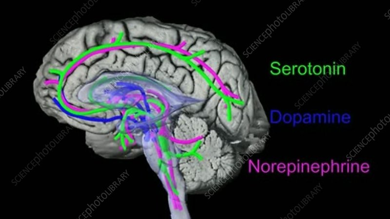
Serotonin acts on the areas of the brain and is shown in green color in the diagram below.


Serotonin is the neurotransmitter for appetite, sleep, memory, happiness, mood, vomiting center, sexual arousal and body temperature maintenance by its actions on the forebrain, brainstem and cerebellum. Lack of serotonin produces depression, anxiety and other psychological dysfunctions.

Serotonin receptor.

Serotonin (5-HT) receptors are designated by digits 1 to 7 and 5-HT1 has A to F subtypes; 5-HT 2 has A to C subtypes. The action of serotonin varies according to its binding with the type of receptor. 5-HT1A is mostly related to mood, learning, memory and behavior. 5-HT1B produces vasoconstriction. 5-HT2 signals via activation of phospholipase. 5-HT2A stimulates urinary bladder contraction. 5-HT2B increases pulmonary hypertension. and in general, produces inhibitory actions and opposes 5-HT1A effects. 5-HT2C stimulates appetite. 5-HT3 can produce nausea and vomiting. 5-HT4 increases GI motility. 5-HT5 consolidates memory. 5-HT6 increases depressive mood. 5-HT7 opposes 5-HT6 effects on mood.

Location of Serotonin receptors in the brain.

The Raphe nuclei B1 to B9 are serotonergic. These neurons are most abundant in the reticular formation, the axons of these neurons connect the brain and spinal cord nuclei.

Serotonin has a dominant role in understanding psychiatric disorders and depression. Therapeutic drugs are used extensively to increase the concentration or prolongation of the action of serotonin in the synaptic cleft.

4. Dopamine.

Dopamine is primarily an inhibitory neurotransmitter, which controls agitation and excessive motor actions.

Biosynthesis of L-DOPA (L-dihydroxyphenylalanine).

Biosynthesis is mentioned earlier. DOPA, norepinephrine and epinephrine are collectively known as Catecholamines. L-DOPA can enter the brain but L-Dopamine is blocked by the blood-brain barrier. In the brain in Substantia Nigra, L-DOPA is converted to L-Dopamine by decarboxylate and Vitamin B6 as a cofactor. After release at the nerve terminals, it is quickly metabolized to methyl dopa by catechol methyl-O- transferase.

Dopamine receptors and action.

Receptor subfamily

Location

Action

Therapeutic potential

Central

D1 and D2

substantia nigra and striatum

motor control

agonists - Parkinson's disease

D1 and D2

limbic cortex and associated structures

information processing

antagonists - schizophrenia

D2

anterior pituitary

inhibits prolactin release

agonists - hyper- prolactinemia

Peripheral

D1

blood vessels

vasodilatation

agonists - congestive

D1

proximal tubule cells

natriuresis

heart failure

D2

sympathetic nerve terminals

decreases release

hypertension

Dopamine has both CNS and outside CNS effects.

In the brain, dopamine modulates and smooths skeletal muscle movement signals. In the limbic system, it organizes emotional memory formation and associated mood elevation.

Outside the CNS, Dopamine is produced locally and acts on surrounding cells only (action is called paracrine effects). Dopamine increases heart rate and BP by its direct action on blood vessels and kidneys. It is suspected that Primary hypertension is an abnormal function of dopamine in the kidneys. In the pancreas, it decreases insulin production, on the GI tract decreases motility, acts to protect GI mucosa and boosts local cellular immunity. In the kidneys, the dopamine role has been studied with interest because it appears dopamine acts as a natriuretic hormone.

Diseases involving movement disorders, like Parkinson's disease and its subtypes, Restless leg syndrome and following minor strokes are treated with DOPA and drugs blocking the action of catechol O methyltransferase.

5. Gamma Amino butyric acid (GABA).

GABA is derived from Glutamate. Both Glutamate and GBA are neurotransmitters of the central nervous system. Glutamate is a neuro exciter and is widely distributed in the brain, mostly in small sized neurons. GABA is an inhibitor NT. GABA neurons are present in the Limbic area of the brain, from these nuclei GABA connects many areas of the brain. A diagram below shows the areas of influence of GABA. 


Chemistry:


Brain neurons produce GABA from glutamine by decarboxylation by the enzyme decarboxylase. The reaction is a rate limiting reaction. GABA concentration in the brain is high.

Mechanism of action of GABA:

GABA has two isoforms- Ionotropic (GABA A) and metabotropic (GABA B) forms. Two separate genes on two chromosomes encode the production of isoforms. GABA has two metabolites Homocarnosine and pyrrolidinone and both these chemicals are anticonvulsants. GABA prevents hyperpolarization and reduces incoming and outgoing signal strength. GABA A receptors are present all throughout the brain. GABA B receptors are formed by the fusion of two G-protein molecules.

Blood-brain- barrier prevents GABA from entering the brain but Glutamine is free to enter. Food containing glutamine are soybean, brown rice, chestnut, mushroom, tomato, spinach, cabbage and cauliflower.

Fluctuation of the levels of GABA has been linked with Autism, Parkinson's disease and anxiety.

Therapeutic drugs having GABA like action are used as pain control medication but the therapeutic action of these medicines does not match the theoretical possibilities it promises.


6. Histamine.

Histamine is well known as a mediator of allergic reactions. It is present in the mast cells of the skin and in the basophils of blood. It enhances inflammatory reactions by increasing vascular permeability and the production of mucus.

As an NT is not that well known. In the brain, it acts via H1, H2 and H3 receptors.

Action in the brain results in wakefulness, alertness and quickens reaction time.

Distribution of Histamine containing cells.

In CNS: Hypothalamic region of the brain.

Outside the brain: The nose, mouth, feet, skin, and feet have a good number of mast cells. In the stomach, chromaffin cells produce and release histamine.

Chemistry: Histamine is produced from the amino acid Histidine by the enzyme histidine decarboxylase.

Enzyme histidine N-methyltransferase and diamine oxidase degrade histamine.

Receptors. Histamine receptors are H 1 to 4.

H1 receptors. Dendrons (short arms of neurons) of the Tuberomammillary nucleus terminate in the dorsal raphe and locus coeruleus. These cells regulate sleep-wakefulness cycles, body temperature, appetite, coordinate endocrine hormones and cognition.

H2 receptors. Basal ganglia, hippocampus and detente nucleus have histamine 2 receptors. Modification of motor activities is the main function. In the stomach H2 receptors on the parietal cells when bind with histamine increase HCl acid production.

H3 receptors. The precise location is not worked out. The action appears to counteract acetylcholine, serotonin and norepinephrine actions.

H4 receptors. H4 receptors in the brain have not been detected.

The realm of antihistamine drugs is increasing with new discoveries of histamine receptors and their role in the etiology of diseases.

 8. Other NTs.

Glycine.


Glycine is an amino acid, is present in every living cell and besides performing metabolic functions it also acts as a neurotransmitter for incoming somatic sensations like pain and touch, vision and auditory sensation. Glycine also acts on strychnine-sensitive receptors and produces inhibitory action and modulates excitatory neurotransmission of glutamine in glial cells.

The final word.

Neurotransmitters are chemical messengers synthesized by neurons and delivered at the synaptic cleft in order to ferry the nerve impulse to the next neuron or to target cells. In the cerebral cortex, the prime NT is acetylcholine. In the autonomic nervous system,  ACh is also the sole NT for both parasympathetic ganglia and postganglionic nerve terminals. In the sympathetic nervous system, ACh is NT for the sympathetic ganglia but epinephrine is the prime NT for postganglionic nerve terminals. In the Limbic nervous system, multiple NTs interact, modify, enhance, or inhibit nerve impulses.

NTs are also classified as excitatory or inhibitory. The same NT may act as both excitatory and inhibitory depending on the receptor it binds. The molecular structure of NT must fit with receptors like pieces of Logo fit with another - a Levo (l) or Dextro (d) from; an aliphatic chain or ring form, makes that difference. And finally, even minor NTs have major effects on the body.

************************************


Wednesday, December 14, 2022

Connective Tissue & Mixed Connective Tissue Disease

 Connective Tissue and Mixed Connective Tissue Disease.

                             PKGhatak, MD

Connective tissue is the most abundant tissue in the human body. It is present underneath the skin, in the lungs, in the mucus membranes of GI and GU tracts, and in the fatty tissue and in the coverings of the brain. The connective tissue fibers are made up of two proteins- Collagen and Elastin. Collagen is present in the tendons and elastin dominates in the lungs and skin. The connective tissue (CT) is a loose collection of cells in the middle of a water rich extracellular matrix. Blood, bones, cartilage, adipose and reticular tissues are special forms of connective tissues.

Embryology of CT.

CT is derived from the mesoderm. A sheet of cells in the developing fetus folds into two tubes ectoderm and endoderm and in between tubes the remaining cells from the mesoderm. The embryonic mesoderm is known as Mesenchyme. From the mesenchyme, the connective tissue, muscles, bones, cartilage, blood vessels and lymph tissues develop. The mesoderm cells undergo various stages of development, from the initial flat sheet of cells to tube formation and back to sheet form. CT cells migrate to the ectoderm and develop into the subcutaneous tissue of the skin; cells migrate to the endoderm and develop into the submucous tissue in the GI tract and interstitial cells in the lungs. The developing mesoderm receives cells from the yolk sack, these cells become Microglial cells of the brain. The end result is connective tissue containing blood and lymph vessels and nerve fibers.

This basic embryology is helpful to understand various manifestations of Connective Tissue Disease (CTD).

Connective tissue disease.

There are a number of Autoimmune CTDs are simply identified as Connective Tissue Diseases. These diseases are Systemic lupus erythematosus (SLE), Scleroderma, Sjogren syndrome, Polymyositis, Rheumatoid arthritis (RA) and Mixed connective disease.

The laboratory screening test for connective tissue disease is ANA (anti nuclear antibody) test. The test is expressed as I:80 to 1: 640 dilutions. However about 15% of the population have a positive test without ever having any disease, these ANA tests are positive only in lower dilution (1:80).

The mixed connective disease (MCTD), as the name indicates, has some of the characteristics of other CT diseases and was previously named Overlap Syndrome.

MCTD (mixed connective tissue disease)

MCTD is a rare autoimmune disease; it is triggered by an unidentified agent. The antibodies generated are directed against collagen fibers wherever they are present. Those cells turn out excess collagen which causes the thickening of skin and fibrosis of internal organs. To describe a full spectrum of MCTD, a synopsis of common manifestations of the other CTDs is presented here.

1. Lupus Erythematosus (SLE).

SLE is a systemic disease that affects many organs and is a steadily progressive illness. Deaths are due to renal or respiratory failure if treatment is not available. However, not every patient follows the progressive course, many just have skin or liver disease only, while others may have skin and lung involvement. The lesions produced by SLE on the individual organs are summarized below. -

Skin. The term erythematosus of SLE is taken from a butterfly red colored skin rashes develop over the bridge of the nose and on checks. Other skin lesions are coin sized dry skin patches on sun exposed skin, resembling psoriasis. And subcutaneous lesions with rolled up raised margins localize lesions are seen on the neck, front of the chest and exposed areas of arms.

Joints. The finger joints of both hands and feet are commonly involved. The stiffness, swelling and joint pain are usual symptoms.

Kidneys. Glomerulonephritis is common. Nephritis is due to immune-complex induced damage to the capillaries of glomeruli. This leads to hypertension, renal insufficiency and renal failure.

Pulmonary. Pleurisy is the most likely to develop when the lungs are involved. Pulmonary fibrosis, pulmonary hypertension and respiratory insufficiency lead to respiratory failure. At times small vessel vasculitis produces pulmonary infiltrates and patchy areas of consolidation.

Liver: Lupus hepatitis is a known feature, often just the liver enzymes are elevated and in others, more liver damage and jaundice develop.

Heart. Sterile pericarditis, myocarditis, coronary artery disease and conduction abnormalities are usual findings. In addition, cardiac valve damage and sterile vegetative emboli to the brain may happen.

Brain. Encephalitis, small vessel strokes, transient ischemic attacks. delirium, psychosis, anxiety and seizures may develop.

Eye. Blurred vision, exudative macular detachment, cystoid macular edema, and ptosis may develop.

Hematology. Anemia, thrombocytopenia, leukopenia, lymphadenopathy and splenomegaly are seen.

Coagulation. Prolongation of coagulation due to lupus-anticoagulant can cause serious problem in certain situations.

Confirmatory serology for SLE. In SLE the ANA test is invariably positive. Several antibody tests are also positive. But the confirmatory test is anti-Native double stranded DNA ( sdDNA). This test is the gold standard diagnostic test of SLE.

2. Systemic Sclerosis (SS), and Scleroderma.

It is a rare disease, one in a million per year is seen in cold climate countries, still less prevalent in tropical countries.

SS is characterized by diffuse fibrosis of the skin and fibrosis of the internal organs. But not every patient has the same degree of involvement. All patients have positive ANA in high titers, and 70 % of them have positive Anti SCL 70 antibody tests, this test now known is known as the Anti topoisomerase I test. This test is positive in about 50 % of cases of diffuse SS.  When the lesions are confined to limited areas of the skin, the term Scleroderma is used and in 20% of cases, this test is positive.

 The following variants of SS are important.

a. CREST syndrome. CREST stands for calcinosis, Raynaud's phenomenon, esophageal dysfunction, sclerodactyly and telangiectasia. The skin lesions in CREST Syndrome are limited to the distal extremities, face and neck.

b. Diffuse disease. The thickening of the skin with areas of increased pigmentation and patchy areas of depigmentation is present all over the body including the trunk. A test that measures antibodies against Topoisomerase I titer becomes positive in over 50 % of patients.

 In contrast, in diseases limited to the skin and few other places, a different antibody test - The Antocentromere antibody test becomes positive in over 50 cases. About 20% of patients with limited SS have anti-topoisomerase I positivity. In 14 % of cases of limited. the SS U1-RNP antibody test is positive; it indicates the presence of Pulmonary fibrosis.

c. Raynaud's phenomenon. Intense blanching of fingers and toes during exposure to cold is associated with pain. Anti U 11 and U12- RNP antibody tests in Raynaud's is associated with GI, Pulmonary involvement and higher mortality.

When SS produces fibrosis in the lungs, kidneys and GI tract the disease produces pulmonary insufficiency, renal insufficiency, hypertension, reflux esophagitis and hypomotility of GI tract including the esophagus respectively. In renal crisis due to uncontrollable hypertension and renal failure in systemic sclerosis, Anti RNA polymerase III antibody test becomes positive.

 3. Sjogren syndrome. Dryness of mouth and eyes and their consequences are the presenting symptoms. The lachrymal glands and salivary glands typically show lymphoid foci with the destruction of glandular elements and fibrosis. ANA is positive in over 95%of cases and anti SS-A and SS-B positive in 65% of cases. SS is often associated with symptoms of RA, biliary ductal inflammation, Hashimoto thyroiditis, polyarteritis and pulmonary fibrosis.

4. Polymyositis and myopathy. Patients typically present with muscle weakness and muscle pain. Muscle enzymes- CPK and Aldolase levels are high indicating muscle necrosis and inflammation. Dermatomyositis is a variant of myositis. Dermatomyositis presents as bilateral "heliotropic rashes”. Another subvariant is Antisynthetase syndrome which is characterized by muscle disease associated with non-erosive arthritis, Raynaud's phenomenon, hyperkeratosis along the palmar and radial aspects of the fingers, pulmonary fibrosis and the presence of anti-Jo-1 antibodies.

5. Rheumatoid arthritis (RA). It is a common disease (about 1% of the population), in contrast with the previous illness mentioned under CTD. It is also different from the rest in having a negative ANA test in 80 % of cases. The blood tests for RA are Rheumatoid Factor (present in all cases), and anti-CCP antibodies (cyclic citrullinated peptides) present in 80 % of cases. Clinical features of RA are dominated by symmetrical polyarthritis of small joints of fingers and toes, The 1st (proximal interphalangeal) joint arthritis at the beginning of the illness is very characteristic. Swelling, pain and morning stiffness of hands and red discoloration of the overlying skin point to an early diagnosis. As the disease advances other small joints like wrist joints, upper cervical spine, temporomandibular, and sternoclavicular joints are affected. Low grade fever, loss of weight and other clinical and laboratory tests follow the general features of CTD. X-rays of the hands show Erosion of bones at the joint faces and osteoporosis. These findings are also distinct features of RA. Extra articular features are subcutaneous nodules over the bony prominence, pleural effusion (small amount), pulmonary fibrosis, pericarditis, vasculitis and enlarged spleen.


                                  Mixed Connective Tissue Disease.

 Most patients with MCTD are young females between 25 -30 yrs. of age. In Native Americans of Minnesota, the incidence is 6 per100, 000 and 2/100,000 in white females. The onset of the disease is insidious and begins with arthralgia of the fingers of both hands which become white, numb and painful on exposure to cold. Later finger joints swell and become painful. Raynaud's phenomenon and myalgia are common symptoms. All MCTD patients carry HLA-DRB1 positive markers in their white blood cells (lymphocytes & monocytes). The blood levels of U-1 RNP (Ribonuclear Protein) antibodies in high titers (over 1:125) or over 25 U are seen in over 95% of patients with MCTD. MCTD is a slowly progressive disease but in some patients, the progression may be arrested for years, in others the disease shows features of one or other connective tissue disease mentioned above. However, the initial phase of MCTD resembles RA but soon the disease takes a course resembling scleroderma and Raynaud's features dominate. Though dryness of the eyes is present, other typical features of Sjogren syndrome are absent. Later, as GI, Pulmonary, and Renal symptoms develop the diseases acquire SLE like illness, but the CNS is very rarely affected. Pulmonary fibrosis is the most devastating aspect of MCTD and death is generally from respiratory failure or pulmonary infection.

Treatment. 

The management of MCTD is based on individual patients because no two patients are having the same system involvement.

The general approach is to use well known immune suppression drugs and symptomatic therapy for individual organ system dysfunction and degree of disability. Disease modifying agents are added and watched for side effects and therapy is modified based on progress over adverse effects. 

Prognosis. Over all prognosis of MCTD is favorable over SLE and SS. A 10 year survival rate of MCTD is 80%. Those who develop Pulmonary fibrosis have the worst prognosis.

********************************************
Thursday, December 8, 2022

Science of Taste

                                      Science of Taste

                                  PKGhatak, MD

Gastronomy scientists held the view for a long time that humans have only salt, sweet, sour and bitter taste perceptions. A Japanese scientist Kikunae Ikeda in 1907 proved there is a 5th taste in humans. He called that Unami taste. He demonstrated meat, shellfish, fish, mushroom, tomatoes, and vegetable proteins when simmered in water for a long time, produce an amino acid that triggers the Umami taste (in the west it is called the savoy test). That chemical is MSG (monosodium glutamate). MSG is commercially manufactured and used by the food industries worldwide. People of South Asian countries believed that Hot is an independent taste “(called Jahl by locals) but science rejected their claim till Vijay Lyall found receptors for Hot in the taste buds.

The tongue surface is covered with filiform papilla. These papillae contain TRPV (transient receptor potential vanilloid family) receptors. The primary function of it is to protect the tongue from hot temperatures and these receptors are also activated by capsaicin. Alcohol potentiates response and lowers the threshold for heat activation from 42C to 34C. Vijay Lyall and his colleague discovered modified TRPV1 receptors on Taste buds. 

Menthol and mint give a cold sensation in the mouth due to the presence of TMPM8 protein which triggers cold receptors. In hot chili peppers, the presence of TRPV1 in capsaicin binds with hot receptors in the mouth. Black peppers contain Piperine molecules that also bind with the same TRPV1 receptors. The list is growing – recently calcium, dryness of food, fattiness, heartiness and numbness of food receptors have been identified.

The 2021 Nobel prize in medicine was awarded to Ardem Patapoutian and David Julius of California for their discovery of the molecular basis of the perception of hot, cold and mechanical force. With this Nobel prize award, all the skeptics holding the notion that only 5 tastes are perceived in humans, melted away.

The French chefs always knew alcoholic beverages magnify taste perception and some fine French restaurants serve, a different type of wine, before bringing in the next course. If one orders desserts in a French restaurant, the waitress will bring a small dish of sorbet for cleansing the palate before bringing gateau and the assortment of petites gateaux. Children love ice cream but many of them refuse to drink milk. That led to the discovery that cold multiplies the sweet sensation of sugar. If you do not believe it, just place a few fresh grapes in the refrigerator and when they are frozen, pop one or two in your mouth. And enjoy the explosion of sweet grapes inside the mouth.

That is not all. In Sanskrit it says – Ghranena Ardha Vojanam. ঘ্রাণেন অর্ধ ভোজনম  । (smell of food imparts half of the pleasantness of eating). The same also works for visual, auditory and touch sensations on taste. In a simple sentence – The Taste of food is more than just the anatomy of Taste.

Now have a look at the Anatomy of Taste.

In the nineteen century, two German scientists Greorg Meissner and Rudolf Wagner discovered taste buds among the papilla of the tongue. A taste bud is made up of tall taste cells, Gustatory cells, each cell is supplied with an afferent fiber of the 1st order neuron of the visceral chemosensory neuron of the Vagus nerve (nucleus tactus solitarius). The top of the cells ends in hair like microtubules and all of them are bunched together and appear at the opening of the taste bud. The taste producing molecules of food attach to the respective microtubules and the nerve impulse is generated and is conducted along the nerve to the sensory nucleus. The taste buds present at the back of the throat, tonsils and palate are innervated by the glossopharyngeal nerve and the rest of the path is shown in the diagram.


From the Vagus nucleus, the sensation reaches the ventroposterior nucleus of the Thalamus. The 3rd order neuron carries the information to the Taste area of the cerebral cortex.

                                               Taste buds.

The revised taste bud types are 7, namely sweet, sour, bitter, salty, umami, cool and hot. An adult person has about 10,000 taste buds in the mouth; some have more and they become food super-critics, others have fewer and they will eat anything placed before them. A human child in utero develops taste buds and at birth, sweet and bitter taste buds are fully functional. In adults, a cell of the taste bud lives for about 10 days and is continuously replaced by new cells of taste buds. As an example, all of us burned our tongues by hot coffee and remembered how the food tasted bland for a few days, then the normal taste returned. Most of the taste buds are present on the surface of the tongue, however, the entire mouth cavity has taste buds but density varies from place to place. In old age, the number of taste buds decreases to about 5,000.

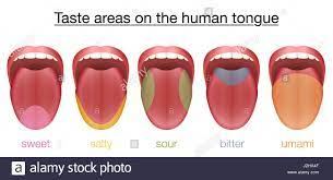
The diagram below shows areas of the tongue with one dominant taste bud but not at the exclusion of other varieties.


From Left to right- sweet, salt, sour, bitter and umami.

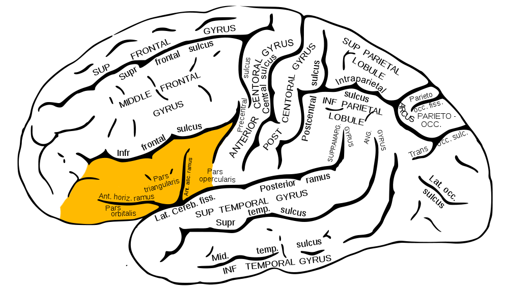
                                                  Taste center.

The insular cortex and inferior frontal gyrus are two parts of the taste center. Within the centers, there are neuronal groups engaged in association, connection with smell, touch, visual, tactile and emotional, hypothalamus, thalamus, Hippocampus and other areas of the brain. These interconnections are responsible for the development of memory of taste, food association with place and time, likes and dislikes of certain food in an individual.


 *******************************************

Saturday, December 3, 2022

Muscle Disease

                        Muscle Disease.

                     PKGhatak, MD


The human body has three types of muscles.

1.The Striated skeletal muscle.

2. Cardiac muscle is also striated but it works involuntarily. Cardiac muscle fibers are joined together without partitions and contract in unison. It is known as the syncytial muscle.

3. Smooth muscle has no striation and is a non-voluntary muscle. It is present in the wall of arteries, Gastrointestinal and genitourinary tracts and sphincters,

When a muscle is mentioned without any qualification it is the skeletal muscle. Skeletal muscle disease is easier to understand if the basic structure and function of muscle are understood.

Basic muscle structure.


Muscles are a collection of muscle cells, usually referred to as muscle fibers. Muscle fibers are long and cylindrical and arranged in bundles and the end of fibers are attached to bones or joint capsules. Muscles are very vascular and are equipped with abundant mitochondria and multiple nuclei. The motor nerve fibers to muscles originate at the anterior horn cells of the spinal cord. Skeletal muscles contract under voluntary control and in abnormal conditions they can also contact their own but such contractions result in shivering or muscle twitching. Respiratory muscles work incessantly without conscious effects and also on demand. The detailed arrangement of the muscle bundle is shown above diagram.

Function of muscle.

Muscles shorten by contraction and result in ambulation or joint movements and the heat generated by muscle action increases body temperature.

How muscle contacts.


The motor nerve transmits nerve impulses at the neuromuscular junction. The impulse propagated along the sarcolemma to the entire length of the muscle. As a result, calcium ions from the sarcoplasmic reticulum are released and the released calcium iron enters into the muscle cells. This generates an action potential. Calcium ion binds with a protein called Troponin which pulls Myosin molecules towards Actin molecules. Actin and myosin molecules slide against each other causing shortening of muscle fibers. This action requires energy and is supplied by ATP. At the end of muscle contraction, the process is reversed and muscle fibers return to the relaxed state.

Diseases of the skeletal muscle.

From the above paragraph, it is clear that muscle disease may arise from the muscle fiber itself and is called Primary Muscle disease. Some other diseases may damage muscle fibers or their functions and these are Secondary muscle diseases.

Primary Muscle Disease.

Primary muscle diseases are 1. Hereditary Mitochondrial muscle diseases and 2. Congenital Myopathies.

 1. Hereditary Mitochondria muscle diseases.

Inherited defective genes that encode energy producing enzymes when passed to a child the disease manifests at birth or later in life. The muscles are weak and disease is initially detected in eye muscles and then progresses through the entire body.

A. Leigh syndrome. The cytochrome enzyme system is defective in Leigh syndrome and eye muscle weakness develops in infancy. Weakness of the whole body soon follows. Associated heart abnormalities and low position of ears and high forehead is a distinct facial features. The child hardly can reach 5 yrs. of age.

B. Keans-Sayer Syndrome. The mutated gene encodes Oxidative phosphorylation. A child may normally develop to early teens then develops drooping eyelids, and soon one or both sided intermittent weaknesses develop. Other features are ataxia, deafness, heart rhythm disturbances and hormone deficiencies.

C. Melas syndrome. The defect is in the NADPH enzyme system. Muscle weakness begins at about age 40 yrs. Seizures, mental changes, migraine, lactic acidosis and strokes develop.

 2. Myopathy.

The main symptom is muscle weakness, other less common symptoms are muscle cramps, stiffness and walking difficulties.

1. Inherited Muscular Dystrophy.

The general term for this entity is Muscular Dystrophy. Progressive muscle weakness is due to muscle degeneration. In the early stage of the disease, regeneration of muscle is seen but later lost. Various from of disabilities develop and death is generally from respiratory failure.  Muscular dystrophies have several clinical types based on the onset and progression of the disease and the genetic defects.

Clinical Types.

      Duchenne Muscular Dystrophy

     . Becker Muscular Dystrophy. ...

  • Congenital Muscular Dystrophy. ...

  • Myotonic Muscular Dystrophy. ...

  • Limb-Girdle Muscular Dystrophy. ...

  • Facioscapulohumeral Muscular Dystrophy. ...

  • Emery–Dreifuss Muscular Dystrophy. ...

  • Distal Muscular Dystrophy.

Duchenne Muscular dystrophy is more familiar to people of the US due to Jerry Lewis MDA Labor Day Telethon. The inheritance is sex link X chromosome in recessive mode, only male child is affected by this genetic disorder. 1 in 3 - 5 thousand male children is affected. In recent years a new class of drug has been approved for the treatment of Duchenne dystrophy. The Antisense Oligonucleotides drug acts as a bridge over the missing exons (functioning genes) like a Band-Aid that produces the protein needed for the repair of muscles. The latest approved drug is a small-molecule Sunitinib.

Secondary Muscle disease:

1. Acquired Muscular Dystrophy.

Many diseases can damage and destroy skeletal muscle. Diseases or agents producing myopathy can be broadly grouped as Endocrine and Metabolic, Drug induced, Inflammatory, Prolonged illness, Neurological, Collagen vascular, Autoimmune and Unknown causes.

One common feature of this group of myopathies is the rapid onset. Inflammatory myopathy requires muscle biopsy to identify a specific entity. Autoimmune and other systemic diseases are identified from clinical and laboratory tests.

2. Dermatomyositis and Polymyositis are often secondary to a malignant process or a viral induced autoimmune disease. The skin rashes of Dermatomyositis appear around the eyes and over the knuckles and elbows and are reddish in color. The visible capillaries over the rough fingernail folds are an important diagnostic clue. In Polymyositis large groups of muscle are affected and weakness of shoulder and pelvis girdles is an important feature.

3. Inclusion body myopathies (IBM) are a common muscle disease of elderly people. Progressive weakness of fingers, wrists, muscles on the anterior aspect of the thigh and legs develop over time and atrophy of muscles follows. Antibodies to Cytosolic 5- nucleotidase-1A, when present indicates an autoimmune component. Congo-red stained rimmed vacuoles and areas of muscle necrosis surrounded by the inflammatory cell are seen in muscle biopsy. Treatment is unsatisfactory.

4. Collagen vascular disease myopathy is also common. Muscle weakness and atrophy in muscles of the face neck and neck in Scleroderma is a distinct feature. Systemic lupus and Rheumatoid arthritis and mixed connective tissue diseases also have muscle weakness and different degrees of muscle atrophy.

 5. Neurological and Neuromuscular disease.

Cerebrovascular accident, otherwise known as stroke, is a good example of how muscle undergoes wasting, stiffness, hyperreflexia and loss of function. These symptoms are due to the loss of inhibitory influence of upper motor neurons (UMN) on lower motor neurons (LMN). Prolonged disuse leads to muscle atrophy and contracture.

Spinal cord injury.

In spinal cord lesions, none of the muscle abnormalities above the site of spinal cord injury show any weakness. At the site of injury, it produces complete loss of muscle function and loss of resting tone of muscles. Below the site of injury, muscles show features of Upper Motor Neuron lesion as described under UMN lesion.

Peripheral nerve

Peripheral nerve lesion produces complete loss of muscle function innervated by that nerve.

Other systemic diseases.

Multiple sclerosis. Parkinson's disease. Meningitis and encephalitis and degenerative neuronal diseases produce muscle loss of function depending on the site of the lesion in the brain, spinal cord, or peripheral nerves.

Amyotrophic Lateral Sclerosis (ALS)

ALS is better known in the USA as Lou Gehrig's disease. It is a motor neuron disease involving both upper and lower motor neurons. ALS usually develops in the past middle life and 5 to 10 % of ALS cases are inherited as an autosomal recessive disease. The initial symptom is muscle twitching at rest which can be demonstrated by gently tapping muscles. Weakness of muscle develops gradually and affects all skeletal muscles of the body. The death is from respiratory failure.

Multifocal motor neuron disease may resemble ALS but it is limited to one side of the body and muscles of the fingers and hands are more affected and with treatment, the condition improves. 

6. Endocrine and Metabolic diseases.

Hyperthyroidism produces muscle wasting and weakness of pelvic and shoulder girdle muscles. Hypothyroidism produces weakness of the shoulder and gluteal muscles due to decreased metabolic rate and energy supply to muscles is limited.

Familial periodic paralysis.

Familial periodic paralysis is due to the mutation of genes that regulate sodium and calcium channels of the neurons. This is an intermittent paralytic disease. Muscles are hypotonus, weak and fail to contract during an attack but function normally in between times.

Hypokalemic periodic paralysis is due to the fall of serum Potassium. The symptom usually develops in adolescence precipitated by a rich carbohydrate meal or strenuous exercise. An attack may last hours to days. Generalized weakness develops later in life.

 In contrast, high serum Potassium periodic paralysis starts in early childhood and attacks are more frequent but last a shorter period.

Glycogen storage disease type VII 

This inherited glycogen storage disease produces muscle weakness due to failure of glycogen utilization in the muscles.

7. Neuromuscular junction.

Myasthenia gravis is an autoimmune disease triggered probably by a viral infection. Antibodies bind with the neurotransmitter Acetylcholine and acetylcholine level falls. Depolarization of the muscle is prevented because of a lack of acetylcholine. During rest, some Acetylcholine is generated and the function returns. But on repeated muscle use paralysis develops again. The drooping of eyelids and ophthalmoplegia are initial symptoms. Paralysis of other muscles, notably to head and neck muscles develops slowly. Speech, drinking, and eating becomes problematic and respiratory muscle paralysis follows. Early diagnosis and prompt immunosuppressive therapy can arrest the disease and improve the functional status of muscles.

Eaton -Lambert disease

Eaton Lambert disease is another example of neurotransmitter disease. Reduced acetylcholine release from the presynaptic nerve terminals due to antibodies to voltage-gated calcium channels produces muscle paralysis. Patients with Small Cell Carcinoma of the lung exhibit this paralysis frequently, other lung carcinoma rarely causes this disease. In contrast to Myasthenia, Eaton-Lamber disease affects the muscles of the legs initially. Head neck muscle may be involved later but much less severely, respiratory muscles are spared. Leg muscle function improves with repeated muscle movements.

Guillain-Barre syndrome.

It is due to antibodies damaging the myelin sheath and axon of nerve fibers. Leg and arm muscles are primarily affected. Because peripheral nerves are mixed nerves, numbness and tingling also develop.

8. Drugs causing muscle paralysis.

 Action on the respiratory center of the brain.

Opioids, Fentanyl and other semisynthetic opioid derivatives have taken so many lives in recent years. The depressive effects of these compounds make the respiratory center insensitive to rising CO2, falling blood pH and hypoxia. In the end the respiratory muscles stop functioning causing death.

9. Therapeutic muscle paralysis.

 It is extremely unpleasant and stressful for a conscious patient to be intubated and mechanically ventilated. Patients fight for air and try to breathe faster than in the ventilator settings. Muscle-paralytic agent Succinylcholine is used to eliminate the patient's effort and completely take over the ventilation by mechanical means. Succinylcholine causes depolarization of the motor- endplate; and because the drug is not immediately broken down by the enzyme cholinesterase, the depolarization persists and muscles are kept in a paralyzed state as long as needed. Eventually, Pseudocholinesterase, another enzyme, breaks down succinylcholine but at a slower rate.

Many derivatives of Tubocurarine combine with receptors of acetylcholine at the motor endplate and produce partial skeletal muscle paralysis and are used as a remedy for muscle stiffness in Upper neuron paralysis. Antibiotics Neomycin, Streptomycin and Polymyxin can potentiate neuromuscular blocking agents by competitive synergistic effect. Centrally active muscle relaxants Mephenesin, Meprobamate, Benzodiazepines and other CNS depressive drugs can decrease intraneuronal activities and block parasympathetic neurotransmission.

 Statins:

Statin is a widely used medicine as a treatment for lipid abnormalities. Many patients complained of nocturnal leg cramps, leg pain and proximal muscle weakness and getting in and out of an automobile becomes hazardous. Researchers discovered that muscle weakness and related side effects of statin are likely due to its effect on the energy production centers, or mitochondria of muscle cells. The intermediate metabolite Lactone interferes with mitochondrial function. Switching to a water soluble statin or reducing the dose of statin may control symptoms, otherwise, discontinuation of statin is required. Statin also produces narcotizing autoimmune myopathy due to antibodies produced against enzyme HMG coenzyme A. This is less common but stopping statin does not stop muscle necrosis.

10. Myositis. 

Many viruses can infect muscles producing muscle pain. Dengue fever causes worse types of backache.

 Myositis Ossificans is a less common disease.

A damaged muscle heals by having connective tissue from adjacent structures infiltrate the muscle. Occasionally bone generating stem cells enter muscles and produce lumps and these lumps are painful and interfere with muscle function.

 11. Rhabdomyolysis.

Crush injury of muscle and compression injury in Compartment syndrome can produce muscle necrosis.  Released Myoglobin from the damaged muscles by blocking glomeruli capillaries and renal failure.

12. Tumors of the skeletal muscle.

Fortunately, tumors of muscle are rare. Rhabdomyosarcoma is one malignancy, seen in all ages and carries a bad prognosis. 


*****************************************


Wednesday, November 23, 2022

Cartilage and Diseases of Cartilage

 Cartilage and Diseases of Cartilage

       PKGhatak, MD

Gristle, that word may be more familiar than cartilage - the term used in biological science. Cartilage is a connective tissue like bone and tendon, connecting two pieces together forming a joint. Joints have a layer of cartilage covering the bony ends preventing bones from grinding and making the movement smooth and pain free.

Cartilage, bone and muscle originate from the same source called mesoderm. Distribution of cartilage in the body follows the same path as that of bone, except in the respiratory tract cartilage alone provides structural stability. Cartilage does not have nerve supply, so it is pain insensitive and neither does it have blood supply or lymph channels. Cartilage derives its nutrition from synovial membrane secretion. The synovial membrane is the innermost covering of a joint, the secretion also acts as a lubricant.

Structure and types of cartilage.

The cartilage is composed mainly of a dense extracellular matrix. Within the matrix a few sparsely distributed cartilage cells chondrocytes and collage fibers are present. The cartilage cells secrete the matrix. Matrix is made up of water, protein and proteoglycans. Chemically proteoglycan is a derivate of chondroitin sulphate compounded with carbohydrate and protein.

Types of cartilage.


Cartilages are Hyaline, Fibrous and Elastic types based on the amount of fibrous and elastic tissues present.

Hyaline. Cartilage. Hyaline means glass like. Hyaline cartilage is smooth, glistening, semisolid and homogeneous.

The skeletal structure of a fetus is made of hyaline cartilage and later cartilage is turned into bones. But a part of hyaline cartilage remains at the end of bones forming movable joints, called the articular cartilage and also called the growth plate. The longitudinal growth of the skeleton takes place by the rapid expansion of articular cartilage under the influence of the pituitary growth hormone followed by calcification.

Hyaline cartilage is the most abundant of cartilage, the distribution in the body is represented in the above diagram.

Fibrous cartilage. Fibrous cartilage joins ligaments and tendons to bones. The fibrous cartilage is made up of tough bundles of fibers and provides strength to the structure.

Elastic cartilage. The external ears (pinna) and epiglottis are made of elastic cartilage. Elastic cartilage is remarkably resilient and retains its original shape after rough treatment.

Diseases affect cartilage.

Acquired diseases of cartilage are few because of poor blood flow preventing microorganisms from reaching cartilage directly. Infection can spread to cartilage from the adjoining tissues. Damage to the cartilage from sporting accidents and wear and tear of weight bearing joints are the principal causes of disease.

A. Genetic cause of cartilage disease.

Dwarfism.

Under the term dwarfism 400 different conditions or syndromes are included. Dwarfs are discussed under Proportionate Dwarfs and Disproportionate Dwarfs.

Proportionate Dwarf. The adult dwarf is 4 feet tall. The limbs, face, skull and torso are proportional. The growth and development of a dwarf child is delayed and mental capacity is low. This is due to a lack of adequate Pituitary Growth hormone.

Disproportionate Dwarf. The limbs are shorter, the torso is average size, the fingers are short and the middle and ring fingers are widely placed. The disproportionate dwarf has a large head and face and the bridge of the nose is flat. The patients have normal mental capacity and intelligence. Under this group, several distinct entities are included. Among all causes of dwarfism Achondroplasia is the most common, occurs 1 in 15,000 to 40,000 births.

Achondroplasia is an inherited autosomal dominant disease. The mutant gene is located in chromosome number 4 and the fibroblast growth factor 3 receptor encoding gene (FGF3) is abnormal. The mutant gene has a 100% penetration. The adult achondroplasia individual is 4ft 6 inches tall, the torso and head are normal, and all four limbs are short. Sometimes, the reverse is true – short torso and normal limbs. The mental faculties are normal in both instances.

Pseudo-achondroplasia is a partial form of achondroplasia. Only the arms are shorter but the height is normal. The gene mutation is located in the Oligomeric matrix protein encoding gene.

 Diastrophic Dysplasias (DD) and Spondyloepiphyseal (SAA) diseases. 

The abnormalities are due to an autosomal recessive mode of inheritance. DD patients are shorter than average people and any growing bone may be affected.  In SAA, the spine and long bones are shorter and in addition, they have visual and auditory abnormalities. A defective gene that codes Parathyroid Hormone related Protein (PhrP) is transmitted by autosomal recessive mode often associated with this condition. Many other congenital dwarfisms in combination with other organ abnormalities are known but they are just too many to mention here.

Most of the work in the field of dwarfism was conducted by Dr. Victor Mckusick of the Johns Hopkins University, Baltimore, MD.

Turner's syndrome. It is an important congenital disease and it is not rare. A female child with only one copy of the X chromosome. The newborn can develop fully and appear normal at birth. At age 5 the small stature becomes noticeable. It is due to the arrested cell division of the growth plate. Other distinguishing features are a webbed neck and failure of development of female characteristics and heart abnormalities.

B. Autoimmune disease of cartilage.

Relapsing Polychondritis is considered as an autoimmune disease, characterized by recurrent inflammation and degermation of any or all the cartilage of the body, often the external ear is the most prominent lesion. Pain, redness, swelling and loss of function of the involved cartilage are common symptoms. Shortness of breath and wheezing develop due to the cartilage of conductive airways showing inflammatory changes and the cartilage is various stages of damage. Rapid respiratory failure and death may soon follow. 

Trecheobronchopatheia osteochrondoplastica.

Localized or diffuse submucosal calcified nodules overlying tracheobronchial rings is the main lesion. Patients are symptom free or have recurrent hemoptysis and shortness of breath.

Normal wear and tear and degernation.

Osteoarthritis is the most prevailing disease of the cartilage. The incidence of osteoarthritis (OA) is 1 in 4 adults. Overuse of weight bearing joints and genetic predisposition are the causes of OA. Obesity, gait disorder (acquired or congenital), osteoporosis, osteomalacia, lack of physical activity, metabolic disorders, and prolonged use of steroids are additional risk factors.

The name osteoarthritis is a misnomer because the lack of blood supply to the cartilage is the principal reason for the delayed repair of cartilage. When bone ends are exposed bones, the damaged bone cells release cytokines and initiate inflammation.

Understanding the basic avascular nature of the cartilage, damaged or diseased cartilage cannot be restored to health by taking vitamins, calcium, magnesium, chondroitin sulfate, proteins and other micronutrients, no matter the amount, because those will not simply reach the cartilage. If there are documented deficiencies then taking them will help bones and other tissue but not the cartilage.

For further information see the previous blog dated Nov.3,2022.

Loose bodies in joints. Osteochondritis Dessicants produce fragmentation of articular cartilage and pieces of cartilage fall in the joint space. These loose bodies interfere with joint movements and may lock up a joint and may require medical intervention to unlock the joint.

C. Cartilage damage from inflammatory joint diseases.

Rheumatoid arthritis, Juvenal rheumatoid arthritis, ankylosing spondylitis, SLE and other collagen vascular diseases secondarily produce damage to the cartilage. To add to this list are - Frostbites, insect bites, sunburns, and congenital syphilis that affects external ears. Granulomatosis with polyangiitis involves airways. Nosal cartilage is often infected with leprosy, Leishmania, and fungi. Epiglottis is affected by amyloidosis and sarcoidosis.

Pseudogout. Calcium monophosphate dehydrate crystal (calcium pyrophosphate) deposit disease is otherwise called pseudogout because similar to gout symptoms. Usually, people between 45 to 60 years old and both sexes are affected. Knees are mostly affected joints but all joints including the spine may be involved. Damage to cartilage is secondary to the chronic relapsing nature of the illness.

D. Inflammatory disease of cartilage.

Infective chondritis. Ear cartilage piercing may introduce infection into cartilage directly; usually, infection of ear cartilage spreads from infected subcutaneous tissues. Pain, swelling and fever follow. Pain is severe and the risk of necrosis is considerable due to the absence of subcutaneous space in between cartilage and skin. Occasionally infection spreads to blood and septicemia develops.

Costochondritis. The joints at the anterior ends of the ribs and sternum can be infected by viruses or other agents. Pain is felt in the chest. Chest pain produces anxiety of the patient mistaking the pain for a heart attack. The illness is self limited and leaves no lasting bad effects.

Tietze syndrome. A similar costochondral joint pain and in addition the joints are swollen. It is usually seen in younger people. The 3rd and 4th costochondral joints are most affected but any joint including neck and shoulder joints may be affected. The cause is unknown but repeated microtrauma may be responsible. Symptoms of Tietz syndrome last longer, at times for years. The swelling may not regress even when the pain disappears. Treatment is not very satisfactory.

E. Traumatic cartilage damage:

The external ear, nose and larynx are more prone to damage from various ways like wrestling matches, whereas, the right and left meniscus of the knee are tourn in sports related injuries.

The most life-threatening injury to cartilage is the fracture of laryngeal cartilage from cloak-holding by law enforcing officers and in professional wrestling events. Once the laryngeal wall becomes frail, it acts as a check valve preventing air movement and respiratory failure only hours away. An artificial tracheal opening is necessary for the long term solution. Choak-holding should be outlawed.

Knee menisci fracture.

When the foot is planted firmly on the ground and the body is twisting, the shearing force of the femoral condyle tears a piece of meniscus. In old people with worn out menisci minor physical activities may produce a tear. Previous minor tear predisposes a major injury in otherwise healthy athletes. The lateral 3rd of the meniscus has adequate blood supply but the inner 2/3 has none. The chance of complete healing is remote in a complete meniscus tear and treatment is surgical.

In recent years cartilage transplants have been tried with encouraging results. An outline of these methods are as follows-

1. Microfracture. Multiple small holes are drilled in the bone and bone marrow underneath the damaged cartilage. Bone marrow cells enter the cartilage through the small openings and start forming new fibrocartilage at the damaged site but not articular cartilage.

2. Osteochondral Allograft / Mosaicplasty. A small piece of healthy cartilage with attached bone is removed from the patient's non-weight bearing joint and inserted beneath the damaged cartilage through a small drilled hole. The new cartilage that forms are articular cartilage. The graft survives for a long time. The procedure is limited because cartilage can be harvested only from a few joints.

3. Osteochondral Allograft. In this procedure, cadaveric cartilage with attached bone is used like in the previous procedure. After initial healing, the new cartilage wears away sooner than expected.

4. Autologous Chondrocyte implant. Articular cartilage is harvested from the patient. The cartilage cells are allowed to grow in the laboratory. When the desired number of chondrocytes is grown, the new cartilage cells are transplanted into the damaged joint.

5. Matrix Autologous Cartilage implant. In this case, the cartilage cells are grown on a special collagen mesh, then the mesh containing new chondrocytes is implanted.

***********************************

Whipple's disease

  Whipple's disease: Whipple's disease is a bacterial infection of jejunum manifest as malabsorption of fat, fat soluble vitamins...