Endoscope
PKGhatak, M.D.
ghatak3@gmail.com
The introduction of the minimal invasive surgery, also known as Endoscopic Surgery, has improved the surgical outcome in terms of patients' postoperative pain and discomfort and the operative results.

The light travels in a straight line and that was the reason surgeons had to look straight down through an opening made by cutting a wide area of the skin, or through a natural opening. This allowed the surgeon to identify tissues before cutting and stitching them back together after the operation.
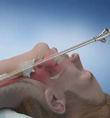
By strategically placing mirrors, reflected images were examined, eg, the larynx and vocal cords, by the ENT surgeons by using an indirect laryngoscope, which some physicians are still using today. The examination of the lung airways, or the esophagus and stomach, was a major undertaking. The patient's head and neck had to be brought in line with the rigid bronchoscope or esophagogastroscope. And in a good number of cases, the patients lost a tooth or two and suffered bruises on the lips and tongue. Thanks to the scientists for inventing fibrotic fibers and the birth of the flexible endoscopy.
A short history of this development is recalled here.
In 1806, Dr. Phillip Bozzini of Germany used a rigid instrument to examine the stomach of a patient. An improved “open-tube” endoscope was designed and used by Dr Anonin Jean Desormeaux of France.
To shine a light from a burning candle down the tube inside the stomach was a major handicap. Sir Francis Cruise of Ireland developed an electric bulb in 1865, and soon electric bulbs were incorporated in such instruments.
Semiflexible scopes.
A German physician, Rudolf Schindler, working with George Wolf, designed the first semiflexible Gastroscope in 1932. The distal end of the scope was flexible and had a system of lenses, and the proximal end of the scope was rigid and made of metal.
Fiber optics:
Light was used to signal between ships at sea, and not much for other types of communication. In 1840, Daniel Collodon and Jacques Banet proved that light could travel along a curved path of a water jet. Many decades later, it became known that light could also travel along a bent quartz rod. In 1940, doctors used illuminated plexiglass tongue depressors. A medical student, Heinrich Lamm, in Germany, demonstrated image transmission through a bundle of optical fibers. In 1054, an article in the Journal Nature, authors A. van Heel, H.H. Hospkin, and Narinder Kapnay explained the science of transmission of images along a fiber optic cable by internal reflection.
Hirschowitz and Pollard of the University of Michigan visited Knapnay and discussed the application of fiberoptics to endoscopy. Three years later, they produced their fiber-optic gastroscope and Hirschowitz introduced the gastroscop on himself. In 1960, Hirschowitz received the patent for the gastroscope, and the American Cystoscope Manufacturing Inc. started producing a fiber-optic gastroscope.
Internal Reflection:
The fiber optic cable has an inner core made of pure silica glass or plastic fiber, the diameter of which is 1/10 of a human hair. The core fiber is surrounded by glass fibers, called the cladding. The optical properties of the inner core fiber and the cladding are different.
The core fiber has a higher refractive index, allowing light to travel more slowly. The light travels along the core by bouncing off the wall of the cladding repeatedly onto the core and keeps the light signal inside. The total internal reflection allows light to travel with minimal loss of signals over a long distance at high speed, even when the cable is twisted or bent.
In 1970, Corning Glass researchers R. D. Maurer, D. Keck, and P. Schultz developed fiber-optic wire with optical losses low enough for wide use in telecommunications.
The Nobel Prize:
The Nobel Prize for Physics in 2009 was awarded to C.K. Kao, W.S. Boyle, and G.E. Smith for their work in fiber-optic and semiconductors. The Nobel committee remarked that the three scientists helped shape the foundation of modern networked society.
Endoscopy surgery:
Technical expertise for minimal invasive surgery by endoscopy surgery requires special dexterity, advanced knowledge in anatomical structures from multiple lines of sight and their relationship with one another in a limited field of vision of the surgeons, in addition to several special instruments.
Three groups of instruments are essential.
Endoscope and with all its accessories.
- Accessory instruments for Endoscopy surgery
Excellent illumination.
Imaging equipment and monitors.

.