Saturday, November 25, 2023

Oriental Sore / Cutaneous Leishmaniasis.

  Cutaneous Leishmaniasis

PKGhatak, MD.


Oriental sore is present in a wide area of the world. The majority of the cases are from the New world countries - Brazil, Bolivia, Peru, Panama, countries around the Mediterranean sea. CL is also prevalent in the Old world countries- the Middle East and Central Africa.

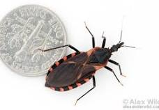
CL is a parasitic disease of the skin and the mucous membranes of the nose, mouth and throat. The parasite is Leishmania which belongs to the Trypanosomastide family and Kenetoplastids order. CT is transmitted to humans by Sandflies.


Incidence of CL.

About 600,000 to 1 million of new cases of CL occur each year. The mortality is few in number and comes from secondary bacterial or, fungal infections but the morbidity is a serious problem.

Anatomy of Leishmania.

Leishmania is an unicellular organism, exists in two different anatomical forms. In the insect vector, leishmania is an oblong flagellated form called Promastigotes reside in the proboscis of the sandfly.. A small round non- flagellated form called Amastigote is ingested by the sandfly from humans during feeding. In the gut of the sandfly, the parasites change to flagellated form.

The nucleus of leishmania is present in the center of the cell; in front of the nucleus in the cytoplasm, a mass of mitochondrial DNA is present called Kinetoplast. From the Kinetoplast a flagellum originates and extends outside. In the cytoplasm, a single stand of mitochondrion and Golgi apparatus are also present.

Leishmania have a unique DNA and glycosomes. It was believed that Leishmania did not propagate by sexual union but recent studies provided contrary evidence that amastigotes exchange nuclear material during binary fission. Leishmania has 34 to 36 chromosomes and during cell division, the chromosomes do not condense.

Life Cycle of Leishmania.

The flagellated Promastigotes infect humans. Sandfly bites introduce the Promastigotes in the human victims. The macrophages, neutrophils and phagocytes engulf the parasite but are unable to break down its surface membrane. Inside the cells, the Promastigotes change into Amastigote. In the macrophages, the amastigotes multiply rapidly to such a number that the cell bursts open and the newly released amastigotes infect other tissues.

Inside the sandfly.

The sandfly takes a blood meal and swallows macrophages loaded with amastigotes, inside the gut the macrophage wall breaks down releasing the amastigotes. Amastigotes transform into Promastigotes and divides and move to proboscis of the sandfly.

 20 different species of leishmania produce CL., and 90 different species of sandflies are the vectors. The important Leishmania species in the old world are L.infuntum, L.donovani, L.major, L.aethiopia. In the New world's species are L.mexicana, L.infuntum, L.guyanensis, L.amazonensis, L.braziliensis and L.panamanensis.

The Phlebotomy sandfly is the vector in old world and Lutzomyia sandfly in the new world. A species of sandfly harbor only certain Leishmania species and that limits one type of CL lesion confined to a local area and absent from adjoining village or locality.

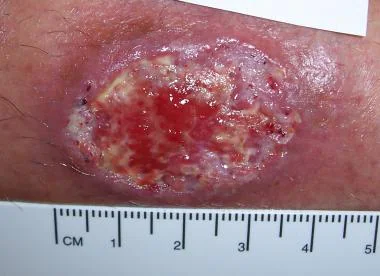
Development of the Skin lesions and Clinical Types.

The sandfly bites on the face, forearms, hands and legs. The bites are painful. A red papule develops at the bite site which turns into a nodule. Several days or weeks later, the nodule ulcerates. This is the beginning of the chronic indolent Oriental sore.

The subsequent healing or progression of CL depends on (a) Leishmania species.(b) victims' immune status- both the innate and acquired immunity. (c) Intercurrent immune diseases. (d) suppressed immunity by drugs which lowers cellular immune functions or delays and diminishes immune globulin synthesis.

Clinical types of CL. (a) Limited Cutaneous Leishmaniasis.(b) Recidivans Leishmaniasis.(c) Mucocutaneous Leishmaniasis. (d) Anergic Diffuse Dermal Leishmaniasis. (e) Post Kala-azar Dermal Leishmaniasis.

Clinical features.

(a) Limited Cutaneous Leishmania. The initial lesion does not progress to ulcers because the victims have good levels of immunity. The lesions heal spontaneously with scars, and patients develop permanent immunity.

(b) Recidivans Leishmaniasis. The initial lesion heals with scars, and months or years may pass without further disease activities. Then new skin lesions begin to appear at or, near the scars. The process goes on for a while, and ultimately the skin lesions heal. Generally, no medication is required.

 (c) Mucocutaneous Leishmaniasis. It is the most devastating form of CL. The initial lesions develop around the nostrils, mouth and eyes. Satellite lesions may develop in the throat. The mucosal lesions produce tissue necrosis and usually penetrate into the deeper layers of tissues and erode the muscles, cartilages and bones. Without medical therapy no recovery is possible. The WHO recommends that all Mucocutaneous Leishmaniasis should be treated and the WHO provides guidance and medications in participating countries.

 (d) Anergic Diffuse Dermal Leishmaniasis. The patients are immune deficient and usually have IHV infection. The initial lesions are papules and nodules but lesions do do ulcerate. Because of the lack of immune cellular response, multiple lesions appear, usually on the face, arms and buttocks. Later, the entire body is covered by several forms of skin lesions – plaques, papule and nodules. The appetence resembles nodular leprosy, however, no nerve damage occurs in this form of leishmaniasis. Treatment with medications is not very effective and where initial success is achieved, the recurrence of new lesions is common.

 (e). Post Kala-azar Dermal Leishmaniasis. Visceral leishmaniasis is better known as Kala-azar. 2 to 20 years after the cure of Kala-azar some patients develop hypopigmented macules, nodules, plaques and erythematous plaques on the face and other areas of the body. The lesions are recurrent and may persist for years.

Diagnosis of CL.

Biopsy. The demonstration of amastigotes within the macrophages is the standard diagnostic method. If there is no blood in the smear and stained with Geismar or Wright stains, a trained person can identify amastigotes without difficulty. An alternative to a biopsy is the aspiration of clear serum from the lesions. This method is equally sensitive. However, in certain forms of CL the parasite numbers are few. So more and more reliance is shifting towards antibody detection by ELISA or antigen DNA detection by PCR test.

Treatment of CL.

The drug treatment is standardized by the WHO and the CDC in the USA. Few cases of CL were detected in the returning veterans from Afghanistan and Syria. In addition to standard therapy, each country has a host of local treatment protocols which are not effective but popular among the locals. Some of them are Cryotherapy, Heat therapy at 40 to 42 degrees C, Paromomycin topical preparations, infiltration of the ulcers with the solution of Sodium Stibogluconate, and urea 10 -15 % solutions. Oral dapsone, Oral Allopurinol and Oral antifungal agents

Standard therapy.

Pentavalent Antimony agents. 

Two commercial preparations are available -Sodium stibogluconate ( Pentostam) and Meglumine Antimonate (Glucantime). Both these two are given intravenously for 10 to 20 days

Liposomal amphotericin B. It is an antifungal drug and is also effective in leishmaniasis but expensive.

Oral Pentostam,

 Oral Miltefosine. It is a wide spectrum antibiotic also effective against Leishmania. Initially, it was approved for cancer therapy.

Various combinations of drug therapy are practiced based on the leishmania species and the degree of tissue damage.

Prognosis.

Mucocutaneous and Anergic diffuse dermal leishmaniasis have poorer prognosis because of destructive facial lesions and disfigurements. Mortality is negligible as such but secondary infection may lead to septicemia and deaths.

Vaccine: No commercial vaccine is produced for Leishmania disease. Russia made a vaccine using attenuated L.major and used in Russian endemic areas with 50 % success in controlling new skin lesions, but the vaccine does not protect against infection from other species than L.major, and also has some troublesome side effects.                           

*************************************************


Tuesday, November 21, 2023

Chagas Disease


Chagas Disease

American Trypanosomiasis.

PKGhatak,MD.


Chagas disease is named after Dr. Carlos Chagas of Brazil. Trypanosoma cruzi causes Chagas disease in South America and Trypanosoma gambiese causes Sleeping Sickness in Africa. Several species of Leishmania are pathogenic to humans and both Trypanosoma and Leishmania belong to one family.

In 1909, Dr. Chagas investigated an unknown illness that was common in the rural areas of Brazil. He found a blood sucking insect feeding on humans and leaving behind feces and urine near the wound. The excreta of the kissing bug were loaded with the infective form of the parasite. The victims, while sleeping rubbed the feces and contaminated the wound and the parasite also entered the body through the conjunctiva of the eyes.

Trypanosoma cruzi can infect in other ways - Contaminated food and drink by vectors' feces and urine often cause local endemics, contaminated blood transmission and organ transplantation are less common.  Mother to child transmission during pregnancy is a major concern and the children in subsequent pregnancies are also infected.

The victims developed illness in two phases – 1. The initial or the first stage. 2. The chronic stage.

The initial stage.

The initial illness is a mild local inflammation and the regional lymph adenitis and in a few instances only mild systemic symptoms. Occasionally, a furuncle, called Chagoma, develops at the wound site. The illness is self limited. About 50 % of new infections may remain symptom free.

In the 2nd or Chronic phase.

Years may pass without any signs of illness, and then patients present with symptoms of serious heart, gastrointestinal and neurological diseases.

The parasite Dr. Chagas discovered was a flagellated Protozoa in the blood of the victims. He conclusively proved the pathogenicity of the protozoa by recovering the same organism is susceptible animals. Dr Chagas named the parasite Trypanosoma cruzi to honor his mentor Dr. Cruz.

The Life Cycle of the Protozoa Parasite:

The organism is a motile, unicellular organism having one flagellum. The protozoa have 5 stages of development and two methods of propagation - one by binary fission and the other by sexual union. The different stages of life of the protozoa are named according to the point of attachment of the flagellum to its body or the absence of the flagella. The Greek word for the flagella is mastigot. The infective form that circulates in the blood of the victims is called Trypomastigotes.

Inside the bug.

In the midgut of the insect, the trypomastigotes transform into Epimastigotes, having the flagellum attached to the end of the body. Here epimastogotes multiply, and the epimastogotes enter the hindgut and transform back to Trypanostigotes, And infect humans when the bug takes a bloody meal.

In the victim.

The Trypomastigotes  transform into a round small unicellular organism inside the victims' cells. The parasite has no visible flagella and is called Amastigotes. Amastigotes multiply by binary fission. Amastigotes transform into trypomastigotes and burst open the victim's cells. The released Trypomastigotes infect more tissue, and also enter the bloodstream and lymphatics and are widely distributed in the body. Some of the Trypomastigotes switch back to Amastigotes  divide again and keep on multiplying by binary division. And the cycle repeats again and again.

Besides humans, small animals like monkeys, armadillos and dogs are also infected.

The insect vector. 


The bug that transmits Chagas disease is a Triatomine bug, a variety of Reduviid bug, locally known as the Kissing bug, as they find it on the faces of victims.

There are several species of the Triatomine bug, one species is active in a certain locality or another. The bug is active at night. They come out of cracks of the wall, fall from the ceiling or just crawl on the bed from outside. The bugs bite on the faces of humans (part not covered during cold nights). A recent report says oral route of infection, in recent days, has been the dominant path of infection of Chagas disease.

Chagas Disease:

The incubation period is short.

The initial illness is mild and self limited, and often remains asymptomatic.

The chronic phase of Chagas disease is a protracted severe symptomatic disease of the cardiovascular, gastrointestinal and Neurovascular systems. The mortality rate is high and the disability is severe.

Pathophysiology:

The amastigotes invade muscle cells of the heart and the smooth muscles of the esophagus, and colon and also invade the brain. The inflammatory reactions are mediated by acute phase Cytokines. The initial myocarditis is followed by necrosis of muscles are replaced by fibrous tissues. The cardiac and smooth muscles of the GI tract become weak and flabby. The heart is the most common organ affected by Chagas disease. Cardiac impulse propagation along the His bundle due to fibrosis manifests as heart block, branch block, cardiac arrhythmia and systemic embolism. Massive dilated cardiomegaly and heart failure are the prominent features of this disease. Megaesophagus leads to difficulty in swallowing solid food, and megacolon leads to chronic constipation and abdominal distention and discomfort.

The CNS infection results in strokes and many other neurological manifestations.

Diagnosis:

The mainstay of diagnosis of Chagas disease is the demonstration of moving Trypomastigotes under the coverslip of a thick smear of blood. Under the trained eyes the movement of the transparent parasite around stationary red cells is not difficult. It is a cost-effective test and practical in the rural communities.

Other confirmatory tests and PCR antigen detection and indirect ELISA antibody detection.

Medical treatment:

Two oral medications, Benznidazole and Nifurtimox are effective in killing the parasite but the drugs must be given early in the acute phase of the disease in children 2 to 18 years of age. The older adults develop toxic side effects more frequently and drug therapy is not recommended in adults. Women of reproductive age are tested and treated with drugs to prevent congenital Chagas disease. 

Benznidazole is an imidazole derivate, actives against intracellular parasites. In Chagas disease, the drug is prescribed for 60 consecutive days. The drug inactivates parasitic enzymes and damages DNA and large protein molecules by generating cation radicals. Common side effects of this drug are various GI symptoms, skin rashes peripheral neuritis, thrombocytopenia and leukopenia, and anemia. It is potentially carcinogenic.

Nifurtimox is a nitrofuran drug. The mode of action of Nifurtimox against the parasite is similar to Benznidazole and it generates radicals through its action of nitrogenous enzymes. The adverse reactions are also like Benznidazole. But when prolonged treatment is necessary, Nitrofurans is not suitable because of its adverse effects which are more intense than Benznidazole. Both drugs are contraindicated in pregnancy.

Surgery: 

In the past various types of cardiac surgery were tried to improve function of the failing frail and flabby heart muscles. In the long run, none of those procedures proved to be beneficial and subsequently abandoned. Heart block is effectively treated with cardiac pacemakers. Only cardiac transplantation is a solution is severe heart failure.

Megaesophagus.

Laparoscopic myomectomy, esophageal mucosectomy are less involved procedures. Heller-Pinotti procedure is a more involved operation and is done in early swallowing difficulties. It is a modified Fundoplication operation.

In advanced cases, Thal-Hatakufu operation is done. In this operation, Esophgogastroplasty is performed. The success of the Thal-Hatakufu operation is moderate.

Megacolon.

A Total Colectomy operation is required with anastomosis with -(a) ileorectal, or, (b) ileoanal, or, simply a colostomy.

Prognosis of Chagas disease depends on the stage of the disease. In the early actives phase, the drug treatment is curative.

The survival in the chronic phase is poor.

Incidence and prevention of Chagas disease.

18 million people in 21 countries of Central and South America are infected with Trypanosoma cruzi and 100 million people are at risk of infection.

 Insecticide is used to control the insect vector. Better housing and public education have been successful in bringing down the spread of Chagas disease.


***************************************************


Whipple's disease

  Whipple's disease: Whipple's disease is a bacterial infection of jejunum manifest as malabsorption of fat, fat soluble vitamins...