Monday, October 24, 2022

Epstein - Barr Virus Infection.


Epstein Barr Virus.

PKGhatak, MD


The Epstein Barr virus was initially called the Burkitt lymphoma virus because the virus was identified in the cells of the Burkitt lymphoma in an African patient in 1964. It is known today as Human Herpes Virus 4. This virus is the most common viral infection among all other herpes virus infections in humans. About 85 % of the world population have serology positive blood for Epstein Barr Virus (EBV), indicating past infection.

EBV virus characteristics.

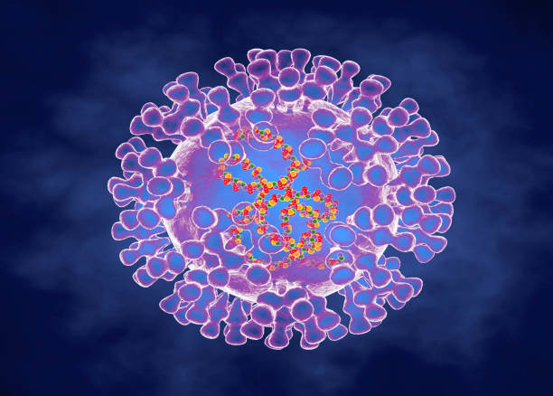
EBV belongs to Gamma-Herpesviridae in the genus group of Lymphocryptoviral. The double stranded genome contains 85 proteins and 50 non-coding RNAs. The viral particle has an outer lipid envelop which fuses with the respiratory cell membrane at the time of infection. The DNA of the virus can turn out 9 different molecules of Entry Proteins, gP 1 – 9, and use them to infect many other cell types of the patient.

The EBV virus, once inside the respiratory cells, multiplies by redirecting the host cell to duplicate virus particles. The virus then leaves respiratory cells and infects B-lymphocytes. Inside the B-lymphocytes, the virus persists for life and goes through 3 cyclic phases, namely active phase, dormant phase and re-infective phase.

A person infected with EBE is unable to clear the virus from his body. A subsection of B-lymphocytes is memory cells. The memory cells continue to turn out immunoglobulins, the initial product is IgM and 2 to 4 weeks later all the products are IgG.

In addition, antibodies are produced against capsular proteins and nuclear proteins.


Why EBV virus lives in perpetuity in humans.

Whenever antibodies are produced in adequate amounts, the virus goes into hibernation within B-lymphocytes and also in some cases in the respiratory epithelial cells. The NK cells, monocytes and macrophages are unable to find the EBV once inside the cells and antibodies are ineffective to neutralize the viral particles. This hide and seek game is played out throughout the life of the patient.

In certain circumstances, the EBV can also infect NK cells, monocytes and macrophages and produces more serious illnesses.

EBV human infection.

Children are the prime target of the virus and about 80 % of human infections take place in children under 2 years old. Most of the infected children remain free of symptoms or had minor symptoms. The only evidence of infection in these cases is positive serology tests.

The next venerable age is teenagers. Teenagers, however, become symptomatic and the illness is known as Mononucleosis or simply Mono and also commonly called a kissing disease. The most remarkable symptoms are the posterior cervical lymph node enlargement and enlarged spleen. Other significant symptoms are sore throat, fever and enlarged tonsils.

Persistent and recurrent activation of EBV.

The EBV can become active within the immune cells, whenever the immune systems are down. The repeated reactivation of the EBV within the immune cells produces Multiple sclerosis and rheumatoid arthritis.

Reactivation within the respiratory epithelial cell leads to Sjogren syndrome and Systemic Lupus Erythematosus (SLE).

The positive ANA, pANCA, cANCA and Rh factor tests are the results of back and forth infections switching cell lines over a time.

In patients with seriously handicapped immune systems, the recurrence of active and inactive stages of EBV leads to B cell Lymphoma and Nasopharyngeal Carcinoma.

Systemic Autoimmune Disease (SADs).

A group of autoimmune diseases has one common factor of connective tissue damage. There are overlapping symptoms between these entities and often some positive serological tests are also common. The diseases are Mixed Connective Tissue Disease (MCTD), Myositis, Rheumatoid arthritis (RA). Systemic Sclerosis (SS), Lupus erythematosus (LE).

Autoimmune vasculitis of various clinical entities is associated with EBV infection.

Serology tests for EBE viral illness.

Mono spot test. And Heterophile antibody test.

Mono spot is no longer recommended for routine use for the diagnosis of mononucleosis in children because of false negative and some false positive results. But the science behind it is solid.

EBV produces antibodies which cross react with RBC antigen of horses and cows. This property of antibodies leads to the name of this test as Heterophile antibody test.

The heterophile antibody test is modified as a Mono slide test, available as a test kit and commonly used in clinics and pediatrists' offices. It is a modified and improved Paul- Bunnell test.

Immunofluorescence tests.

Viral Capsid Antigen (VCA) IgM Test.

The test is performed using antibodies tagged with fluorescence dye mixed with the patient's serum. A positive test is seen in about a week after infection and disappears in 4 to 6 weeks.

VCA IgG Test.

The test becomes positive in 2 to 4 weeks and then declines slightly and remains positive for the rest of the life.

Early Antigen Test (EA) Test.

Anti EA IgG antibodies appear during the acute phase of the infection and disappear in 3 to 6 months. In 20 % the Anti EA IgG may remain positive for a very long time, otherwise, an Anti EAIgG positive test indicates a recent infection. 

EBV Nuclear Antigen Test.

It is also a fluorescence test against viral nuclear antigen that becomes positive in 2 to 4 months after infection and remains positive for the rest of life.

False Positive Heterophile Antibody Test.

False positive heterophile antibody results are seen in Viral hepatitis, Rubella, Toxoplasmosis, HIV/AIDS, Malaria, Lupus and Pancreatic Cancer.

A false negative Heterophile test is extremely rare in adults. Children do not readily produce Heterophile antibodies in the first two weeks following infection.

Blood Test.

The mononucleosis name is due to an increase in monocyte count in the peripheral blood. The total count increases only modestly in the range of 10,000 to 20,000. The lymphocyte count is generally over 50 %. And on the smear, Atypical lymphocytes appear.


*******************************************************

Sunday, October 23, 2022

Monkeypox


Monkeypox.

PKGhatak, MD


Monkeypox virus belongs to an enveloped double stranded DNA virus of the genus Orthopoxvirus in the family Poxviridae and belongs to a subfamily of Chordopoxvirniae. In central and west African jungle the monkeys are the natural host and this was also documented in squirrels, prairie dogs, rats, dormouse and primates.

                               Artists version of a monkeypox virus.

The first human illness was recorded in the Democratic Republic of Congo in 1970 just two years after Smallpox was eliminated there. From time to time local outbreaks were reported by the WHO. Cases were seen in central and west African countries when children were bitten by monkeys.

There are two subclasses of this virus – one in the Congo Basin virus which is more virulent and has a higher rate of transmission, and the other is in West African. At present both varieties are infecting the people of Cameroon.

The first reported case in the USA was in 1980. In 2022 the USA declared monkeypox a public health emergency. Human to human transmission was seen in NYC in the homosexual community. Infection of humans takes place through the exchange of body fluid, respiratory droplets, prolonged close contact, and occasionally by the use of contaminated towels and bed sheets.

The incubation period is 5 to 21 days. The initial symptoms of malice, body aches, fever and lymphadenopathy for 4 -5 days, then the rashes appear on the face and extremities including on the palms and soles. In homosexual individuals, rashes are also present in the genital and anal areas. In contrast to other varieties of pox, monkeypox patients show striking lymphadenopathy.

Pox is known to appear on the lips and mouth, conjunctiva of the eyes. The rashes go through the stages of macules, papules, vesicles and pustules. In three weeks, the scabs of pustules dry out and then drop off. Until all scabs fall, the patient remains a source of infection. The monkeypox illness is a mild disease but in immune deficient people it can produce severe illness. In African countries as high as 10 % population is contracted with infection, and the fatality is generally below 3 %, and in children, the mortality has gone up to 6 %.

Diagnosis of monkeypox is done on samples obtained from the skin of the lesions or the fluid of vesicle/pustule by PCR test to confirm the monkeypox virus. Blood tests are not reliable and other varieties of pox produce the same serology test results.

Monkeypox vaccines.

Two vaccines are available in the US. In post exposure two doses of the Jynneos vaccine are given. Another vaccine ACAM2000 is less commonly given but available on a limited scale. In very high risk groups prior exposure vaccination is approved.

People vaccinated for smallpox have limited protection in high risk groups, for the general public provides 85 % protection against monkeypox.

Antiviral drugs.

In Europe, an antiviral drug, Tecovirimat, originally developed for the treatment of smallpox is approved for monkeypox. In the USA Tecovirimat is approved temporarily on an emergency basis and availability is limited.

*************************************

Cerebro Spinal Fluid

 Cerebro Spinal Fluid (CSF)

PKGhatak, MD

The previously held view of the formation and circulation of the cerebrospinal fluid (CSF) has undergone modifications because current reproducible and non-interventional methods of investigation, like MRI and molecular and biological biology, discovered newer facts that contradicted the past theory.

To update the current thinking, a brief review of the histology of the Choroid plexus, Ventricles of the brain and Arachnoid granulations is necessary.


Choroid plexus.


The Ependymal cells of the choroid plexus secrete the CSF by an active process. The central capillary of the choroid plexus is lined with one layer of fenestrated cuboidal cells. The albumin content of CSF is much lower than plasma. The transport of water across the ependymal cells of the carotid plexus is carried out by separate transport proteins, one for the ventricular side and the other for the capillary side. It is an active process requires energy from ATP and likely also from K / Cl transporter. About 30 % of the total water volume of CSF comes from the inter-neuronal space, generated from the metabolic activities of the nerve cells, like Lymph formation in the other organs. There are to and fro moments of fluid between interstitial fluid and CSF.

Blood Brain Barrier.

This special characteristic of the blood vessels of the brain is due to the tight seal between the endothelial cells of the capillaries and supplemented by perivascular feet of astrocytes surround the basement membrane. In two locations this barrier is absent namely the choroid plexus and posterior pituitary gland.


Arachnoid Granulation:

Arachnoid granulations are collections of capillary networks kept suspended into the large veins of the subarachnoid space. The anatomical structure suggests arachnoid granulations are filters - a one way passage of CSF into the venous blood, getting rid of waste products and excess water. But that is not the case. Arachnoid granulation plays a minor role in this respect.

The perineural space along the cranial nerves and spinal nerves and the perivascular space known as Virchow Robin spaces are the main routes of drainage and filter of CSF into the general lymphatic system. This is specially evident in the Olfactory nerve as it travels through the cribriform plate of the nose.


Circulation of CSF:


The circulation of CSF in the brain and spinal cord.

The circulation of the CSF is an active process by the synchronous motion of cilia lining the surface of the ventricles and the central canal of the spinal cord. The CSF exits the brain through two openings of Luschka in the hindbrain and circulates in the subarachnoid space. The mechanism of a steady state of CSF formation, circulation and drainage has undergone many modifications. The circulation is pulsatile in nature due to and fro movement of the CSF. The interstitial fluid exchange with CSF is a major component of circulation. A continuous bidirectional fluid exchange at the blood brain barrier produces flow rates. The general lymphatic system of the body is the main route of drainage of waste products of the brain and an entry point of the immune system in the brain.


******************************


Whipple's disease

  Whipple's disease: Whipple's disease is a bacterial infection of jejunum manifest as malabsorption of fat, fat soluble vitamins...