Wednesday, November 16, 2022

Leishmania

                                                                    Leishmaniasis

                                                         PKGhatak, MD


Leishmania is a flagellated protozoan of the genus Leishmania. Of the several species of Leishmania, about 20 species are infectious to humans. The flagellated tiny protozoa enter human victims from the regurgitated gut contents of female sandfly bites when the fly probes for a blood meal. There are about as many varieties of sandflies as there are leishmania species. The infectious form of the parasite is called Promastigotes. Promastigotes bind to the receptors on the skin macrophages. The parasites drop their individual flagella and enter the macrophage and continue to live inside the macrophage and at this stage, the parasite is called Amastigotes and the amastigotes multiply and infect more macrophages; eventually, macrophages of the skin, reticulo-endothelial tissue and viscera are loaded with parasites. 

The human body is unable to mount an immune defense against leishmania because the parasite successfully thwarts both innate and acquired, cellular and humoral arms of the immune system. Thymic 1 cells (TH1) secrete IL-12 which transforms native T-cells into TH1 cells. TH1 cells produce TNF gamma (tissue necrosis factor gamma) and NK cells (natural killer cells). These are the resistive forces to the body. The parasite switches T-cells to TH2 cells which secrete IL-4 and IL-10 and which protect parasites from being killed.

Leishmaniasis is prevalent around the tropical and subtropical regions. The countries are separated into the Old World and the New World countries. The old world belongs to Central Asia, the Eastern shore of the Mediterranean, the Indian subcontinent, South Eastern Asia, and Eastern African countries. The new world comprises Central American and South American countries. It is estimated that between 1 and 2 million people are newly infected every year, but only a small fraction of them will develop the disease and 20,000–30,000 die each year.

 An increased number of leishmania cases are seen in the last 25 years. This increase is due to globalization and climate change, air travel, sharing contaminated needles between IV drug users, being unable to detect contaminated donated blood, close human interactions with domesticated animals, particularly dogs, which serve as an intermediate host, HIV infection and blood and organ donations.

Clinical feature.

Several factors determine the nature of Leishmania clinical presentation. Besides the leishmania species and sandfly subtype, other factors are climate and the immune status of the individual. The cutaneous, mucocutaneous and visceral forms of leishmania are the main clinical types. Visceral Leishmania is better known as Kala-azar, an Indian word for Black fever. In certain endemic areas, Kala-azar is followed by dermal lesions which outwardly resemble Lepromatous leprosy and are known as post-kala-azar dermal leishmaniasis (PKDL). In Brazil mucocutaneous leishmania is known as Espundia. In the old world, the cutaneous lesions are known as Oriental Sores.

Cutaneous lesions (Oriental Sores).


The incubation period is weeks to months following the fly bite. The skin lesion may be single or multiple, dry or wet. In the new world, a single lesion is usual, multiple lesions are more common in the old world. The lesions may spread by lymphatics and satellite lesions are seen. Initially, a red papule appears and slowly but gradually progresses to a macule and ulcer. In central Asia, Afghanistan and western Pakistan, cutaneous leishmania is the common presentation.

Mucocutaneous. 

Mucocutaneous presentation is mostly seen in Mexico and Brazil. Lesions on the face are associated with ulcerated lesions in the nose, mouth, pharynx and trachea. Ulcers and scar tissues produce deformities and organ dysfunction. The incubation period is 1 to 3 months. Urgent medical treatment is necessary.

Visceral.

At an earlier time, the undivided province of Bengal, India was the epicenter of kala-azar. The incidence has fallen sharply but sporadic cases are seen in Bihar, Nepal, Bangladesh, West Bengal, and the Eastern shore countries of North Africa. The incubation period is 3 to 6 months and may be as long as a year. Fever, weakness, enlarged spleen and liver are followed by a characteristic double spike of fever every day, anemia and pancytopenia, increased skin pigmentation and dryness of the skin. Dependent edema, ascites and secondary infection follow and deaths in 2 years if no treatment is available.

PKLD (post kala-azar dermal leishmaniasis).

Years after visceral leishmania is treated and cured, nodular lesions appear on the face, the trunk and the limbs. The lesions are numerous and have the appearance of leprosy, except for the perception of touch and pain remains. And no spontaneous amputation of digits and toes occurs in leishmania as it happens in leprosy.

Viscerotropic leishmania.

This is a subvariety of visceral leishmania but after initial symptoms, the disease does not progress.

Diagnosis.


The parasite must be demonstrated in the samples taken from a skin biopsy and from the bone marrow biopsy in visceral leishmania.

Treatment.

CDC designated drugs used in the treatment of leishmania as orphan drugs. Anyone can contact CDC for guidance and will be assured of a supply of medications.

In 1920, Dr. Upendranath Bramhachari, a native of Kolkata, India, discovered that when Urea Stibamine was given IV it was very effective in bringing an end to the human misery in Bengal. Initially, the cure rate was 90 % and not many side effects were noted. But in the long run, safer Antimony compounds were introduced. Even today, the medical community of India pays tribute to Dr. Bramhachari for his service to humanity.

Antimonial.  The pentavalent antimony (Sb V) enters macrophages and fuses with the membrane of the Lysosome of macrophages. There it is converted to Sb III (trivalent) compound. Sb III is a poison to the amastigotes.

Miltefosine.  It is an oral anti-leishmania drug. It blocks the lipid metabolism of the parasite. It is also useful in children but has the potential for malignancy. Combining it with Amphotericin B and Paromomycin increases efficiency and lessens side effects.


                                                             Dr. Bramhachari.

***************************************************************************


Saturday, November 12, 2022

Malaria


                                                                      Malaria

                                                           Some important aspects

                                                     PKGhatak, MD


A tiny one-cell parasite infected primates in equatorial Africa, perhaps thousands of years ago. At a later date, the parasite found humans as suitable prey. It was a time before writing developed. We do not know whether those people knew malaria was a parasitic disease. Egyptian, Chinese and Indian physicians left some documents. After a retrospective analysis of those documents, some appeared to be malaria fever cases.

Romans thought the febrile illness was from Mal-Aria (bad air) rising from the swamps; they correctly identified the swamp as the breeding ground of mosquitoes but failed to connect the mosquitoes with the spread of malaria.

That task was completed in 1857 in Kolkata, India by Ronald Ross. He identified the parasite in the female Anopheles mosquitoes. He recovered the same parasite Plasmodium relictum from the blood of birds, linking mosquitoes with birds' malaria. He was awarded the Nobel Prize in 1902 for his groundbreaking research. Subsequently, other researchers discovered the complete life cycle of the malaria parasite - the Plasmodium.

Malaria parasite.

Malaria belongs to the Genus Plasmodium of the Superphylum Alveolate, and the Phylum Apicomplexa. Like all Alveolate, Plasmodium contains a plant plasmid, used in Photosynthesis by the plant. The plasmodial plasmid is a bit different, it is used in extracting a fatty acid, Isoprenoid, from the host cells in order to develop and multiply in the liver and RBCs in humans and in the salivary gland of mosquitoes, and not used in photosynthesis.

Six species of Plasmodium infect humans, namely -

1.Plasmodium falciparum (P. falciparum)

2. P. malariae

3. P. vivax (with identical morphologically P. simiovale and two other related species P. minuta and P. tenue)

 4.P. ovale

5. P. Knowles.


Know the Female Mosquitoes.


Female Mosquitoes are equipped with chemical sensors for Carboxylic acid, a CO2 derivative. Using the sensors, mosquitoes zero in on a person who has higher carboxylic acid in the breath. Female mosquitoes must have a high protein meal (blood) for the development of eggs and laying fertilized eggs on the surface of non-flowing bodies of water, e.g., ponds/swamps, etc.

Follow the parasite.

Mosquitoes' salivary glands are loaded with the infective forms of the parasite, and mosquitoes deposit them when they bite and suck human blood. At this stage, the parasite is known as Schizont (no English word for it). From the skin, the parasites travel to the liver and enter the liver cells. In the hepatocytes (liver cells) the parasite multiplies. The parasites break free from the liver cells and infect RBCs. Duffy antigen receptors on the RBC surface are the anchoring point of the parasite and serve as the entry point into the red cells. At this stage,  the parasite is called Merozoites. In the RBCs, the parasites begin to divide by themselves (nonsexual multiplication) and the new Merozoites very quickly multiply and reach a huge number and the red cells burst open and the merozoites enter into the bloodstream and begin to infect more RBCs. And the cycle keeps repeating until some merozoites change to male and female reproductive units called Gametocytes and wait for the mosquito to bite and draw blood. In the gut of the mosquito, the male and female gametocytes unite and produce their progeny, the schizonts. Schizonts move to salivary glands and wait to infect a new victim.

Time taken for the parasite to complete human RBC cycles.

In the case of P. falciparum, P. ovale and P. vivax the cycle is repeated every 3 days. In P. knowlesi and P. malariae the cycle only a day.

Symptoms of malaria.

Malaria symptoms vary according to the species of Plasmodium.

a. Shaking chills followed by high fever. 

With no warning, a sudden onset of chills and shaking starts. The chattering of teeth, assuming a fetal position and covering the body with all comforters or blankets does not bring any relief of chills till the fever starts. The temperature is usually 104 degrees or higher and weakens, profuse sweating and prostration follow when the fever breaks.

b. Hemolytic anemia, jaundice and renal insufficiency and renal failure.

The broken RBCs release not only the parasites but also the hemoglobin into circulation. Free hemoglobin is an oxidant that damages glomerular capillary endothelium and decreases renal filtration. In recurrent episodes of hemolytic anemia, renal failure develops. Hemoglobin is broken down in the liver into heme and globulin. The heme is a waste product. Light jaundice becomes evident, urine and stool develop darker shades of yellow.

c. Anemia, failure of children to thrive in endemic areas, susceptibility to tuberculosis and HIV infections are common in certain countries in alarming numbers.

d. Enlarged spleen and liver and marked anemia. Parasites induce immune reactions and chronic inflammation and enlargement of organs.

e. Blackwater fever.

Certain patients, generally non-native residents, taking Quinne irregularly for the treatment of malaria develop dark colored urine due to the presence of hemoglobin in urine and chronic renal failure. It is thought to be an autoimmune disease, triggered by the interaction of quinine with plasmodium. The antibodies then attack the kidneys.

Laboratory diagnosis:

Peripheral blood examination.

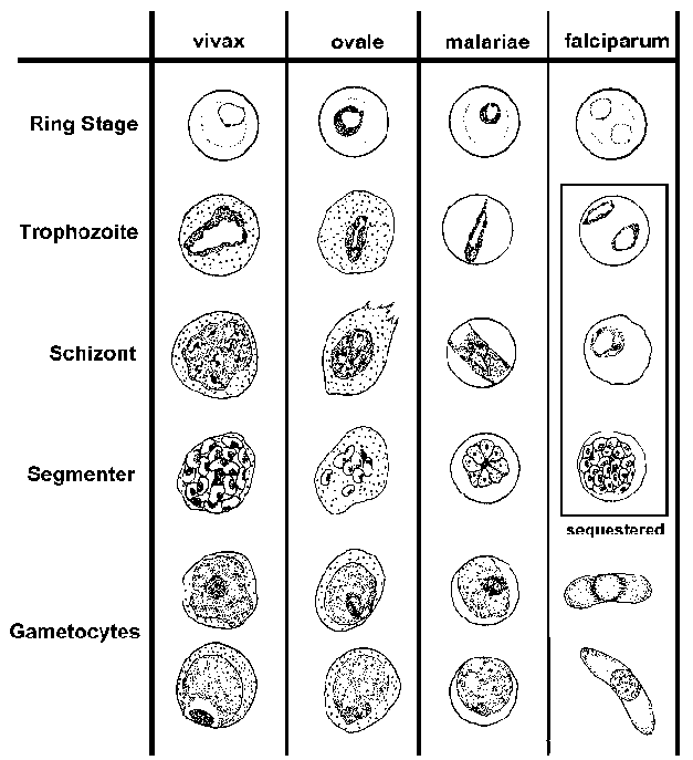
Until the use of direct detection of the antigen of the malaria species by PCR was introduced, the microscopic examination of blood slides for the presence of parasites was the accepted method of diagnosis of malaria.


  Falciparum                         Vivax                     Ovale                      Knowlesi
  • numerous rings
  • smaller rings
  • no trophozoites or schizonts
  • Cresent-shaped gametocytes
  • enlarged erythrocyt-e
  • Schüffner-'s dots
  • 'ameboid' trophozoit-e
  • similar to P. vivax
  • compact trophozoite
  • fewer merozoites in schizont
  • elongated erythrocyte
  • compact parasite
  • merozoites in rosette


Special features of falciparum malaria.

P. falciparum fever is also called malignant malaria and cerebral malaria. Worldwide, 2 million people are infected and ½ million die on yearly basis. The initial symptoms marked by shaking chills last only 30 min, followed by high fever, headaches, and confusion and children develop convulsions and coma rapidly. The fever returns every 48 hrs. Falciparum induces swelling of the RBCs, and the surface membrane protein of the parasite changes to knob like projections and attaches to the endothelium of capillaries. The circulation is compromised and endothelial cell hypoxia induces adhesive properties of the endothelial cells and locally released cytokines, cells facilitate platelet thrombi and the microcirculation of the tissue is cut off. Hypoxic injury of the brain occurs in the basal ganglia and hindbrain. Generalized swelling of the brain produces encephalopathy and various degrees of coma and convulsions.  With early medical intervention, cerebral changes are reversible. The mechanism of acute renal injury is multiple. Free hemoglobin in the circulation oxidizes glomerular capillaries, swelling, edema and piecemeal necrosis of uneven distribution of renal lesions leads to a marked reduction of GFR. The capillary circulation of the renal tubules is severely compromised and results in Acute Tubular Necrosis and renal failure.  Nearly 40 % of adults develop renal failure and cerebral malaria incidence is high in children and the mortality is high.

P. vivax.

Vivax malaria is less virulent than falciparum but deaths are still very high. Currently, southern Africa, Central & South American countries, the Indian continent, and Indonesia & Malaysia peninsular regions report most cases. Fever recurs every 48 hours, the temperature reaches 104F, and the febrile period is about 4-8 hours. Most of the symptoms are from released cytokines, TNF alpha and IL-6. Pulmonary involvement and enlarged spleen often rupture without trauma. Anemia is of chronic nature and secondary infections in endemic areas are not unusual. The parasite may remain dormant in the liver for years and one infection triggers six or more infections. In addition to renal failure, hypoglycemia, low platelet count and bleeding episode, loss of taste and occasionally lack of fever are some of the unusual features.

P. ovale.

Ovale malaria is the mildest form of human malaria.  It is seen in the Indian subcontinent. The fever recurs every 48 hours (tertian fever). Fever begins after 12 days following mosquito bites.

P. malariae. Massive splenomegaly from the abnormal immune response is a feature of P. malariae malaria. Fever recurs every 48 hours intervals. The incubation period is 16 to 60 days. Nephrotic syndrome is due to immune complex formation with malaria antigen and antibody and produces renal failure. 

P. Knowles. It is the malaria of Macaque monkeys; humans acquire malaria due to close contact with macaques. Fever recurs every day (quotidian fever). GI symptoms like hiccups and dysphagia are distinguishing symptoms.  Because of a few cases in the community, robust immunity does not develop. So, the fever continues and results in anemia and weakness.

Asymptomatic Carrier.

In endemic area, some people acquire immunity from repeated malaria infections. Unless their immunity is compromised by intercurrent infections or diseases, they remain malaria free. Once they move to other countries where malaria is rare, they can transmit the parasite to others by donating blood or organs.

Malaria continually develops resistance to drugs.

Plasmodium quickly develops gene mutations and renders drugs ineffective. For example, anti-folate medication becomes ineffective due to a point mutation of the Dihydrofolate dehydrogenase enzyme. Another point mutation involving two transporter proteins. By accruing mutations, the parasite increases the rate of expulsion of antimalarial drugs from the cytosol. Still, another way to develop drug resistance is by gene amplification of those genes mentioned above, making the mechanism work at a higher level, making unmatched therapeutic blood levels.

Defects in the DNA repair genes accelerate mutations, specially in P. falciparum.

Anti-malaria drugs.

Currently 5 groups of antimalarial drugs are available to treat malaria.

1. Endoperoxide – example Artemisinin.

2. 4-Aminoquinolines and related derivatives – Chloroquine, Mefloquine.

3. Napthaquinolies – atovaquone.

4. Antifolate – Pyrimethionine.

5. 8-Aminiquinols – Primaquine.

P. falciparum has developed resistance to every one of the above 5 groups of drugs.

Secondary drugs used in conjunction with the above 5 groups are -

Doxycyclines, Sulfadoxime, Lumefantrine.

New class of Drugs.

Monoclonal antibodies are used subcutaneously, prevent schizonts from attaching to RBCs and subsequently infect liver cells when used in combination with other standard antimalarials. Imatinib, a small molecule anti-leukemia drug, works by inhibiting the Kinase, has also been used recently and has the potential to rapidly clear malaria parasites from blood and liver. A drug, Alisporivir, used in hepatitis C, was found to be effective in Artemisinin-resistant P. falciparum infection. 

Several drugs are used in various combinations like chemotherapy for cancer. WHO's recommendations of drug therapy and chemoprophylaxis are essential to keep informed about the therapeutic challenges posed by rapid drug-resistant malaria spreading from one region to the other endemic area.

                           .

                                             wormwood

 Chinese sweet wormwood, source of artemisinin.

Artemisinin was introduced by Dr.Tu Youyou in the western world, at one time Artemisinin was the mainstay of drug therapy of P. falciparum and was used extensively alone or in many various combinations with other compounds. For his work, he too received the Nobel prize in 2015.

Malaria therapy for neurosyphilis.

Before Penicillin was available to treat syphilis, fever by P. vivax infection induced fever as the therapy for neurosyphilis by introduced by Julius Wagner-Jauregg. In Europe P. knowles was used for this malaria therapy till 1950. For this therapy, he was awarded the Nobel prize in medicine in 1972.

*****************************


Tuesday, November 8, 2022

Heat Intolerence


                                                       Heat Intolerance.

                                                   PKGhatak, MD


Heat intolerance is not a disease; it is a symptom. Some might say, “It is too hot in here,” and others might look at him in surprise because they feel comfortably cold in the same room.

The question is, who is right, or is there a medical explanation for this temperature perception difference between people.

Yes, there is a physiological reason for heat intolerance.

The Heat Center is located in the brain, and is connected with the skin and the internal organ temperature receptors., The Heat Center (HC) is located in the Hypothalamus of the brain, which is connected with the pituitary endocrine gland, which in turn is connected with the sympathetic and parasympathetic nervous systems.

By varying the output, the heat center of the body can maintain a constant core temperature throughout the winter and summer.

Source of body heat.

The human body derives heat from 5 separate sources:

1. Basal metabolism

 2. The specific dynamic action of food,

 3. Physical activities

 4. Shivering 

  5. Non-shivering thermogenesis.

Heat Center regulates temperature settings, but the metabolic process generates heat. It is like a computer; multiple programs are running in the background. The metabolic process continues at all times during the lifetime of the individual. The organ functions continue at a basal, and is called Basal metabolism. In humans, the Basal Metabolic Rate (BMR) is 76 kilocalories/Kg body weight /Hour. This is the main source of body heat.

2. Specific dynamic action of food (SDA). The process of digestion of food generates heat. Protein gives out the most heat, about 20 % of the calories contained in protein food. This is the reason we feel comfortably warm after eating a steak.

3. Physical activities. All forms of physical activities generate heat,

4. Shivering. Exposed to cold and wind, the body begins to shiver. Muscle contractions generate body heat.

5. Non-shivering thermogenesis: It takes place in Brown Fat and muscles. Adrenaline is released at the sympathetic nerve terminal of brown fat tissue and muscle. Adrenaline accelerates the conversion of fatty acids and glucose to heat, ketones, H2O, and CO2.


Excessive body heat and heat intolerance:

1. Menopause is due to falling sex hormone levels. Various symptoms, mostly due to the perception of excess heat produce the symptoms of menopause.

 2. Hyperactive thyroid gland.  Excessive secretion of Thyroxine (hormone of the thyroid gland) increases BMR, generates fine muscle tremors, and increases heart rate and blood pressure. Catabolism of fatty tissues and muscles produces excessive heat.

Drug abuse. Amphetamines, cocaine, antipsychotic, and antidepressant drug use generate a sense of excess body heat.

Anticholinergic drugs. Anticholinergic drugs decrease sweat production and prevent cooling from evaporation and loss of heat. The core body temperature rises.

Caffeine. It is a cardiovascular and neuronal stimulant. It produces small vessel constriction and decreases heat dissipation. A mild increase in temperature is usual in therapeutic doses. In posing, caffeine raises the core temperature. 

Heat stroke. When exposed to high heat in a humid environment, the body fails to dissipate heat by radiation and evaporation. The core temperature rises and derails the functioning of metabolic and enzyme systems. It turns into an emergency situation, and can permanently damage the brain and heart, and even death.

Malignant Hyperthermia. This is another life threatening medical condition. It is an inherited disease inherited as an autosomal dominant trait. exposed to volatile anesthetic agents during general anesthesia, large amounts of calcium leave the calcium stores and enter the skeletal muscle fibers. Increased intramuscular calcium produces sustained muscle contractions. An excessive amount of heat is generated. Blood vessels may contract for a long time and can cut off blood flow, which is called Rhabdomyolysis. The released myoglobin from the necrotic muscle can block the kidney filters, producing metabolic acidosis and hyperkalemia (high serum potassium). Followed by cardiovascular collapse and death even death. 

Multiple sclerosis.  In Multiple sclerosis, the demyelination may directly affect the hypothalamus and the autonomic control center. Excessive heat generation may also be accompanied by derailed heat dissipation because of spinal cord lesions in multiple sclerosis.

Diabetes mellitus can damage the autonomic nervous system, and excessive heat and fluctuation of body heat may result.

Multisystem atrophy of the nervous system. The autonomic center damage is the result of increased heat.

edited May 2025.

**************************************

Friday, November 4, 2022

How We See the Outside World

                                               How we see the outside world.

                                                   The science behind it.

                                              PKGhatak, MD


Looking straight ahead, we see a vast area, horizon to horizon. Using a visual field measurement chart and recording one eye at a time, we find each eye has a visual field of 155 degrees on the horizontal plane and 130 degrees on the vertical plane. By adding the visual fields of two eyes together, our visual field should be 310 degrees but in fact, the visual field is smaller because the nasal field of the two eyes overlaps.

That overlap gives us the dept of vision, meaning, we can judge the distance of an object in front. It is an essential quality for reading, writing, driving, painting, target shooting and other activities which require bringing objects in sharp focus.


The diagram above illustrates visual fields and where the images develop.

The visual field is divided into the Nasal field and the ear (Temporal field. To make a clear distinction between the left and right visual fields, the right-hand side of the visual field is colored red and the blue color for the left.

Note that the nerve fibers carrying the image from the nasal half of the right eye, represented by blue lines, are crossing the midline and going to the left side to join the nerve fibers carrying the image from the temporal half of the left eye. The same is true for the left eye, marked with red color.

It is interesting to note that crossed and uncrossed nerve fibers join and carry images representing the right and left visual fields and not the left or right eyes.

The second point to note is that the nerve fibers make a connection at a nerve center called Lateral Geniculate Body (LGB). The right visual field image goes to the left side and the left visual field image to the right side of the LGB. The stratification of nerve fibers is maintained in the LGB, meaning the temporal fibers remain outer and nasal fibers on the inner nuclei of the LGB.

The third point is that the same stratification is also maintained in the Visual Cortex. The visual cortex is located in the back of the cerebral hemisphere, one on each side in the Occipital Lobe.

Not shown in the diagram, there is upper and lower field stratification on the visual cortex. The lower half of the visual field is represented in the upper part of the opposite cortex, and the upper half in the lower visual cortex.

By knowing the detailed anatomy, the doctors are capable of detecting any segmental visual field loss and treating properly without wasting much time.

How two images merge into one.

This fusion of two images in the visual cortex and perceived as one depends on unison eye movement and convergent reflexes that make image generation on the corresponding points on each retina. When initial images are close but not exactly one, then a further adjustment is made by the eye muscles to focus properly.

Why does an object too close to the eyes appear blurred.

The eyes are situated 2 inches apart. The images of the two eyes are not precisely the same, the right eyes see a little more on the right-hand side and the left eyes see more on the left-hand side. The images fuse completely when the object is at a comfortable distance from the eyes. The disparity grows more and more when the object is brought closer and closer.

How images are formed on the retina.

Light rays travel in straight lines in all directions from an object. The pupil by changing its size brings light rays in focus on the retina. The photosensitive pigment of the retina converts the electromagnetic energy of light into electrical stimuli. Next, the rods and cones cells of the retina perceive the stimuli and then pass them to nerve cells which convert the electrical stimuli into nerve impulses and send out nerve impulses to the brain.

 How the color is represented in the brain.

In the daytime, sun rays are bright but colorless. But we know that daylight hides 7 colors from red to violet (color of the rainbow) in it. The color receptors of the eyes are cone shaped cells, called cones of the retina. Cones are plentiful in the central part of the retina and are only a few in the periphery. Cones are densely packed in the Fovea of the retina. The red, green and blue colors are detected by three different cone cells. The blue color generates maximum brightness of 2.56 to 2.76 eV and the red color is the least, generating 1.65 to 1.90 eV. These three cone cells make a unit known as the Trichrome unit. By various combinations of the impulse generation by the trichrome units, we are able to perceive the entire color spectrum. This is the basis of the printers' color cartridges we are so familiar with.  The pigment of the cone is called Opsin. It is a protein and it is most sensitive to light waves with a wavelength of 550 nm, blue responds to light waves with a wavelength of 450 to 485 nm and green to 500 to 565 nm wavelength.


White and Black color.

There is no black color receptor.  Instead, black, white and gray color perception depends on various shades of gray colors. The pigment of the gray scale is Rhodopsin, and the receptors are known as Rods. Rods are most plentiful at the periphery of the retina and are only sparsely distributed in the central part of the retina. Rhodopsin is most sensitive at light waves of 555 nm.

We see better in the corner of the eyes when light is very faint. There are no cones at the periphery of the retina, which is the reason we are unable to see color in the dark.

Why are black and white images not as clear as a color picture.

Each cone is connected with one neuron of the retina that sends out stimuli to the brain. 5 or 6 Rods are connected with one nerve cell, as a result, color picture has more pixels so to speak, and is sharper and clearer than black and white images.

Upside down and revered sidewise image.

This diagram illustrates why the upside down and left side on the right side image projected on the retina. 


This is a pinhole camera taking a picture of a tree. The same Laws of Optics work for the eyes.

How the Brain rights up images.

A newborn child grabs things with hand and tries to put them in his /her mouth. That is the beginning of the learning process of the brain and by the time the baby is 2yr old the proper interpretation of images by the brain is nearly complete but the learning process of the brain continues for lifelong.

Near and Distant vision.

Refractive errors of one or more components of the eye (cornea, anterior chamber, lens and vitreous) fail to bring light precisely on the retinal receptors and when the light rays fall short- then the condition is known as Near- Sightedness and when the light rays converge beyond the light receptors then it is called Far- Sightedness.

Double vision.

Double vision may be monocular or binocular. Mono-ocular double vision comes from the eyes, or contact lens injury, or foreign body injury of the cornea.

Binocular double vision is due to the failure of mages to merge into one. The defect may lie anywhere from the optic nerve to the visual cortex. Since eyes are extensions of the brain and when an eye is damaged it may not improve unless treatment starts immediately.

A quick way to determine mono-ocular or binocular double vision

The mono-ocular double vision disappears when the eye producing the double vision is closed and the person looks through the other eye. In binocular double vision persists by closing and opening one eye at a time.

In daylight, our world appears in full complements of spectrum colors - clear and beautiful and images up-righted and sides correctly positioned. In the dark the story is different. In fading light, the world appears in various shades of gray, fuzzy and best visible to us by the corner of the eye provided one is neither near-sighted nor far-sighted.


*******************************************************


*** 


Monday, October 31, 2022

Movement Disorder

                                                   Movement Disorder

                                              PKGhatak, MD


Muscles move joints and joints move the body. Movement does not only refer to ambulatory movements but movements of part of the body when the person is stationary. In any movement, two groups of muscles work in a coordinated manner, one group contacts the other group must stay in a relaxed state for the movement to occur. If both groups contact simultaneously, the joint is held in a fixed position and is called spasm.

Well known Movement disorders:

Parkinson's disease, Parkinson syndrome, tremors, ataxia, chorea, tics, myoclonus and spasticity.

Parkinson's Disease (PD) and Parkinson Syndrome:

Parkinson's disease is due to the sickness of Dopamine (neurotransmitter) producing cells located in the Substantia nigra in the hindbrain. Slowly the cells die and many neurological abnormalities develop, including dementia and psychosis.

Movement abnormality in PD.

Slow movement and lack of associated movements, like swinging of arms while walking, are striking features. Increased muscle rigidity of neck, head, limbs and back muscles produces a stooping forward posture; and stiffness of legs and thighs produces a shuffling gait. Tremors of hands at rest produce pill-rolling movement of fingers; later writing, feeding, and grooming become difficult. Loss of balance causes frequent falls. The cause of PD is unknown.

Parkinson Syndrome. This term is reserved for Parkinson like features in patients with known causes, for example, poisoning from heavy metals and pesticides, repeated cerebral concussion, and adverse effects of drugs used in Psychosis, Schizophrenia, Depression, etc.

Parkinson's disease (PD) Subtypes.

Experts classify the movement abnormalities in two catigories.1. Tremor predominant. 2. Postural instability - gait difficulties.

1. Tremor-predominant.

Clinically tremor-predominant PD is of three kinds: Mild, Progressive malignant and intermediate varieties.

a. Mild.

Tremors are mostly seen while trying to work with hands.

B. Progressive malignant PD tremors.

Tremors are a rapidly progressive type. The resting tremors are present during waking hours and in light sleep, but during deep sleep, tremors disappear. Tremors prevent patients from doing most routine work - like cooking, cleaning, or answering telephone calls.

C. Intermediate

The symptoms of this subtype fall between the above two subtypes.

2. Postural instability-gait difficulty.

Due to increased rigidity, the patients assume a stooping posture. There is a lack of spontaneous corrective adjustments of posture. Decreased two-way communication with the cerebellum produces unsteady balance and a slight bump against a chair or wall the patient falls forward on the ground.

Subtypes of Parkinson's disease.

A. Progressive Supranuclear Palsy (PSP).

The most distinctive feature of PSP is recurrent sudden backward fall on attempted walking, however, during and after the fall the patient remains fully conscious.  Pseudobulbar palsy leads to difficulty in chewing and swallowing. Intranuclear ophthalmoplegia produces abnormal eye conjugate movements when looking downwards. The patient is unable to look upwards and on attempts to look upward produce horizontal nystagmus. The upper eyelids are kept half-closed. PSP patients have no rigidity and tremors unless the disease is far advanced.

B. Restless Leg Syndrome (RLS).

Patients develop an uncontrollable urge to move. Symptoms may be intermittent, and the degree may vary. Restlessness may also be felt in the arms, neck and head. Sitting on a chair for a long period, driving an automobile and sleeping at night become difficult. Patients also describe a strange sensation of insects crawling under the skin, pins and needles, throbbing and aching pain deep in the legs and lower body.

The use of Dopaminergic drugs -  Pramipexole, Ropinirole and Rotigotin  produce some relief in the early phase of the disease. Later L-dopa may be helpful.

C. Lewy body dementia.

Abnormal muscle movements are generally periodic, bizarre, uncontrollable and purposeless. Apathy and profound loss of memory lead to total dependence on others for activities of daily living.

Other Neurological Conditions.

Neurological conditions produce muscle disorders, for example, Multiple System Atrophy (MSP), Cortico-Bulbar syndrome, Low pressure hydrocephalous, Huntington disease, Wilson disease, Tourette syndrome, Motor stereotypies and Multi System Atrophy.

Multi System Atrophy. 

The impairment of the autonomic nervous system produces a loss of spontaneous adjustment of BP with postural changes and with physical activities. Postural hypotension is common. Of the GI symptoms, constipation and difficulty in swallowing are common. Men may develop erectile dysfunction and difficulty in voiding urine. Olivopontocerebellar atrophy produces stiffness and incoordination of movements. loss of balance and slowness of movements.

Corticobulbar syndrome.

Degeneration of neurons in the motor and sensory cortex produces symptoms of poor muscle coordination, slowness of thought processing, language difficulty and speech abnormality. Increased and prolonged increased muscle tone produces stiffness of posture.

Huntington disease.

Huntington disease is an autosomal dominant inherited condition. Progressive severe dementia, obsessive compulsive disorder, mania, and bipolar disorder are major components. Writhing slow dance like movements, jerky movements of limbs, (called Chorea), language and speech abnormalities are present. Slow eye movements, muscle rigidity, impaired gait, balance and posture resemble PD. 

Wilson disease. It is a recessive mode of inherited disease involving excess copper accumulation in the brain and other vital organs. The muscle disorder includes tremors, difficulty in swallowing and speech and trouble in ambulation. Cirrhosis of the liver at an advanced stage is common. Copper accumulation at the periphery of the cornea of the eye produces a distinct diagnostic feature called the Kaiser Flacherie ring.

Tourette syndrome. It is an inherited disease, but the precise genetic abnormality is not known. Involuntary movements commonly involve the face and uncontrollable vocal sounds followed by the arms, legs, or trunk. Vocalizations of embarrassing words called Coprolalia often accompanied by obscene jesters, Copropraxia, make patients difficult to socialize with. The symptoms usually begin in childhood and spontaneously may disappear when older.

Motor stereotypes.

These are fixed repeated movements with no useful purpose, appear at the same predictable location and time. Movements last a minute or longer and occur several times a day. Typical motor stereotypes are thumb sucking, lip biting, hair twirling, teeth grinding and head banging. Symptoms start in childhood and rarely persist in adult life. Sometimes more complex motor dysfunctions involving hand and arm movements, like waving arms and wiggling fingers in front of the face and closing and opening hands.

Essential tremors. In this disorder, rhythmic involuntary fine shaking movements of hands generally appear in midlife. Tremors mostly occur when the patient is in the company of others and performing normal tasks like drinking a cup of coffee or buttoning a shirt. Tremors may also happen in the head, producing nodding of the head as yes-yes or no-no jesters.

Spasticity. Prolonged muscle contractions lead to increased tone of muscles that interfere with normal walking or performing activities of daily living. Spasticity is a feature of cerebral stroke, Multiple sclerosis, spinal cord injury, spina bifida and severe brain injury.

Dystonia. Dystonia is an involuntary muscle spasm producing twisting motion of the limbs or trunk producing abnormal posture and positions. An example is Writer's cramp. The onset of dystonia is gradual and always follows a specific muscle action. Muscles fail to relax in between contractions preventing smooth functioning.

Ataxia. Ataxia means uncoordinated muscle actions, mostly noticeable in arms and legs. Cerebellar ataxia is more common. In the younger age group, viral infections, tumors, cysts and vascular disruption in elderly people are common causes.

Tardive Dyskinesia (TD). Tardive means late onset. Dyskinesia is defined as difficulty in performing voluntary functions. Muscles of the face, tongue and upper extremity show repeated involuntary movements like grimacing, lip smacking, pursing of lips, eye blinking, and rapid involuntary arm, torso and finger movements. Long term use of anti-Schizophrenia, Anti choline and anti-depression drugs use are associated with TD. Discontinuation of drugs improves symptoms if done early.


*****************************


Monday, October 24, 2022

Epstein - Barr Virus Infection.


                                                          Epstein Barr Virus.

                                                   PKGhatak, MD


The Epstein Barr virus was initially called the Burkitt lymphoma virus because the virus was identified in the cells of the Burkitt lymphoma in an African patient in 1964. It is known today as Human Herpes Virus 4. This virus is the most common viral infection among all other herpes virus infections in humans. About 85 % of the world population have serology positive blood for Epstein Barr Virus (EBV), indicating past infection.

EBV virus characteristics.

EBV belongs to Gamma-Herpesviridae in the genus group of Lymphocryptoviral. The double stranded genome contains 85 proteins and 50 non-coding RNAs. The viral particle has an outer lipid envelop which fuses with the respiratory cell membrane at the time of infection. The DNA of the virus can turn out 9 different molecules of Entry Proteins, gP 1 – 9, and use them to infect many other cell types of the patient.

The EBV virus, once inside the respiratory cells, multiplies by redirecting the host cell to duplicate virus particles. The virus then leaves respiratory cells and infects B-lymphocytes. Inside the B-lymphocytes, the virus persists for life and goes through 3 cyclic phases, namely active phase, dormant phase and re-infective phase.

A person infected with EBE is unable to clear the virus from his body. A subsection of B-lymphocytes is memory cells. The memory cells continue to turn out immunoglobulins, the initial product is IgM and 2 to 4 weeks later all the products are IgG.

In addition, antibodies are produced against capsular proteins and nuclear proteins.


Why does the EBV virus live in perpetuity in humans.

Whenever antibodies are produced in adequate amounts, the virus goes into hibernation within B-lymphocytes and also in some cases in the respiratory epithelial cells. The NK cells, monocytes and macrophages are unable to find the EBV once inside the cells and antibodies are ineffective to neutralize the viral particles. This hide and seek game is played out throughout the life of the patient.

In certain circumstances, the EBV can also infect NK cells, monocytes and macrophages and produces more serious illnesses.

EBV human infection.

Children are the prime target of the virus and about 80 % of human infections take place in children under 2 years old. Most of the infected children remain free of symptoms or had minor symptoms. The only evidence of infection in these cases is positive serology tests.

The next venerable age is teenagers. Teenagers, however, become symptomatic and the illness is known as Mononucleosis or simply Mono and also commonly called a kissing disease. The most remarkable symptoms are the posterior cervical lymph node enlargement and enlarged spleen. Other significant symptoms are sore throat, fever and enlarged tonsils.

Persistent and recurrent activation of EBV.

The EBV can become active within the immune cells, whenever the immune systems are down. The repeated reactivation of the EBV within the immune cells produces Multiple sclerosis and rheumatoid arthritis.

Reactivation within the respiratory epithelial cell leads to Sjogren syndrome and Systemic Lupus Erythematosus (SLE).

The positive ANA, pANCA, cANCA and Rh factor tests are the results of back and forth infections switching cell lines over a time.

In patients with seriously handicapped immune systems, the recurrence of active and inactive stages of EBV leads to B cell Lymphoma and Nasopharyngeal Carcinoma.

Systemic Autoimmune Disease (SADs).

A group of autoimmune diseases has one common factor of connective tissue damage. There are overlapping symptoms between these entities and often some positive serological tests are also common. The diseases are Mixed Connective Tissue Disease (MCTD), Myositis, Rheumatoid arthritis (RA). Systemic Sclerosis (SS), Lupus erythematosus (LE).

Autoimmune vasculitis of various clinical entities is associated with EBV infection.

Serology tests for EBE viral illness.

Mono spot test. And Heterophile antibody test.

Mono spot is no longer recommended for routine use for the diagnosis of mononucleosis in children because of false negative and some false positive results. But the science behind it is solid.

EBV produces antibodies which cross react with RBC antigen of horses and cows. This property of antibodies leads to the name of this test as Heterophile antibody test.

The heterophile antibody test is modified as a Mono slide test, available as a test kit and commonly used in clinics and pediatricians' offices. It is a modified and improved Paul-Bunnell test.

Immunofluorescence tests.

Viral Capsid Antigen (VCA) IgM Test.

The test is performed using antibodies tagged with fluorescence dye mixed with the patient's serum. A positive test is seen in about a week after infection and disappears in 4 to 6 weeks.

VCA IgG Test.

The test becomes positive in 2 to 4 weeks and then declines slightly and remains positive for the rest of the life.

Early Antigen Test (EA) Test.

Anti EA IgG antibodies appear during the acute phase of the infection and disappear in 3 to 6 months. In 20 % the Anti EA IgG may remain positive for a very long time, otherwise, an Anti EAIgG positive test indicates a recent infection. 

EBV Nuclear Antigen Test.

It is also a fluorescence test against viral nuclear antigen that becomes positive in 2 to 4 months after infection and remains positive for the rest of life.

False Positive Heterophile Antibody Test.

False positive heterophile antibody results are seen in Viral hepatitis, Rubella, Toxoplasmosis, HIV/AIDS, Malaria, Lupus and Pancreatic Cancer.

A false negative Heterophile test is extremely rare in adults. Children do not readily produce Heterophile antibodies in the first two weeks following infection.

Blood Test.

The mononucleosis name is due to an increase in monocyte count in the peripheral blood. The total count increases only modestly in the range of 10,000 to 20,000. The lymphocyte count is generally over 50 %. And on the smear, Atypical lymphocytes appear.


*******************************************************

Sunday, October 23, 2022

Monkeypox


                                                             Monkeypox.

                                                  PKGhatak, MD


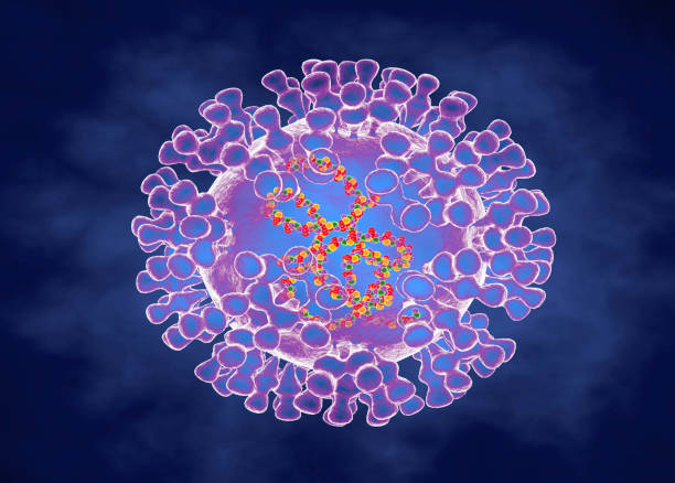
Monkeypox virus belongs to an enveloped double stranded DNA virus of the genus Orthopoxvirus in the family Poxviridae and belongs to a subfamily of Chordopoxvirinae. In the central and west African jungle, the monkeys are the natural host and this was also documented in squirrels, prairie dogs, rats, dormouse and primates.

                                                 Artist's version of a monkeypox virus.

The first human illness was recorded in the Democratic Republic of Congo in 1970, just two years after Smallpox was eliminated there. From time to time local outbreaks were reported by the WHO. Cases were seen in central and west African countries when children were bitten by monkeys.

There are two subclasses of this virus – one in the Congo Basin virus which is more virulent and has a higher rate of transmission, and the other is in West African. At present both varieties are infecting the people of Cameroon.

The first reported case in the USA was in 1980. In 2022, the USA declared monkeypox a public health emergency. Human to human transmission was seen in NYC in the homosexual community. Infection of humans takes place through the exchange of body fluids, respiratory droplets, prolonged close contact, and occasionally by the use of contaminated towels and bed sheets.

The incubation period is 5 to 21 days. The initial symptoms of malaise, body aches, fever and lymphadenopathy for 4 -5 days, then the rashes appear on the face and extremities, including on the palms and soles. In homosexual individuals, rashes are also present in the genital and anal areas. In contrast to other varieties of pox, monkeypox patients show striking lymphadenopathy.

Pox is known to appear on the lips and mouth, the conjunctiva of the eyes. The rashes go through the stages of macules, papules, vesicles and pustules. In three weeks, the scabs of pustules dry out and then drop off. Until all scabs fall, the patient remains a source of infection. The monkeypox illness is a mild disease but in immune deficient people, it can produce severe illness. In African countries as high as 10 % of the population is infected, and the fatality is generally below 3 %, and in children, the mortality has gone up to 6 %.

Diagnosis of monkeypox is done on samples obtained from the skin of the lesions or the fluid of vesicle/pustule by PCR test to confirm the monkeypox virus. Blood tests are not reliable and other varieties of pox produce the same serology test results.

Monkeypox vaccines.

Two vaccines are available in the US. In post exposure two doses of the Jynneos vaccine are given. Another vaccine ACAM2000 is less commonly given but available on a limited scale. In very high risk groups prior exposure vaccination is approved.

People vaccinated for smallpox have limited protection in high risk groups, for the general public provides 85 % protection against monkeypox.

Antiviral drugs.

In Europe, an antiviral drug, Tecovirimat, originally developed for the treatment of smallpox, is approved for monkeypox. In the USA, Tecovirimat is approved temporarily on an emergency basis and availability is limited.

*************************************
Cerebrospinal Fluid

                                              Cerebrospinal Fluid (CSF)

                                             PKGhatak, MD


The previously held view of the formation and circulation of the cerebrospinal fluid (CSF) has undergone modifications because current reproducible and non-interventional methods of investigation, like MRI and molecular and biological biology, discovered newer facts that contradicted the past theory.

To update the current thinking, a brief review of the histology of the Choroid plexus, Ventricles of the brain and Arachnoid granulations is necessary.


Choroid plexus.


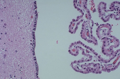
The Ependymal cells of the choroid plexus secrete the CSF by an active process. The central capillary of the choroid plexus is lined with one layer of fenestrated cuboidal cells. The albumin content of CSF is much lower than plasma. The transport of water across the ependymal cells of the carotid plexus is carried out by separate transport proteins, one for the ventricular side and the other for the capillary side. It is an active process that requires energy from ATP and likely also from K / Cl transporter. About 30 % of the total water volume of CSF comes from the inter-neuronal space, generated from the metabolic activities of the nerve cells, like Lymph formation in the other organs. There are to and fro moments of fluid between interstitial fluid and CSF.

Blood Brain Barrier.

This special characteristic of the blood vessels of the brain is due to the tight seal between the endothelial cells of the capillaries and supplemented by perivascular feet of astrocytes surround the basement membrane. In two locations this barrier is absent namely the choroid plexus and posterior pituitary gland.


Arachnoid Granulation:

Arachnoid granulations are collections of capillary networks kept suspended into the large veins of the subarachnoid space. The anatomical structure suggests arachnoid granulations are filters - a one way passage of CSF into the venous blood, getting rid of waste products and excess water. But that is not the case. Arachnoid granulation plays a minor role in this respect.

The perineural space along the cranial nerves and spinal nerves and the perivascular space known as Virchow Robin spaces are the main routes of drainage and filter of CSF into the general lymphatic system. This is specially evident in the Olfactory nerve as it travels through the cribriform plate of the nose.


Circulation of CSF:


The circulation of CSF in the brain and spinal cord.

The circulation of the CSF is an active process by the synchronous motion of cilia lining the surface of the ventricles and the central canal of the spinal cord. The CSF exits the brain through two openings of Luschka in the hindbrain and circulates in the subarachnoid space. The mechanism of a steady state of CSF formation, circulation and drainage has undergone many modifications. The circulation is pulsatile in nature due to and from movement of the CSF. The interstitial fluid exchange with CSF is a major component of circulation. A continuous bidirectional fluid exchange at the blood brain barrier produces flow rates. The general lymphatic system of the body is the main route of drainage of waste products of the brain and an entry point of the immune system in the brain.


******************************


Friday, October 21, 2022

What Cerebellum Does


                                           What Cerebellum Does

                                        PKGhatak, MD


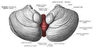
The human cerebellum occupies a small space at the back of the head, just above the neck, and it is a part of the hindbrain. Sensory information from joints, tendons and soft tissues from the limbs and trunk travel to the cerebellum. The motor stimuli coming from the cerebrum motor cortex, make connections with the nuclei of the cerebellum before reaching the lower motor nuclei of the spinal nerves and the cranial nerves. This interconnection of the motor cortex with the cerebellum results in a smooth sustained movement of the body, able to maintain the head position in space and in relation to the rest of the body and keep balance without falling. And that much knowledge people had till 1990.


Now the cerebellum is a subject of intense research for a variety of reasons.

Research in developmental biology documented three separate stages of growth of the cerebellum in humans. As the forebrain is added, the cerebral cortex, which has increased the mental faculties, knowledge gained about self, mind, higher state of consciousness, language, and development of culture has made humans as humans. But the cerebellum did not lag behind.

The growth of the cerebellum is more spectacular than the cerebral cortex. Cerebellum weighs 150 grams and contains 70 billion neurons, whereas the cerebral cortex weighs 1300 grams but contains only 16 billion neurons. The surface area of the cerebellar cortex is 1500 square centimeters, which is about 80 % of the surface area of the cerebral cortex. The cerebellar cortical folds are arranged like pages of a book - lots of surface area in a small space.


Compared with the cerebral cortex the layout of neurons and their connections in the cerebellum is much simpler and more uniform. Cerebellum receives input and sends out output to the same side of the body that is also a contrast with the cerebrum which controls the opposite side of the body. The central part of the cerebellum is old, but the lateral parts, the cerebellar hemispheres, kept pace with the growth of the cerebrum.

Cerebellum is the site of the formation and storage of working memory and the integration of automated sequences of thoughts and actions. This results in developing behavioral patterns, speech, dexterity of complex movements, a skill needed for technological advances and language development.

In association with the cerebrum, it forms kinetic memory, plans coordinated muscle movement patterns, retains reflex memory for self preservation and executes normal speech.

Cerebellum with two way communication with the thalamus and basal ganglia and receives direct input from the spinocerebellar tract and maintains balance and a stable head position both in states of body movements and at rest.

Vestibular sensory information is closely monitored by the cerebellum, that information is used to control conjugate movements of eyes, prevent double vision and reflex eye movements.

Rapid eye movement (REM) sleep includes Implicit memory involving perception and body movements. Implicit memory uses cerebellar retained memories.

*********************************


Archive

  Archive. P.K.Ghatak, MD 2025  March. Laser Endoscpoe 2025 February. Use of Ultrasound in Medicine MRI 2025 January. Bacteriology and Rober...