Neurotransmitters
PKGhatak, MD
Nerve
cells communicate with other nerve cells, muscles, and glands, or
internal organs by chemical
molecules. Even though the nerve impulse travels along the nerve fibers as an electrical impulse, at the end of the nerve terminal, a
tiny gap exists, that gap is
bridged by a neurotransmitter,
Neurotransmitters
(NT) are chemical messengers that carry forward electrical signal from neurons to postsynaptic neurons or cells of the target organ and
produce the intended action. NT must meet the following criteria. NT must
be synthesized by the neuron, found in the presynaptic nerve
terminal, must produce depolarization and propagation of the nerve
impulse. NT must be quickly removed either by enzymatic degradation, diffusion or reuptake by the nerve terminal.
From
the time the first neurotransmitter (NT) Acetylcholine was discovered in 1921 by Otto
Loewi of Germany, many more NTs have been added and today well over
100 NTs are known. Not all chemicals fulfill all the above criteria, however, all of them produce depolarization and transmission of nerve
impulses. Chemically some of the NTs are simple gases like nitrous oxide
(N0), carbon monoxide (CO) and complex protein molecules, like
pituitary adenylate cyclades activating peptide, are included in the newer
class of NT.
In
this review, only those NTs abnormality that produces significant changes in the body are discussed. These are-
1. Acetylcholine, and Biological
amines, 2. Epinephrine, 3. Noradrenaline, 4. DOPA and 5. Serotonin.
Inhibitory
neurotransmitter – 6. Gamma-aminobutyric acid (GABA) and 7. Histamine.
1. Acetylcholine
(ACh)
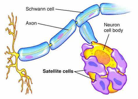
Acetylcholine
is released at synaptic junctions by the neurons of the central
nervous system, as shown in the diagram.
Acetylcholine
is synthesized by the nerve cells and then stored in vesicles at the
nerve terminals waiting to be rereleased at the synaptic cleft.
Chemistry
of ACh.
ACh is
synthesized from Acetyl CoA and Choline. Acetyl CoA is the end
product of glucose metabolism; choline is either locally produced
by the nerve cells from another amino acid serine or taken up from
the blood. Chemically, choline is a Quaternary ammonium compound, acts almost like a vitamin in humans, except that
it can be obtained from another amino acid. Eggs, organ meat, fish,
milk and beans are good dietary sources of choline. Excess choline can
cause low BP, weakness, increased sweating, fishy body odor, liver disease and cardiac problems. A deficiency of choline causes non-alcoholic fatty
liver, muscle damage and hyperhomocysteinemia (high Homocysteine
blood level).
Neurons
take up free choline via the 1.Na+/Choline transporter,2. a degradation product of ACh by the enzyme Acetylcholinesterase from the synaptic cleft and, 3. also from blood as shown in the diagram below.
Synthesis
of ACh.
Acetyl
CoA + Choline = Acetylcholine, by the enzymatic action of
acetylcholine-transferase.
Degradation
of ACh into Acetyl CoA and Choline by enzyme Acetylcholine esterase.
Action
of ACh.
Autonomic
nervous system.
(a).
Parasympathetic division. It acts both on ganglia and postganglionic
endings and its effects are -
Eyes -
Constriction of pupils and fall of intraocular pressure.
Glands
- Increases secretion of GI glands,
Bronchi-
Constriction of bronchi, increased mucus secretion.
Smooth
muscles of GI tract – increased motility, spasm and colicky pain,
contraction of gall bladder and defecation.
Heart
and arterioles. Slowing of the heart rate and vasodilation of
arterioles.
Urinary
bladder. Precipitate micturition.
(b).
Sympathetic Division - It acts only on the sympathetic ganglia and the Adrenal medulla and causes high BP and increased heart rate.
Central
Nervous system. It causes restlessness, insomnia, tremors, dysarthria
and convulsions.
Voluntary muscles. Contraction of muscles and fasciculation.
ACh has Nicotinic and
Muscarine effects. Nicotinic receptors stimulate the ganglia of both
sympathetic and parasympathetic divisions and the adrenal medulla. It also
stimulates parasympathetic postganglionic nerve terminals. Nicotinic receptors stimulate the Neuromuscular junction of the skeletal muscles.
Muscarinic
receptors are present in the parasympathetic nervous system and signal secretion
from glands and smooth muscle contractions. Muscarine has no action on
the skeletal muscles and does not act on the brain cells.
Diseases, due to autoimmune disease and some unknown causes, decrease the functions of ACh: these conditions are - Myasthenia gravis, Eaton Lambert syndrome, Guillain-Barre syndrome and Ascending paralysis in febrile children.
When used as a drug - it temporarily produces muscle paralysis and is used every day in surgery to ensure a better surgical outcome. Patients on mechanical ventilators at times require muscle paralytic drugs to prevent patients from struggling to breathe.
2. Biological
amines:
Walter
Bradford Cannon of Massachusetts, USA, in 1932 discussed the properties of
adrenaline and used this phrase - Flight or Flight
response.
Epinephrine
and Noradrenaline.
In Europe, these two neurotransmitters are known
as Adrenaline and Noradrenaline. Both Adrenaline and Noradrenaline
are hormones. Norepinephrine (noradrenaline) is the neurotransmitter of the sympathetic division of the autonomic nervous system.
Biosynthesis
of Norepinephrine.
L-Phenylalanine,
an amino acid, is the source of Dopamine and Epinephrine, the intermediate steps and enzymes involved in this process are as follows.
L-Phenylalanine to L-Tyrosine by enzyme Amino acid Hydroxylase.
L-Tyrosine to L-Dopa. (1-3,4 -Dihydroxyphenylalanine) by the enzyme amino acid
Hydroxylase.
L-
Dopamine to Norepinephrine by enzyme Betahydroxylase.
Norepinephrine to Epinephrine by the enzyme N-Methyltransferase.
Noradrenaline
is the transmitter for all the neurons of the brain and spinal cord of the sympathetic division of CNS,
and outside the CNS, for the sympathetic postganglionic neurons.
Noradrenaline increases blood pressure vessels due to increased tone. It produces bronchodilatation in the airways of the lungs and relieves nasal congestion. The difference in action is based on alpha and beta
receptors and their subtypes.
Origin
of noradrenergic neuron.
These neurons originate from the locus
coeruleus, tegmentum and dorsal medullary group.
Action
of Noradrenaline and Epinephrine
|
Effect on
|
Norepinephrine
|
Epinephrine
|
|
Heart rate.
|
Slowed
|
increased
|
|
Force of cardiac contraction
|
No effect
|
increased
|
|
Cardiac output
|
No effect
|
increased
|
|
Cardiac irritability
|
increased
|
Much increased
|
|
Systolic BP
|
Rises
|
rises
|
|
Diastolic
|
Rises
|
falls
|
|
Vascular bed in muscles
|
constriction
|
dilatation
|
|
Vascular bed skin & viscera
|
contraction
|
contraction
|
|
Vascular resistance in the heart
|
increased
|
decreased
|
|
Glucose metabolism
|
unchanged
|
increased
|
|
Bronchial smooth muscles
|
No effect
|
relaxed
|
|
Intestinal muscles
|
relaxation
|
relaxation
|
|
Intestinal sphincters
|
constriction
|
relaxation
|
|
Pregnant uterus
|
Increased contractions
|
Contraction lessened
|
|
Capillary permeability
|
No effect
|
reduced
|
|
|
|
|
|
|
|
|
Epinephrine is a lifesaving drug in an acute allergic reaction and anaphylactic shock producing laryngeal edema and death due to airway obstruction. Adrenaline has multiple applications in everyday medical practice and is too numerous to list here.
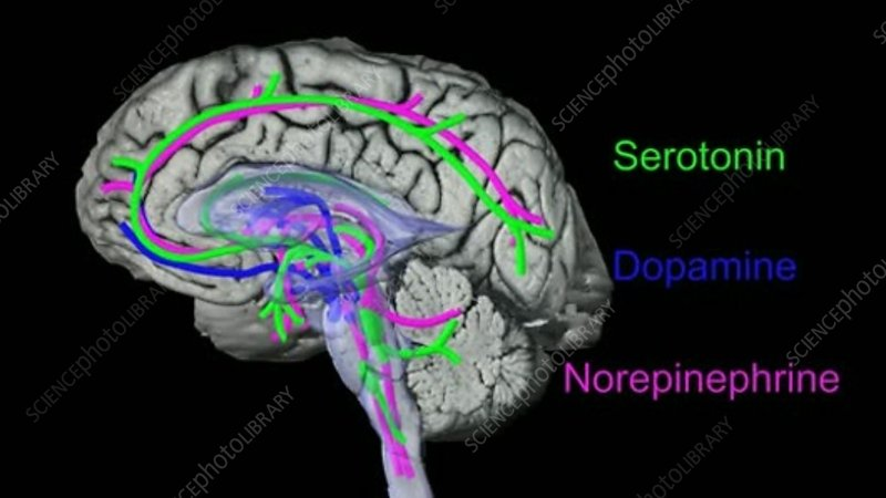
3. Serotonin.
Serotonin chemically is 5-Hydroxytryptamine (5lHT). It is formed from the amino acid Tryptophan to 5-Hydroxytryptophan by enzyme Trytophanhydoxylase (adding “OH” group) and then to 5-Hydroxytryptophan (serotonin) by enzyme Aromatic amino acid decarboxylase and coenzyme pyridoxal phosphate (removing
COOH group). 5-HT is present in large
amounts in the GI tract and the brain neurons and platelets.
Serotonin acts on the areas of the brain and is shown in green color in the diagram below.
Serotonin is the neurotransmitter for appetite, sleep, memory, happiness, mood, vomiting center, sexual arousal and
body temperature maintenance by its actions on the
forebrain, brainstem and cerebellum. Lack of serotonin produces
depression, anxiety and other psychological dysfunctions.
Serotonin
receptor.
Serotonin
(5-HT) receptors are designated by digits 1 to 7 and the receptors of 5-HT1 are assigned - A to F. The receptors of 5-HT 2 have A to C subtypes. The action of serotonin varies
according to its binding with the type of receptor.
5-HT1A is mostly
related to mood, learning, memory and behavior.
5-HT1B produces
vasoconstriction. 5-HT2 signals via activation of phospholipase.
5-HT2A stimulates urinary bladder contraction.
5-HT2B increases pulmonary hypertension. and in general, produces
inhibitory actions and opposes 5-HT1A effects.
5-HT2C stimulates appetite.
5-HT3 can produce nausea and vomiting.
5-HT4 increases GI motility.
5-HT5 consolidates memory.
5-HT6 increases depressive mood.
5-HT7 opposes 5-HT6 effects on mood.
Location
of Serotonin receptors in the brain.
The
Raphe nuclei B1 to B9 are serotonergic. These neurons are most
abundant in the reticular formation, the axons of these neurons
connect the brain and spinal cord nuclei.
Serotonin has a dominant role in understanding psychiatric disorders and depression. Therapeutic drugs are used extensively to increase the concentration or prolongation of the action of serotonin in the synaptic cleft.
4. Dopamine.
Dopamine is primarily an inhibitory neurotransmitter, which controls agitation and excessive motor actions.
Biosynthesis
of L-DOPA (L-dihydroxyphenylalanine).
Biosynthesis is mentioned earlier. DOPA, norepinephrine and epinephrine
are collectively known as Catecholamines. L-DOPA can enter the brain
but L-Dopamine is blocked by the blood-brain barrier. In the brain, L-DOPA is converted to L-Dopamine by decarboxylate
and Vitamin B6 as a cofactor. This actions take place in Sustantia Niagra. After dopamine is release at the nerve terminals, it
is quickly broken down by catechol methyl-O- transferase.
Dopamine receptors and action.
|
Receptor
subfamily
|
Location
|
Action
|
Therapeutic
potential
|
|
Central
|
|
|
|
|
D1 and D2
|
substantia
nigra and striatum
|
motor
control
|
agonists -
Parkinson's disease
|
|
D1 and D2
|
limbic
cortex and associated structures
|
information
processing
|
antagonists
- schizophrenia
|
|
D2
|
anterior
pituitary
|
inhibits
prolactin release
|
agonists -
hyper-prolactinemia
|
|
Peripheral
|
|
|
|
|
D1
|
blood
vessels
|
vasodilatation
|
agonists -
congestive
|
|
D1
|
proximal
tubule cells
|
natriuresis
|
heart
failure
|
|
D2
|
sympathetic
nerve terminals
|
decreases
release
|
hypertension
|
Dopamine has both CNS and outside CNS effects.
In the brain, dopamine edits signals that are going out to the skeletal muscle. In the limbic system, it organizes and forms emotional memory in associated mood elevation.
Outside the CNS, Dopamine is produced locally and acts locally (action is called paracrine effects). Dopamine increases heart rate and BP. It is suspected that Primary Hypertension is due to an abnormal dopamine action on the kidneys. In the pancreas, it decreases insulin production, on the GI tract, decreases motility, acts to protect the GI mucosa and boosts local cellular immunity. It appears that dopamine acts as a natriuretic hormone in the kidneys.
Diseases involving movement disorders, like Parkinson's disease, Restless leg syndrome and minor strokes, are treated with DOPA.
5. Gamma
Amino butyric acid (GABA).
GABA is derived from Glutamate. Both Glutamate
and GABA are neurotransmitters of the central nervous system.
Glutamate is a neuroexciter and is widely distributed in the brain, mostly in small sized neurons. GABA is an inhibitor NT. GABA neurons
are present in the Limbic area of the brain, from these nuclei GABA connects
many areas of the brain. A diagram below shows the areas of influence of GABA.
Chemistry:
Brain
neurons produce GABA from glutamine by decarboxylation by the enzyme
decarboxylase. The reaction is a rate limiting reaction.
GABA concentration in the brain is high.
Mechanism
of action of GABA:
GABA
has two isoforms, Ionotropic (GABA A) and metabotropic (GABA B)
forms. Two separate genes on two chromosomes encode the production of
isoforms. GABA has two metabolites, Homocarnosine and pyrrolidinone and both these chemicals are anticonvulsants. GABA
prevents hyperpolarization and reduces incoming and outgoing signal
strength. GABA A
receptors are present all throughout the brain. GABA B receptors
are formed by the fusion of two G-protein molecules.
Blood-brain barrier prevents GABA from entering the brain but Glutamine is free to
enter. Food containing glutamine is soybean, brown rice, chestnut,
mushroom, tomato, spinach, cabbage and cauliflower.
Fluctuation
of the levels of GABA has been linked with Autism, Parkinson's
disease and anxiety.
Therapeutic drugs having GABA-like action are used as pain control medication but the therapeutic action of these medicines does not match the theoretical possibilities it promises.
6. Histamine.
Histamine
is well known as a mediator of allergic reactions. It is present in
the mast cells of the skin and in the basophils of blood. It enhances
inflammatory reactions by increasing vascular permeability and the production of mucus.
As an
NT is not that well known. In the brain, it acts via H1, H2 and H3
receptors.
Action
in the brain results in wakefulness, alertness and quickens reaction
time.
Distribution
of Histamine containing cells.
In
CNS: Hypothalamic region of the brain.
Outside
the brain: The nose, mouth, feet, skin, and feet have a good number of mast
cells. In the stomach, chromaffin cells produce and release histamine.
Chemistry:
Histamine is produced from the amino acid Histidine by the enzyme histidine decarboxylase.
Enzyme
histidine N-methyltransferase and diamine oxidase degrade histamine.
Receptors.
Histamine receptors are H1 to H4.
H1
receptors. Dendrons (short arms of neurons) of the Tuberomammillary nucleus terminate in the
dorsal raphe and locus coeruleus. These cells regulate sleep-wakefulness cycles, body temperature, appetite, coordinate endocrine
hormones and cognition.
H2
receptors. Basal ganglia, hippocampus and dentate nucleus have
histamine 2 receptors. Modification of motor activities is the main
function. In the stomach, H2 receptors on the parietal cells are activated and histamine increases HCl acid production.
H3
receptors. The precise location is not worked out. The action appears to
counteract acetylcholine, serotonin and norepinephrine actions.
H4
receptors. H4 receptors in the brain have not been detected.
The realm of antihistamine drugs is increasing with new discoveries of histamine receptors and their role in the etiology of diseases.
8. Other NTs.
Glycine.
Glycine is an amino acid, present in every living cell and besides
performing metabolic functions, it also acts as a neurotransmitter
for incoming somatic sensations like pain and touch, vision and auditory sensation.
Glycine also acts on strychnine-sensitive receptors and produces
inhibitory action and modulates excitatory neurotransmission of
glutamine in glial cells.
The final word.
Neurotransmitters
are chemical messengers synthesized by neurons and delivered at the
synaptic cleft in order to ferry the nerve impulse to the next neuron or
to target cells. In the cerebral cortex, the prime NT is
acetylcholine. In the autonomic nervous system, ACh is also the sole NT
for both parasympathetic ganglia and postganglionic nerve terminals.
In the sympathetic nervous system, ACh is NT for the sympathetic ganglia
but epinephrine is the prime NT for postganglionic nerve terminals.
In the Limbic nervous system, multiple NTs interact,
modify, enhance, or inhibit nerve impulses.
NTs
are also classified as excitatory or inhibitory. The same NT may act
as both excitatory and inhibitory depending on the receptor it
binds. The molecular structure of NT must fit with receptors like pieces of Logo fit with another - a Levo (l) or Dextro (d) from; an aliphatic chain or ring form, makes that difference. And finally, even minor NTs have major effects on the body.
************************************