Sunday, March 12, 2023

Anatomy of the Human Soul

                                                          Anatomy of the Human Soul

                                                                Pineal gland

                                                         PKGhatak, MD


                        Pineal gland.


A tiny endocrine organ in the brain that controls the circadian rhythm in humans is very much in the domain of Philosophers ever since the 17th century French nobleman Rene Descartes called the Pineal gland the site of the human soul. Today his concept of the soul residing in the pineal gland is dismissed, but the interest he generated still persists.

In the lower animal, the pineal gland acts as a light perceptive organ and is referred to as the third eye. But in the higher animals, light perception is the function of the retina. The pineal gland is also known as Conarium, Epiphysis cerebri, and Pineal body.


Descartes and the Pineal gland.

In the first book, Treatise of Man, Descartes describes a kind of conceptual model of man which consists of two ingredients, a body, and a soul. In the end, he, however, says nothing about the soul. The pineal gland plays an important role in Descartes' account. He believes body sensations, imagination and memory originate in the pineal gland and the body moves because the pineal gland directs them to do it. He sees animal spirits transformed into human bodily sensations and higher mental faculties when these animal senses reach the pineal gland via tubes, and threads, and are pressurized in ventricular cavities and directed to the pineal gland by these mechanical means.

The pineal gland, he believes, moves in three ways:

 1. By the force of the soul.

2. By the spirits randomly swirling about in the ventricles

3. As a result of the stimulation of these sense organs.

In his second book, The Passion of the Soul, published in 1649, he describes things other than the body's own parts, which are perceptually present within us, belong to the soul. The soul joins all body parts and so the soul belongs to the whole body. And the pineal gland is the only organ that joins the soul with the body with threads and spirits in the nerves. Descartes does not regard the soul as the principle of life but as the principle of thought. The ultimate and the most proximate cause of passion of the soul is simply the agitation by which the spirits move the little gland in the middle of the brain. [Please see the footnote]

Pine cone


Anatomy.

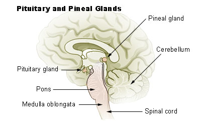
The pineal gland is located in the middle of the midbrain on the roof of the 3rd ventricle and situated below the tail end of the corpus callosum (the body of a major bundle of nerve fibers), in between the two Thalami. The pineal gland looks like a pinecone and so it was named the Pineal gland. The pineal gland is 0.8 mm in size and weighs 0.1 gm and is about the size of a rice grain. This endocrine gland is very vascular, second only to the kidneys (per each unit of mass). The blood-brain-barrier (BBB) is absent here and the hormone is secreted directly into the blood and also in the CSF. The cerebrospinal fluid bathes this gland through a small recess of the 3rd ventricle which continues within the stalk of the pineal gland.


Chemistry of Melatonin.

The pineal gland produces and releases melatonin. Melatonin is N-acetyl 5-methoxytryptamine. The amino acid Tryptophan is the source of melatonin.

Melatonin synthesis.

Tryptophan is converted to 5-hydroxytryptophan by hydroxylation. 5-hydroxytryptophan is decarboxylated to 5-hydroxytryptamine and this product is known as Serotonin.

Serotonin is converted to melatonin in two steps -

Step 1. A rate-limiting enzyme N-acetyltransferase transfers the Acetyl group from Acetyl CoA to 5-hydroxytryptamine and converts it to N-amino-5-hydroxytryptamine.

Step 2. N-amino-5-hydroxytryptamine undergoes methylation. The methyl donor is S-adenosyl methionine and the enzyme catalyzing this reaction is O-methyltransferase. And N-methyl-5-hydroxytryptamine is produced. This molecule is melatonin.

The reactions are shown as follows-

Serotonin + Acetyl CoA → N-Acetyl serotonin. This reaction is catalyzed by an enzyme N-acetyltransferase.

N-Acetyl serotonin + S- adenosylmethionine → N-acetyl 5-ydroxyserotionin.  This reaction is catalyzed by an enzyme O-methyltransferase.

Darkness induces Melatonin synthesis and release.

Darkness causes the release of Norepinephrine from the sympathetic nerve terminals of the pineal gland. The enzyme system is primed by norepinephrine and Cyclic AMP is generated. (cAMP). cAMP activates N-acetyltransferase and melatonin synthesis starts. As melatonin is forming, melatonin is secreted in the CSF and the blood. The pineal gland does not store melatonin in the gland.

If the artificial white light is of a certain strength, the effect of dark on the pineal gland ceases and no melatonin is produced or secreted. During international travel by airlines, the normal dark-light cycle is disrupted and resulting in sleep disturbances.

Breakdown of melatonin.

Melatonin is broken down in the liver by hydroxylation, then conjugated with sulfate and glucuronic acid and excreted in the urine.

Nerve supply of Pineal gland.

Somatic innervation. The 5th cranial nerve sensory nucleus, the Trigeminal ganglion, supplies nerve fibers to the stock and the gland. These fibers contain neuropeptide PACP which are vasoactive compounds. (PACP is pituitary adenylate cyclase acting polypeptide)

Autonomic innervation.

Sympathetic division nerve fibers come from the superior cervical ganglion. The parasympathetic fibers originate from the Otic and Pterygopalatine ganglia

Embryology of the Pineal gland.

In the 17th week of embryonic life, an invagination of the roof of the 3rd ventricle occurs. Initially, the pineal primordium contains Pax6 cells, arranged in a radial manner. After the neural tube fuses, the Pax6 cells rearrange into a rosette formation and then disperse in all directions. All pineal cells are derived from these progenitor Pax6 cells.

An adult pineal gland contains hormone secreting pinacocytes and microglia, astrocytes, and supporting cells. In the adult pineal gland, some progenitor cells remain. Calcium deposit in the pineal gland is common in the elderly, occasionally the entire gland may be calcified.

Role of the Photoendocrine system on the Pineal gland.

The retina of the eyes, supra-chiasmatic cells of the hypothalamus and noradrenergic sympathetic nerve fibers terminate in the pineal gland. Information about light exposure and circadian rhythmic variation is integrated into the pineal gland and regulated melatonin secretion.

Melatonin concentration in the CSF of the 3rd and 4th ventricles is higher than plasma and blood. What effect melatonin has on the neurons of the brain is not known.

Melatonin Receptors.

MT 1 and MT 2 are two types of melatonin receptors in humans. MT1 receptors are present in the suprachiasmatic cells of the hypothalamus, pituitary gland, retina and hypothalamus. When Melatonin binds with MT1, it produces inhibitory effects on the pituitary, and the release of hormones is inhibited and the blood level of Prolactin falls. Through the MT1 receptors, melatonin maintains the circadian rhythmic release of hormones of other endocrine glands.

MT2 receptors are present in the retina. When retinal receptors are stimulated, Dopamine release ceases. It also allows phase shifting of the internal circadian clock to the natural earth clock of the light and dark cycle. Other effects of MT2 receptor activation are increased phagocytosis and enhanced osteoclast activities and vasodilatation.

Hallucinogenic action.

DTM (dimethyltryptamine) is a hallucinogenic compound. Only a small amount of DMT is found in the Pineal gland. This fact might have started the notion that the pineal gland is a psychic center and controller of human behavior. 

Melatonin use.

Melatonin in the USA is an OTC drug (over the counter). It is available in 3 mg tablets, made solely in the laboratory. One melatonin compound. Ramelton is approved by the FDA for the treatment of insomnia, but the results are not consistent.

Indication of use.

Jet lag, Circadian rhythm disorder in the blind, Delayed sleep-wake phase sleep disorder in people who have delayed sleep and delayed wake time than required of them. In insomnia, melatonin reestablishes NON-RAM sleep. It is useful in shift workers and sleep disorders in children.

Adverse effects.

Melatonin is a safe supplement. However, it is a biological amine like Histamine and Dopamine. So, care should be taken when used with epileptic drugs, anti-platelet agents, BP medications, antidepressant drugs, immune modifying drugs, and anti-anxiety drugs.

____________________________________

Rene Descartes (1596 - 1650), a French mathematician, scientist and philosopher. He stated " Je pense, donc Je suis" (I think, therefore I am.)

Footnote: https://plato.stanford.edu/entries/pineal-gland/

********************************************************

Wednesday, March 8, 2023

C - Reactive Protein

                                            C-Reactive Protein (C-RP)

                                         PKGhatak, MD


In an article published in the Journal of Experimental Medicine in 1930, William S. Tillett and Thomas Francis Jr described a serological reaction with a molecule of pneumococcus. A clearer picture emerged from the authors, Abernathy and Avery, in 1941 from their work on the serology of pneumococcal infection describing a flocculation reaction with C-polysaccharide of pneumococcus and a plasma protein. That protein is now known as C-Reactive Protein.

Chemistry of C-RP.

C-RP is a member of the Pentraxin family of proteins. Pentraxin is a ring-shaped planer symmetry protein, with a hole in the center, like a doughnut. This structure gives a high degree of stability to the molecule and resists enzymatic attacks. The name pentraxin came from the Greek Penta Ragos meaning five berries. The molecular weight of C-RP is 115,135 Daltons and each molecule is composed of five identical non-glycosylated polypeptide subunits, each contains 206 amino acid residues. Not more than 15 sugar units are present in each C-RP molecule, but the glucose molecule is not one of them. The amino acids, asparagine, serine, threonine, hydroxylysine, and hydroxyproline, are the only 5 amino acids present, in repeats in a C-RP molecule. The glycosidic linkage between carbohydrate and protein occurs more frequently through oxygen rather than nitrogen.

Site of biosynthesis.

Hepatocytes synthesize C-RP when Interleukin 6 (IL-6) is released from the damaged cells into the blood and IL-6 is carried to the liver. IL-1 beta enhances IL-6 response in the synthesis of C-RP. The C-RP levels may go up 1000 times from a low normal blood level, 0 to 8 mg/L.  Protein molecules are synthesized elsewhere; an enzyme Glycosyltransferase, specific for each type of sugar molecule, catalyzes the attachment of the sugar to the protein moiety.

                                            Pentraxin molecule


Plasma level of C-RP.

Normal plasma levels of C-RP are between 0 to 8 mg/L. The half-life of C-RP is 19 hrs. and remains constant under all conditions of health and disease. The concentration in plasma is solely determined by the rate of synthesis by hepatocytes under IL-6 stimulus, which reflects the intensity of the pathological processes. Once the IL-6 levels fall, the CRP synthesis stops and plasma levels return to the base level in 24 -48 hrs. This property of C-RP makes it an ideal acute-phase protein for-

  1. Screening for physical illness.

  2. Monitoring the response to treatment

  3. Detecting any intercurrent infection in immunosuppressed patients

Highly sensitive C-RP (hs-CRP).

More than 30 years ago, researchers noticed lower than normal levels of CRP can be quantified and used in clinical medicine for cardiovascular risk assessment and prediction of future events. High-sensitive CRP determination is an immunoassay and is reported as mg /L. Interpretation of results of hs-CPR is as follows-

    1.hsCRP less than 1mg/L is not associated with any acute cardiovascular event and does not have increased incidence when followed for 20 years.

  1. hs-CPR 1 to 3 mg/L is a medium risk factor for Coronary events, Stokes and PAD ( peripheral arterial disease).

  2. hs-CRP over 3 mg/L results should be considered a risk factor for the diseases listed in No2., only if other causes of a more common condition like Rheumatoid arthritis, SBE (subacute bacterial endocarditis), or periodontal disease, etc., are eliminated by repeating hs-CRP a week later.

A few special features of C-RP.

Ligand binding molecules.

C-RP has the highest affinity for phosphocholine residue. It binds readily with small molecules of ribonuclear proteins. C-RP binds with histone, apoptotic cells (programmed cell death) and oxidized LDL (low density lipoprotein).

Complement Activation.

C-RP-ligand complex binds with the C1q complement of the classic pathway. In the process, complements C1, C3 and C4 are completely used up. This complex only minimally activates C3 complement and does not activate the complement of the alternate pathway C5 to C9. The outcome of activities is the initial innate response to tissue injury or infection and promotes the opsonization of cellular debris from the inflammation site and stimulates healing.

Autoimmune disease.

The first reported evidence came from works on the blood of Rheumatoid Arthritis (RA) patients.

C-RP deposits are present in the nuclei of the cells of the synovial membrane. The intensity of RA correlates well with plasma C-RP levels and has replaced ESR (erythrocyte sedimentation rate) for monitoring of disease activities and follow up of patients under treatment. Ulcerative colitis shows a similar pattern. But SLE (systemic lupus erythematosus), Scleroderma and Polymyositis show no close relationship with plasma levels of C-RP and disease activity. This is explained based on the works of certain patients who are unusually susceptible to pneumococcal infection and have very low C-RP in plasma. They have a polymorphism of the gene which encodes Guanine and Thymine ( G &T) nucleotides in the Intron of the gene, which accounts for a low plasma C-RP level.

Kidney.

C-RP binds with the immune complex deposited on the basement membrane of the glomeruli in several varieties of glomerulonephritis. In an acute renal transplant rejection episode, C-RP binds with renal tubular epithelial cells and endothelial cells of the peritubular capillaries in the kidney interstitium.

A short summary of the C-RP function.

  1. C-RP is an acute phase protein, though not specific for any particular disease, nevertheless is a useful clinical tool for the determination of the disease activities and follow up.

  2. C-RP activates the classic complement pathway and has an inhibitory effect on the activation of the alternate complement pathway.

  3. hs-CRP is a predictor of the future coronary event. Prognostic indicator of Cardiovascular events, Strokes and PAD (peripheral arterial) disease.  

  4.                          

  5. Taken from NIH publication.
  6. **************************************

Saturday, March 4, 2023

Liver Cancer

                                                        Liver Cancer.

                                                     PKGhatak, MD


Liver Cancer is a deadly disease. Cancers of the liver are of two main categories, namely, Primary Liver Cancer and Metastatic or Secondary liver cancer.

Primary Liver Cancer:

The liver is made up of about 15 thousand individual lobules. Each lobe is a structural and functional unit of the liver. Liver cells, Hepatocytes, when they turn cancerous, are called Hepatocellular cancer. The liver has a dual blood supply - the portal and systemic circulatory systems. A bile duct network drains the bile from the liver. 

The endothelial cells of the blood vessels and bile duct cells can turn cancerous. Cancers of different cell lines differ from each other in many aspects. In addition, the liver is richly supplied with immune cells and malignant tumors may develop from immune cells also.


 A. Hepatocellular cancer.

Hepatocellular cancer is by far the most common cancer of the liver. The annual incidence of hepatocellular cancer in the USA is 35,000. Native Americans have the highest incidence among all ethnic groups. Older Americans have more cancers than the younger age group. The incidence of hepatocellular cancer in East Asian countries is the highest in the world. A very rare form of cancer is occasionally seen in very young children in the hepatic stem E-MB  and MEM-HB cells.

Risk factors.

Hepatitis B and hepatitis C are viral infections and proven causes of liver cancer. A fungal toxin, Aflatoxin, is carcinogenic. Alcoholic liver cirrhosis and Non-alcoholic fatty liver disease (NASH) can turn cancerous. Cigarette smoking and alcohol are risk factors. Muscle-building androgenic steroids and oral contraceptive pills are additional risk factors. Liver cysts and cysts associated with polycystic disease of the kidney are also risk factors. Very rarely, cancerous cysts are seen in the use of anabolic steroids and birth control pills.

In addition, hereditary metabolic diseases like Hemochromatosis (iron), Wilson disease (copper), alpha 1 antitrypsin deficiency ( enzyme), porphyria cutaneous tarda, and glycogen storage disease are known to cause Hepatocellular cancer.

Symptoms.

A small and early liver cancer does not produce any symptoms. A patient with growing cancer develops loss of weight, loss of appetite, upper abdominal fullness, and abdominal pain. At this stage, the diagnosis of liver cancer leads to a better outcome. But most patients come to the doctors when they develop jaundice and dark-colored urine; at this stage, the disease has progressed too far for a good outcome.

Diagnostic tests.

Alpha fetoprotein in serum is elevated, and Liver function tests show elevated ALT over AST and high bilirubin. Ultrasound is a very useful tool to detect tumors. CT scans and needle biopsies under ultrasound guidance give a definite diagnosis.

Treatment.

In most instances, a complete resolution of liver cancer is not possible because of the advanced stage of cancer at the time of diagnosis. In suitable cases, local or regional resection of the liver is possible. The liver is a remarkable organ in its capacity to grow back to full size if it is free. In selected cases, a lobectomy or total hepatectomy followed by a liver transplant is done. Immunotherapy, chemotherapy, and several ablative therapy methods are available.

Prognosis:

In general, this is most disheartening. 5-year survival is less than 20%, and in advanced cases, it is less than 3 %.

 Carcinoma of the Bile Ducts.

The medical term for cancer of the bile ducts is Cholangiocarcinoma (CLC). CLC is less common than hepatocellular carcinoma. However, people who have habits of eating raw fish have an alarmingly high rate of CCL. 


The diagram above shows bile ducts inside the liver and also outside. The outside bile duct for this discussion is dealt with into two separate categories, namely Hilar and  Common bile duct cancers.

The life cycle of a Liver Fluke.

CLC (cholangiocarcinoma) is the second most common liver cancer in the world, but the incidence in the USA is much less than hepatocellular carcinoma. But overall, CLC is increasing in all countries, including the USA. In the USA, the Hispanic ethnic group has a higher CLC. In the world, Thailand has the highest rate of  CCL, about 40 in 100,000 people, followed by China, Japan, and other East Asian countries, with a clear association with Liver fluke infestation of the biliary system. In the Mekong River basin countries, Liver fluke infestation is between 30% to 70 % of the population, and the death rate from CCL is 3% of all deaths.

Risk factor.

Primary sclerosing cholangitis is a precancerous condition; a congenital bile duct disease called Choledochal cyst disease has a high rate of CLC. Ulcerative colitis and Crohn's disease have a higher incidence of CLC. Infestation of the liver fluke is a risk, as mentioned above. Both liver cells and the bile duct system developed from the same progenitor cell in the embryonic stage of development. As a result, the non-specific risk factors mentioned under hepatocellular cancer (alcoholic liver cirrhosis, NASH, diabetes, etc.) are also risk factors for CLC.

 A. Intrahepatic CLC.

Intrahepatic CLC is the least common among the three CLCs. Initially, CLC  is not distinguishable from hepatocellular carcinoma; the diagnosis is made only after a liver biopsy. Patients remain asymptomatic in the early stage. Occasionally diagnosed by chance when ultrasound or abdominal CT scans are done for other reasons. The prognosis at this stage is very good with surgery and chemotherapy. However, the majority of patients seek medical attention because of the development of jaundice. Diagnosis is relatively easy by ultrasound and fine needle biopsy. A standard care protocol in the USA hospitals uses a multidisciplinary approach and provides partial or complete removal of the liver and liver transplantations.

B. Hilar CLC.

Hilar CLC is the most common of CLC, accounting for about 70% of CLC. Pathologically, these are adenocarcinomas. Because of their location, jaundice develops earlier than in other liver cancers. The patient seeks medical attention because of worsening jaundice, dark urine and light stool, loss of appetite, and weight loss. Laboratory tests confirm obstructive jaundice. CEA and alpha-fetoprotein are positive. Diagnosis requires a fine needle biopsy. Curative surgery is not possible in the majority of cases. Initial therapy is draining bile by inserting a stent in the common bile duct, draining into the duodenum, done during an ERCP examination, however, a transcutaneous bile duct stent can also be done. 

C. Common bile duct CLC.

The risk factors are the same as the above group and an additional risk factor is an abnormal opening of the pancreatic ducts.

Abnormal Pancreatic Duct.

Anatomical variations of pancreatic ducts are common. Occasionally, one or both pancreatic ducts may join the common bile duct, rather than opening into the ampulla. In this circumstance, chemical inflammation of the duct from pancreatic enzymes leads to fibrosis and stricture, and also carcinoma

Obstructive jaundice is the presenting symptom of common bile duct CLC. In clinical practice, obstructive jaundice is common. The causes of obstructive jaundice are as follows.


                                                   Taken from NIH publication.

Among the benign causes, gallstone and bile duct inflammation are most common, followed by common bile duct stricture, and Mirizzi syndrome. Gallbladder cancer leads the malignant causes followed by cancer of the periampullary region. 12 % of cases are due to cholangiocarcinoma.

                              Mirizzi syndrome in a diagram.

Diagnosis and treatment. 

Initial diagnostic workup is no different from other types of CLC. Initial treatment is a stent placement during ERCP or can be placed transcutaneously. The definitive treatment is the Whipple procedure or a modified Whipple operation. The prognosis is encouraging with hepatic transplants and immunotherapy.

 [ See Chronic pancreatitis blog, dated April 9, 2022]. 

Childhood Liver Cancer, Hepatoblastoma, is seen in children less than 3 years of age. It is more common in premature and underweight newborns. The primary cause of it is unknown. Several inherited conditions are associated with hepatoblastoma. Resection of the liver when performed early produces the best outcome.


edited: May 2025.

************************************************


Sunday, February 26, 2023

Food Poisoning


                                            Food Poisoning

                                          PKGhatak, MD


Food and drinks can be contaminated with microorganisms if not handled properly. Certain bacteria grow in the leftover food kept outside the refrigerator and produce toxins. When people eat leftover food, these toxins produce an illness, which is called food poisoning.

In general, such a definition of food poisoning is not universally followed. Viral gastroenteritis, cholera, amebiasis, typhoid fever and others are also included in food poisoning. It is better to address those illnesses as food borne illnesses rather than food poisoning.


Bacterial Toxin.

Bacteria can grow over a wide range of temperatures, from 4 degrees C to 60 degrees C,  provided moisture and nutrients are present. A growing bacterial colony produces toxins.

Toxins produced by bacteria are in two groups-

A. Exotoxin. Endotoxins are secreted by bacteria in the food. Exotoxins can be heat resistant or destroyed by heating.

B. Endotoxin. Endotoxins are present in the bacterial bodies and released in food when the bacteria die or are killed by antibiotics.

Preformed toxins in food produce symptoms in 1 to 6 hrs. after ingestion, whereas, bacterial infection and toxin generation take over 8 hrs. and then symptoms develop. Bacteria commonly responsible for food poisoning are Staphylococcus, Bacillus, Clostridium, E. coli and Vibrio species.


Staphylococcus aureus food poisoning.

S. aureus readily grows when food is left on the kitchen countertop. Symptoms start within 1 to 6 hrs. after eating food. Reheating food before eating does not destroy the bacterial toxin. Nausea and vomiting are common symptoms and start abruptly. Campy abdominal pain and diarrhea follow. The illness is self limited and patients recover in 24 hrs.


Bacillus cereus.

Reheated fried rice is often the source of poisoning. Intense nausea and vomiting start within 1 to 8 hours after eating food. The toxin can be detected in stool and leftover food.

Symptoms last for 12 hrs. It is also a self limited illness.


Clostridium botulinum.

Clostridium botulinum is a soil bacterium. Canned vegetables not properly washed and low oxygen environment in a sealed can are ideal for the production of toxins. Another source is natural honey. The ingested toxin produces nerve paralysis and the illness is known as Botulism. Though the symptoms start days after ingestion of the poison, botulism is due to the toxin present in food and not a bacterial infection.

Botulism. The sudden onset of fluctuant and intermittent but severe muscle paralysis in a healthy person should alert the physicians of botulism. Botulism toxin prevents the release of a neurotransmitter, Acetylcholine, at the neuromuscular junctions.

Symptoms generally develop 3 days after ingestion of the toxin and continue to progress further for another 3- 4 days. Paralysis of eye muscles produces double vision, and eyelid paralysis causes drooping eyelids. Muscles of swallowing and speech muscles are also paralyzed, producing difficulty in eating and drinking, and a nasal voice. In severe cases, the muscles of the limbs may be paralyzed. Identifying the toxin in unused portions of food and serum helps in the diagnosis. An electroencephalogram(EEG) is also useful.

Antitoxins are available to reverse the effects of toxins. Hospitalization and close monitoring of respiration and blood oxygen and CO2 are essential parts of the management of botulism. Public health authorities must be informed.


Clostridium perfringens.

This spore-forming Clostridium is found in soil and feces. Uncooked beef, poorly cooked poultry and fish are the source of toxins. Between 6 to 24 hours after eating, symptoms of severe diarrhea and abdominal cramps develop. The illness can last one to several days. The initial onset may be from preformed toxin, but subsequently, the bacteria multiply in the gut epithelium and continue to generate enterotoxin and produce symptoms.

Enterotoxin producing E. coli.

This entity falls in between preformed toxin and bacterial infection and perhaps, both are operative.

Animals and humans harbor E. coli in their large guts. When food and drinks are contaminated with feces, severe watery diarrhea and abdominal cramps develop within a day or two after ingestion. It is a common cause of food poisoning among international travelers. Antibiotic fluoroquinolones are effective therapy.

Vibrio parahaemolyticus.

This vibrio usually contaminates shrimp, crab and shellfish. Eating raw oysters or lightly cooked seafood produces a sudden onset of abdominal pain and diarrhea within 2 to 48 hrs. Nausea, vomiting and diarrhea last for 2 to 5 days.

Proper hydration is required to prevent dehydration.


************************


Thursday, February 16, 2023

Peptic ulcer

                                                     Peptic ulcer disease.

                                              PKGhatak, MD


When ulcers develop in areas of the stomach normally bathed by acid and pepsin, those ulcers are called peptic ulcers. All sections of the stomach and the first part of the duodenum come in contact with acid and pepsin. And occasionally in an incompetent lower esophageal sphincter, the lower end of the esophagus develops ulcers due to gastric reflux. 


Typically, peptic ulcers are found in the first part of the duodenum and the Pylorus.

It is estimated that about 4 million new cases are detected annually worldwide. In the USA, about 5 to 8 % of the population have peptic ulcer disease. In the past several decades, the incidence of peptic ulcer fell but in recent times, the incidence has remained steady.

Gastritis and gastric erosion are included in Gastric ulcer diseases because the mechanism of development of these is the same as ulcers.

Why do people develop Ulcers of the stomach.

The immediate cause of peptic ulcer is excess acid and protein digestive enzyme pepsin production by the cells of the mucous membrane of the stomach.

Risk factors of peptic ulcer disease.

1. Infection of Helicobacter pylori (H. pylori).

2. Cigarette smoking.

3. Alcohol abuse. 

4. Chronic use of aspirin or non-steroidal anti-inflammatory drugs (NSAID).

5. Anxiety and stress.

6. Use of systemic steroids.

Peptic ulcer.

The common practice today is to detect peptic ulcers by endoscopy. Any area of the stomach and duodenum that appears abnormal under endoscope is usually biopsied and the histology of the tissue is examined. Gastritis and ulcers due to H. pylori can be detected and if any doubt exists, a biopsy should be enough to exclude cancer.

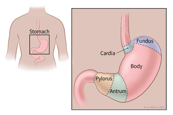
Peptic ulcers are small, less than 1 cm in diameter, have clear margins and a clean base. Cancers are usually located in the cardia and fundus of the stomach, usually large, and the margins are heaped up, the base is necrotic.


Panel A: are benign lesions. Panel B: is a cancer of the stomach.


A brief review of the characteristics of the gastric mucosa.

The inner lining of the stomach is called the mucous membrane. The diagram below shows the relative thickness of the stomach wall and a cutout section shows the different cells in the mucous membrane.


The function of the cells of the mucous membrane.

1. Parietal cells. These cells produce a strong inorganic acid - hydrochloric acid. The pH of the stomach is 1 to 2. Parietal cells also produce the Hematinic principle, a glycoprotein, which is essential for Vitamin B12 absorption.

2. Chief cells. Chief cells secrete an inactive form of protein digestive enzyme, Pepsinogen. Pepsinogen at pH 1 or 2 is converted to an active enzyme, Pepsin. And pepsin remains active in pH 1 to 8.

                                location of endocrine cells, stained brown.

3. Endocrine cells. The endocrine cells are G-cells, ECL-cells, and D-cells.

G-cells secrete Gastrin directly into the bloodstream. (that is the nature of all endocrine glands and cells). Gastrin is brought back to the mucosa by circulation. Gastrin stimulates the growth and development of parietal cells and enterochromaffin-like cells (ECL) cells.

ECL-cells produce and store Histamine and secrete histamine. Histamine is a potent stimulant for acid secretion. In addition, ECL cells secrete chromograninA derived peptides, pancreastatin and other peptides.

D-cells. D-cells secrete Somatostatin. It is an inhibitory hormone. It decreases the release of gastrin and decreases the motility of the stomach and intestine. Also delays the release of other peptides from ECL cells.

5. Goblet cells and mucus secreting glands. Goblet cells secrete mucus. The mucus has a very significant function. The mucus is of two layers, one of which is adherent to the cell layer and the free layer on the top of the adherent mucus layer. Both layers are about 1 mm thick and chemically are glycosylated mucin peptides. It contains a high concentration of bicarbonate ion [HCO3-]. HCO3 is immediately available to neutralize any HCl (hydrochloric acid) that comes in contact with the mucus layer.

The mucus barrier acts as a cushion on which gastric juice floats. It is like an atom or a proton kept separated from the copper tube and confined to a narrow zone by powerful electromagnets in a cyclotron (atom smashers).

Acid production in the stomach.

The parietal cell contains the Carbonic Anhydrase enzyme. This enzyme greatly accelerates the reaction between H20 +CO2 -->H2CO3, taking place within the cell cytoplasm. H2CO3 then dissociates into H ion [H+] and HCO3 ion [HCO3-]. Energy dependent [Na+]/[H+]  pump  (commonly called Proton pump)     expels  [ H+] into the stomach lumen and in exchange [K+] enters the cells. [HCO3-] is carried from the cells into the blood by a cation exchanger transport protein and it brings back [Cl-] inside the cells. [Cl-] enters the gastric lumen via Cl channels. [H+] unites with [HCO3-] because opposing charges attract each other and form HCl.

Factors accelerate Acid production.

The Vagus nerve is the secretory motor nerve for the GI tract. As soon as food enters the stomach, vagal stimulation produces Acetylcholine. Acetylcholine directly stimulates gastric acid secretion and indirectly by increasing gastrin release.

G-cell.

Stimulate acid production via gastrin.

Histamine.

Histamine is a potent stimulant for vasodilatation, increased glandular secretion, and other properties. In the stomach, the Histamine is produced and released by ECL- cells, nerve cells and mast cells. Histamine 2 receptors are present on the surface of the Parietal cells. Once histamine combines with the H2 receptors, acid production increases. Histamine is the main regulator of acid secretion influenced by local conditions.

D- cell.

D-cell produces Somatostatin. It inhibits acid production and decreases gastric motility and other inhibitors control beyond the stomach.

Conditions favor peptic ulcer.

Smoking.

Cigarette smoke contains Nicotine. Nicotine decreases Prostaglandin generation and makes gastric mucosa vulnerable to ulcer formation.

Alcohol.

Alcohol is a poison to cells when it comes in direct contact with cells. Alcohol destroys the mucus coat of the stomach, making it easier for alcohol to come in contact with the mucosa. An excessive amount of alcohol produces gastritis and chronic use produces ulcers. Once peptic ulcers are formed, even a small amount of alcohol damages cells and prevents ulcer healing and causes pain.

Aspirin.

Aspirin on ingestion turns into salicylate. Salicylate is toxic to cells and produces erosion of surface cells, disrupts the H+/K+ pump and loss of K+ from the cells. Hemorrhagic gastritis and gastric ulcers are common adverse effects of aspirin.

Since aspirin inhibits the enzyme cyclooxygenase permanently for the life of affected cells, adverse effects persist even after aspirin is withdrawn.

NSAID.

Non-steroidal anti-inflammatory drugs are commonly prescribed (e.g. Advil) for joint and bone injuries, post orthopedic surgery, and all varieties of arthritis and are also available as OTC (over the counter drugs). Peptic ulceration and gastritis have increased in recent years, going against the trend seen in several past decades.

NSAID inhibits the COX-1 enzyme. COX-1 is another enzyme needed for Prostaglandin synthesis. The effect of NSAID is however temporary, once the drug is stopped, the cells can recover.


Helicobacter pylori (H.pylori).


H.pylori is a gram negative, highly motile, unipolar-multi flagellated, microaerophilic organism. The body of H. pylori is spiral and so it is called Helicobacter. Scientists believe H. pylori infected the stomach of humans 2,5000 years ago and is still infecting humans. More than 50 % of the world's population is already infected, and 100 % of people in Africa and Asia have H. pylori chronic gastritis, although 80% are asymptomatic. Most people are infected in early childhood by the oral-oral route and the fecal-oral route. Once infected, 80 % of the bacteria are present in the mucus of the gastric lumen and 20% in the mucus secreting cells of gastric epithelium but absent in the parietal cells, endocrine cells and the chief cells of the epithelium.

The bacterium has developed a unique mechanism of evading the immune defense of the victims and continually inventing ways to resist antibiotics. The bacterial wall has several layers of envelopes of different proteins and in addition, has several adhesion proteins and these proteins keep the bacteria firmly attached to the cells. 

Detection.

Breath test.

The human body does not have urea splitting enzyme urease, but H. pylori contains urease that breaks down Urea into CO2 and ammonia. A tablet containing 13C  urea and citric acid is taken by mouth and the expired air is collected and analyzed. Presence of C13- CO2 in expired breath indicates H. pylori infection.

Biopsy.

Patients present with symptoms of peptic ulcer usually undergo endoscopic examinations. Biopsy from the suspected areas is taken. H.pylori is easily detected, if present.

Treatment.

Because antibiotic resistance is common, the antibiotic regimen is often changed. A commonly used regimen is mentioned and also a reference to the 2022 recommendation is provided.

Antibiotics:

Amoxicillin and one of the following two antibiotics: Macrolides and Metronidazole (an anaerobic bacterial antibiotic). Antibiotics are combined with Bismuth subnitrate. Bismuth forms a complex with the bacterial wall, inhibits multiple bacterial enzymes resulting in decreased synthesis of adherence protein molecules and deprives H. pylori source of nutrition.

Acid suppression.

Pantoprazole. It blocks the proton pump and acid secretion comes to a total stop.

Histamine receptor blockers. It acts by competitive binding with H2 receptors of the parietal cells and blocks the action of histamine in producing acid.

Antacids. Commonly used aluminum oxide and calcium carbonate as acid neutralizers are not therapeutically sufficient in the treatment of peptic ulcers.

____________________________________________________

https://www.gastroenterologyandhepatology.net/archives/june-2022/helicobacter-pylori-treatment-regimens-a-us-perspective/

(Copy & paste on your browser.)

*******************************************

Thursday, February 2, 2023

Iron Deficiency Anemia.

                                                             Iron Deficiency Anemia.

                                                       PKGhatak, MD


 Anemia is defined by the WHO as having less than 12 gm /dL of hemoglobin in women and less than 13gm /dL in men. By this criterion, 33 % of the world's population has anemia and half of the cases are due to iron deficiency. Children below 5 years old and women of childbearing age are particularly susceptible to iron deficiency anemia in sub-Saharan Africa and South Asia. Iron deficiency anemia is the number one nutritional disorder in the world.

Iron deficiency anemia develops when the iron store is depleted and the daily loss of iron exceeds the daily iron intake. The consequences of iron deficiency anemia are many, chief among them are dysfunctions of several systems of the body, and notably among them are limitation of physical work, reduced energy production, depressed immune function, recurrent infection, poor digestive function, and various neurocognitive functions. The diagram below is an outline of iron utilization. 


Iron containing proteins in humans.

Four general categories of proteins contain iron: (1) heme proteins (e.g., hemoglobin. (2) mononuclear iron proteins (e.g., superoxide dismutase), (3) diiron-carboxylate proteins (e.g., ribonucleotide reductase, ferritin), (4) iron-sulfur proteins (e.g., aconitase), Hemoglobin is the most abundant iron-containing protein in humans.  More than one-half of total-body iron is contained within hemoglobin.

Basic physiology of Iron in humans.

Iron is a very reactive metal and combines readily with oxygen, when it reacts with H2O2 (hydrogen peroxide) it generates oxygen radicals. Oxygen radical sickens living cells and in the end, kills cells. To protect tissue from its harmful effects,  a protein, ferritin, combines with iron. Moving in and out of cells requires conversion of ferric to ferrous state and must pass special areas of the cell membrane called gates. Once inside the cells, iron is converted back to ferric state and in the blood and inside the cells iron remains combined with ferritin.

The generation of RBC is called erythropoiesis. Demand for erythropoiesis comes from: (a)Tissue oxygenation, (b)Erythrocyte turnover, (c) Blood loss from hemorrhage.

(a)Tissue oxygenation remains more or less stable in health.

(b) Approximately 20 mL of old erythrocytes die daily, and 20 mg of iron is recovered from the dead RBC.  The immediate source of iron for erythroblasts is mono or diferric transferrin, found in high concentrations in plasma. The sources of diferric transferrin are the gut (diet), macrophages (recycled iron), and the liver (stored ferritin iron). the diet. 

2.5mL of whole blood contains 1.0 mg of iron. 1.0 mg of iron is absorbed from the diet daily and 20 mg of iron from recycled erythrocytes is available to support erythropoiesis. Once iron stores are depleted, dietary and recycled erythrocyte iron is not usually sufficient to compensate for acute blood loss. In a normal person, less than 2 mL of blood is lost daily in the stool.  But this tiny amount of blood in stool does not give a positive occult blood test. A minimum of 60 mL/d is needed for a positive occult blood test to detect the presence of blood in the stool. Women in childbearing age lose additional blood due to menstruation. Repeated nosebleeds and regular blood donation lower body stores of iron.

Iron in pregnancy:

It is estimated that about 1.2 gm of iron is required from conception through delivery. The breakdown is as follows: (i)Mother's erythrocyte mass should increase from 350 to 450 mL. And that needs 450mg of iron (ii) Cessation of menstruation saves 600 mg of iron (iii) A full tern newborn has 280 mg (iv) Placental loss 90 mg.

At birth, the fetal red cell mass is 50 mL/kg. (Compared with 25–30 mL/kg in adults). Even in anemic mothers, the fetal ferritin levels remain 10 times higher than the maternal ferritin, indicating nature's preferential treatment of a growing child over the mother.

Common causes of iron deficiency in poor countries.

1. Poor diet. Red meat, organ meat and egg are good sources of iron. Poor people can hardly provide meat or eggs on a regular basis. Bush meat in Africa, at one time adequately supplied the local population, but increased demand by the growing urban population has dried up this source. Vitamin C improves iron absorption, but tea and coffee interfere with absorption. Inorganic iron absorption requires multiple mechanisms,  but infections and inflammation depress iron absorption by hepcidin inhibitory action. 

2. Malaria. Malaria is a special circumstance. Intravascular hemolysis from the effects of parasites and Blackwater fever produces severe anemia. In addition, malnutrition depresses iron absorption.

 3. Hookworm. It is an intestinal parasite, that is a significant source of GI blood loss in millions of people in South Asia and Africa. Defecation in the open, and walking in bare feet in flooded fields make it possible for hookworms to enter the body. Each worm takes 0.3 to 0.5 mL of blood from the upper small intestine and produces anemia. [ see footnote ]

4. Gastrointestinal factors. Suppressed gastric acid secretion from taking a proton pump inhibitor or gastric mucosal atrophy from gastritis, prevents the release of iron from organic iron in food. Tropical sprue and other duodenal pathologies hinder iron absorption from the gut. Liver diseases may decrease ferritin production and in chronic infection, excess hepcidin decreases GI iron absorption. And finally, Helicobacter infection of the stomach produces gastritis and causes decreased iron absorption.

A look at the peripheral blood in iron deficiency anemia.


Blood of iron deficient anemia.


                                       Blood of a normal person.

The characteristic features of iron deficiency anemia are

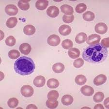
The RBCs are microcytosis (small sized cells) and hypochromic (pale). In addition, a combination of increased red cell distribution width (RDW), decreased red blood cell (RBC) count, decreased MCH (mean corpuscular hemoglobin), and decreased mean cell volume are manifested. Red cells are pale in the center, smaller in size and the shape is variable and easily identifiable on blood smears. Because of non-steady state hemopoiesis, the RDW shows wider variation and is usually over 15%.  This is marked in contrast with the Sickle cell trait. The red cells in the sickle cell trait are also small and pale in the center and the hemogram appears similar to iron deficiency but RDW remains in the normal range due to the fact that hemopoiesis is steady because the iron stores in the body are full and so also the of serum iron and ferritin saturation index.  In iron deficiency, the serum ferritin level is 15 μg/L or below.  In research centers, the iron concentration of the reticulocytes, erythroblasts, the bone marrow and the liver is determined, but in clinical practice, those tests are not needed for diagnosis.  Iron deficiency causes increased release of soluble transferrin (transport protein) from erythroblasts. Therefore, ratios of soluble transferrin receptor and ferritin are used to detect iron-deficient erythropoiesis. 

Treatment of iron deficiency anemia,

Replacement of the iron stores is the first priority. Oral iron preparations are of two kinds. Inorganic and inorganic iron. Inorganic iron preparations are affordable but difficult for patients to tolerate because they produce constipation, nausea and anorexia. Organic iron preparations are easier to tolerate but expensive. In previous generations, intravenous iron therapy was problematic because of frequent hypersensitive reactions. The improved formulation has made rapid correction of anemia possible.

Advances in Iron knowledge.

 The most significant advance is the discovery of hepcidin. Hepcidin expression is highly variable and influenced by a circadian rhythm. That knowledge should improve dosing regimens.  When iron therapy does not improve iron deficiency anemia, TMPRESS6 gene mutation is suspected. In normal conditions, the TMPRESS6 gene provides instructions for the Matriptase-2 protein molecule, which is a part of the controlling mechanism of hepcidin production in the liver.

________________________________________________________________________________________

Footnote:


1. Larva entering by piercing skin of feet---> enter venous blood and carried to the right heat chambers, then to pulmonary capillaries----> larva moving out from lung alveoli to airways and crawls up the trachea----> coughed up and swallowed----> enters the stomach and settles in upper small intestine---- sucks blood, matures, mates, releases eggs----> egg containing feces deposited in open fields and the cycles continue.

********************************************************************


Sunday, January 29, 2023

Cardiomyopathy

                                                Cardiomyopathy.

                                         PKGhatak, MD

Cardiomyopathy is a weakened condition of the Heart Muscles and the heart chambers increase in size and subsequently the heart fails. It is suspected that 1 in 5,00 adult people may have cardiomyopathy without being aware of it. People seek medical attention only when cardiomyopathy becomes symptomatic. The initial symptoms are shortness of breath and a marked decrease in energy level to carry on the usual work.

There are wide varieties of conditions that lead to cardiomyopathy and based on pathophysiology the disease is classified as Dilated, Hypertrophic, Restricted, and Arrhythmogenic Cardiomyopathy.

Cardiomyopathy also happens due to inherited defects of genes, producing structural changes in the heart muscle or metabolic defects, which cause accumulation of iron and other material in the heart muscles.

 I. Dilated Cardiomyopathy:

The heart is enlarged and the muscles are stretched and become thin. Thinned out heart muscles fall behind in pumping blood out of ventricles into the circulation. Accumulated blood causes congestion of the lungs, and produces shortness of breath and in other organs. Dilated cardiomyopathy is diagnosed by clinical examination and confirmed by a chest x-ray and echocardiogram. 

 X-ray criterion is the transverse diameter of the heart exceeds 50% of the chest diameter in PA view.

A few common causes of dilated myopathy.

1. Virus infection.

Viral myocarditis of a mild nature happens frequently. In most cases, myocarditis resolves spontaneously without any health consequences. The common viruses causing these conditions are Parvovirus B-19, Coxsackie A & B, HIV, Covid-19, Cytomegalovirus, Epstein-Barr virus and Adenoviruses.

Damage to heart muscles in viral infection happens in two stages. In the initial phase, the inflammatory cells infiltrate the heart muscle. Immunocytes release acute phase inflammatory cytokines namely IL-1 beta, TNF alpha, and Interferon alpha and these agents should be sufficient to avert a full blown inflammation, but in some cases, they fail. In the second phase, about 7 to 14 days after the initial symptoms, T-cells accumulate. Vasoconstriction and release of Perforin molecule create multiple pores like apertures in heart muscles. Immunocytes release excess amounts of  IL-1, IL-6, CCL5, and TNF beta. Interferon decreases B-Cell activities and facilitates T-Cell activation. Cytokines continued to appear in the heart in response to the presence of viral particles in the myocardium. Because of the close resemblance of the viral antigen to muscle protein, the cytokines make mistakes one for the other. and damages the heart muscle.

A diagnosis of viral cardiomyopathy requires endocardial biopsy and identifying viral antigens. Heart failure and other organ insufficiencies are treated with medications. A cardiac transplant is a definitive treatment.

2. Chagas Disease:

Chagas disease is due to a protozoan parasite, Trypanosoma cruzi. It is an important cause of dilated cardiomyopathy in the Americas. In Central and South American countries, the Chagas disease is endemic. It is estimated that 5 million people are suffering from Chagas disease with an annual rate of infection of 200,000 and annual deaths of 10,000.

About 40,000 people were identified with Chagas disease in the USA among the South American migrant communities in 2019.

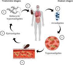
The parasite is carried by a bug, Triatoma infestans, commonly known as the kissing bug. The bug bites humans during sleep and leaves a fecal deposit at the site. The contaminated puncture wound, usually on the face or forehead, is the entry point of the parasite. Local inflammation and adenopathy may or may not produce symptoms. About 5 % of victims develop myocarditis. In the chronic phase, 40% develop dilated cardiomyopathy from the continued presence of parasites at the heart and resultant immune inflammation, tissue necrosis and scars replacing heart muscles. Cardiac conduction abnormalities are common and later heart failure predominates.

The diagram shows the life cycle of T. cruzi.

3. Alcoholic Myopathy.

People are well aware of liver disease due to alcohol; however, many will be surprised to learn about 25 % of all cardiomyopathy. Alcohol acts as an important factor in causing cardiomyopathy. About 2% of heavy alcohol users risk cardiac complications. Colored people have more risks of cardiomyopathy than light colored people. Families with Alcoholdehydrogenase deficiency are more likely to have cardiomyopathy. Alcohol is a Mitochondrial poison. As mitochondrial functions falter, acetaldehyde accumulates in the cells and oxygen radicals form as a consequence and the combined effects of these produce cell deaths. Cardiac muscles are replaced by weak scar tissue, and cardiomyopathy develops in the same way as described earlier. There are no distinctive histological characteristics of alcoholic myopathy, however enlarged and disorganized sarcoplasmic reticulum, fat and glycogen deposition and dilated intercalating discs are seen under electron microscopy in addition to various stages of diseased and disappearing mitochondria.

4. Chronic coronary arterial disease and Hypertension.

These two diseases are silent killers of heart muscles. Acute coronary events produce damage to heart muscles, even when emergency Angioplasty is successfully performed and helps to salvage most of the ventricular muscles, some loss is unavoidable. Chronic insufficiency produces almost identical damage but the pace is slow and takes place over a much longer time. Scar formation eventually leads to dilated myopathy and heart failure.

Hypertension produces ventricular muscle hypertrophy initially. If left untreated, the hypertrophic muscles eventually flatten out. A flabby enlarged heart fails. Hypertension is usually associated with coronary artery disease and induces cardiomyopathy but in the absence of coronary artery disease, hypertension alone is an independent cause of cardiomyopathy.

5. Diabetes mellitus.

Diabetes mellitus produces microvascular changes. The exact statistic is variable based on the nature of the inquiry but consistently higher than the controlled groups.

6. Thyroid disorder.

Both hypothyroidism and hyperthyroidism produce Cardiomyopathy but the mechanism is different. Hypofunction of the thyroid decreases metabolism in general and specially in the heart, delaying cardiac muscle repair and replacement. That leads to an enlarged flabby heart and often a viscous pericardial effusion. In hyperthyroid conditions, the cardiac muscles are overworked due to sinus tachycardia, increased demand for more cardiac output and associated hypertension. As the heart fails, the cardiac chambers dilate.

II. Hypertrophic Cardiomyopathy.

Hypertrophic cardiomyopathy (HCM) is an autosomal dominant inherited disease. One copy of the mutated gene is all that is needed for HCM to manifest. Of the several genes responsible for HCM. These gene mutations are common- MYH7 and MYPPC3 gene; other mutated genes are MYPBC3, TNNT2, TNN13, TPN1, MLC2, and MLC3. 1 in 700 people in the Americas are carriers of these genes and about 75,000 people have HCM at a given time. But most are unaware of the presence of mutant genes, and unfortunately, a sudden cardiac arrest may be the first sign of it. 

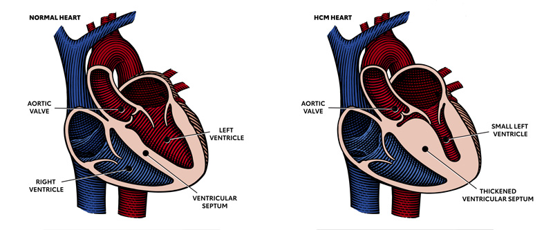
                                  Normal heart                    HCM heart

In the right-hand picture please note the thickened partition between the ventricular cavities. That part blocks the path of blood going out into the aorta during the ventricular ejection phase. In addition, the Mital valve is displaced and often deformed producing mitral insufficiency and reducing left ventricular ejection fraction further.
Calcium iron enters the myocardium in excess amounts and binds with actin-myosin. The cardiac impulse conduction path and coronary arterial system are altered. These are additional causes of abnormal heartbeats, the most serious one is ventricular tachycardia and it is often fatal. The initial symptoms are delayed till the teenage years, and a sudden collapse during a sporting event is usual. Diagnosis is made on clinical examination and confirmed by an echocardiogram. Treatment of HCM is multipronged.  Beta blocks and calcium channel blockers are effective as initial therapy. A recently approved drug, Mevacamten, is a new class of drug and is a reversible cardiac myosin ATPase inhibitor. It reduces the formation of actin-myosin cross-bridges. This action counteracts the inherited defect of this enzyme. The drug promotes energy savings in the myocardium and reduces outflow tract obstruction.
The definitive therapy is surgery. It involves removal of the defective parts and repairs. Technically known as Septal myomectomy, an alternative surgery is Alcohol septal ablation. In failed cases, a cardiac transplant is the only option. Genetic counseling is an essential part of therapy and all close relatives of newly diagnosed HCM patients should be examined for the presence of HCM by examination and echocardiogram.

 III. Restrictive Cardiomyopathy.

Restrictive cardiomyopathy is much less prevalent and accounts for only 5 % of all cases of diagnosed cardiomyopathies. This entity also consists of varied clinical conditions. The common pathophysiology is that abnormal proteins or abnormal cells accumulate between the muscle fibers of the ventricular wall, making the ventricular muscles less pliable and muscles fail to stretch fully. Returning blood accumulates in the atrium and causing both atria to dilate. The cardiac output falls and biventricular failure develops. In addition, the stagnant blood in heart chambers may clot and produce systemic and pulmonary embolism, which are additional features of restrictive cardiomyopathy.

Restrictive cardiomyopathy is a disease of the older generation. Shortness of breath and cardiac arrhythmia are presenting symptoms, additional symptoms are marked loss of weight, fatigue and the effects of arterial embolism like strokes, renal infarction, or limb ischemia.

1. In the Western world, the common causes of restrictive cardiomyopathies are Amyloidosis, Systemic sclerosis, Sarcoidosis and Hemochromatosis. Incidence is increasing in cases of post radiation therapy for malignancy and cancer treatment with Adriamycin.

 2, Primary Endomyocardial Fibrosis and Restrictive Cardiomyopathy.

Taking the world as a whole, Primary Endomyocardial Fibrosis (EMF) is the most prevalent in this group and affects 12 million people. Most cases are seen in equatorial Africa and less frequently in tropical and subtropical Asia and in South America. EMF is somewhat similar to Loeffler eosinophilic endocarditis fibrosis seen in Non-tropical countries. The role of the eosinophil in EMF is still debated, some believe that eosinophils infiltrate the heart muscles due to dead and dying cardiac muscles. Others believe cytokines released by eosinophils produce myocardial necrosis and fibrosis.

The primary EMF is inherited as an autosomal dominant trait with variable penetration. Mutation of genes encoding Sarcomeric proteins - Troponin I, Troponin T alpha & beta, cardiac actin-myosin heavy chain are responsible for this disease.

[  For more information see footnote  ]

IV. Arrhythmogenic Cardiomyopathy.

Arrhythmogenic cardiomyopathy is suspected to be present in 1 in 5,000 people, many of whom are asymptomatic. It is an inherited autosomal dominant condition. Mutation of at least 13 genes is identified. These genes are called Desmosomal genes and also called PKP2 genes. They provide instructions for making components of cell structures called Desmosomes. Under normal conditions, desmosomes keep the muscle fibers of the heart bound together so that all the heart muscles receive cardiac impulses from the sinus node in an orderly fashion and the ventricles can contract in unison.

In this disorder, the cells of the myocardium detach from each other and die. The damaged heart muscles are replaced by fibrous tissue and disrupts cardiac impulse transmission, leads to arrhythmia. The right ventricle is the prime site of this pathology, but since 2008, the same pathology was identified in the left ventricular wall.

PKP2 gene mutated people develop symptoms between 20 and 30 years of age. Sudden syncopal attacks are a usual presentation. Cardiomegaly and heart failure follow the same patterns as other forms of cardiomyopathy. PKP2 gene mutation was detected in 60% of cases, the other genetic abnormality is under investigation.

____________________________________

Footnote:

For further reading, please look at blogs dated -

 1. Amyloidosis …. dated August19, 2000.

2. Hemochromatosis.......Oct 18, 2020.

 3. Radiation pneumonitis...... January 10, 2021.

 4. Connective tissue & MCTD...... Dec 14, 2022.

 5. Sarcoidosis. This is a multisystem disease of unknown cause. Lung lesions resemble pulmonary tuberculosis, and extensive lymph node engagement is a distinctive feature. Many vital organs like the heart, eyes, and liver are commonly affected. Diagnosis requires tissue biopsy demonstration of non-caseating granuloma like TB but no acid-fast organisms are present. Other important lab findings are elevated serum YKL-40, ACE and IL-2R.

 6. Adriamycin. Adriamycin is a potent cardiotoxic drug. It permanently damages heart muscles and produces fibrosis.

*********************************************

Archive

  Archive. P.K.Ghatak, MD ghatak3@gmail.com 2025  March. Laser Endoscpoe 2025 February. Use of Ultrasound in Medicine MRI 2025 January. Bact...