Thursday, December 8, 2022

Science of Taste

                                                            Science of Taste

                                                     PKGhatak, MD

Gastronomy scientists held the view for a long time that humans have only salt, sweet, sour and bitter taste perceptions. A Japanese scientist,  Kikunae Ikeda, in 1907 proved there is a 5th taste in humans. He called that Unami taste. He demonstrated meat, shellfish, fish, mushrooms, tomatoes, and vegetable proteins when simmered in water for a long time, produce an amino acid that triggers the Umami taste (in the west it is called the savoy test). That chemical is MSG (monosodium glutamate). MSG is commercially manufactured and used by the food industries worldwide. People of South Asian countries believed that Hot is an independent taste “(called Jahl by locals) but science rejected their claim till Vijay Lyall found receptors for Hot in the taste buds.

The tongue surface is covered with filiform papilla. These papillae contain TRPV (transient receptor potential vanilloid family) receptors. The primary function of it is to protect the tongue from hot temperatures and these receptors are also activated by capsaicin. Alcohol potentiates response and lowers the threshold for heat activation from 42 °C to 34 °C. Vijay Lyall and his colleague discovered modified TRPV1 receptors on Taste buds. 

Menthol and mint give a cold sensation in the mouth due to the presence of TMPM8 protein which triggers cold receptors. In hot chili peppers, the presence of TRPV1 in capsaicin binds with hot receptors in the mouth. Black peppers contain Piperine molecules that also bind with the same TRPV1 receptors. The list is growing – recently calcium, dryness of food, fattiness, heartiness and numbness of food receptors have been identified.

The 2021 Nobel prize in medicine was awarded to Ardem Patapoutian and David Julius of California for their discovery of the molecular basis of the perception of hot, cold and mechanical force. With this Nobel prize award, all the skeptics holding the notion that only 5 tastes are perceived in humans, melted away.

The French chefs always knew alcoholic beverages magnify taste perception and some fine French restaurants serve a different type of wine, before bringing in the next course. If one orders desserts in a French restaurant, the waitress will bring a small dish of sorbet for cleansing the palate before bringing the gateau and the assortment of petites gateaux. Children love ice cream but many of them refuse to drink milk. That led to the discovery that cold multiplies the sweet sensation of sugar. If you do not believe it, just place a few fresh grapes in the refrigerator and when they are frozen, pop one or two in your mouth. And enjoy the explosion of sweet grapes inside the mouth.

That is not all. In Sanskrit it says – Ghranena Ardha Vojanam. ঘ্রাণেন অর্ধ ভোজনম  । (smell of food imparts half of the pleasantness of eating). The same also works for visual, auditory and touch sensations on taste. In a simple sentence – The Taste of food is more than just the anatomy of Taste.

Now have a look at the Anatomy of Taste.

In the nineteenth century, two German scientists Greorg Meissner and Rudolf Wagner discovered taste buds among the papillae of the tongue. A taste bud is made up of tall taste cells, Gustatory cells, each cell is supplied with an afferent fiber of the 1st order neuron of the visceral chemosensory neuron of the Vagus nerve (nucleus tractus solitarius). The top of the cells ends in hair like microtubules and all of them are bunched together and appear at the opening of the taste bud. The taste producing molecules of food attach to the respective microtubules and the nerve impulse is generated and is conducted along the nerve to the sensory nucleus. The taste buds present at the back of the throat, tonsils and palate are innervated by the glossopharyngeal nerve and the rest of the path is shown in the diagram.


From the Vagus nucleus, the sensation reaches the ventroposterior nucleus of the Thalamus. The 3rd order neuron carries the information to the Taste area of the cerebral cortex.

                                               Taste buds.

The revised taste bud types are 7, namely sweet, sour, bitter, salty, umami, cool and hot. An adult person has about 10,000 taste buds in the mouth; some have more and they become food super-critics, others have fewer and they will eat anything placed before them. A human child in utero develops taste buds and at birth, sweet and bitter taste buds are fully functional. In adults, a cell of the taste bud lives for about 10 days and is continuously replaced by new cells of the taste buds. As an example, all of us burned our tongues by hot coffee and remembered how the food tasted bland for a few days, then the normal taste returned. Most of the taste buds are present on the surface of the tongue, however, the entire mouth cavity has taste buds but density varies from place to place. In old age, the number of taste buds decreases to about 5,000.

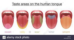
The diagram below shows areas of the tongue with one dominant taste bud but not at the exclusion of other varieties.


From Left to right, sweet, salt, sour, bitter, and umami.

                                                  Taste center.

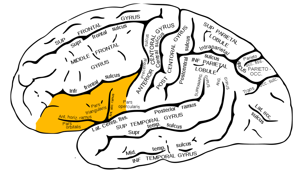
The insular cortex and inferior frontal gyrus are two parts of the taste center. Within the centers, there are neuronal groups engaged in association, connection with smell, touch, visual, tactile and emotional, hypothalamus, thalamus, Hippocampus and other areas of the brain. These interconnections are responsible for the development of memory of taste, food association with place and time, likes and dislikes of certain food in an individual.


 *******************************************

Saturday, December 3, 2022

Muscle Disease

                                                          Muscle Disease.

                                                   PKGhatak, MD


The human body has three types of muscles.

1. The Striated skeletal muscle.

2. Cardiac muscle is also striated but it works involuntarily. Cardiac muscle fibers are joined together without partitions and contract in unison. It is known as the syncytial muscle.

3. Smooth muscle has no striation and is a non-voluntary muscle. It is present in the wall of arteries, the Gastrointestinal and genitourinary tracts and the sphincters,

When a muscle is mentioned without any qualification, it is the skeletal muscle. Skeletal muscle disease is easier to understand if the basic structure and function of muscle are understood.

Basic muscle structure.


Muscles are a collection of muscle cells, usually referred to as muscle fibers. Muscle fibers are long and cylindrical and arranged in bundles and the end of fibers are attached to bones or joint capsules. Muscles are very vascular and are equipped with abundant mitochondria and multiple nuclei. The motor nerve fibers to muscles originate at the anterior horn cells of the spinal cord. Skeletal muscles contract under voluntary control and in abnormal conditions, they can also contract on their own but such contractions result in shivering or muscle twitching. Respiratory muscles work incessantly without conscious effort and also on demand. The detailed arrangement of the muscle bundle is shown in the diagram above.

Function of muscle.

Muscles shorten by contraction and result in ambulation or joint movements and the heat generated by muscle action increases body temperature.

How muscle contacts.


The motor nerve transmits nerve impulses at the neuromuscular junction. The impulse propagated along the sarcolemma to the entire length of the muscle. As a result, calcium ions from the sarcoplasmic reticulum are released and the released calcium ions enter into the muscle cells. This generates an action potential. Calcium ion binds with a protein called Troponin which pulls Myosin molecules towards Actin molecules. Actin and myosin molecules slide against each other causing shortening of muscle fibers. This action requires energy and is supplied by ATP. At the end of muscle contraction, the process is reversed and muscle fibers return to the relaxed state.

Diseases of the skeletal muscle.

From the above paragraph, it is clear that muscle disease may arise from the muscle fiber itself and is called Primary Muscle disease. Some other diseases may damage muscle fibers or their functions and these are the Secondary muscle diseases.

Primary Muscle Disease.

Primary muscle diseases are 1. Hereditary Mitochondrial muscle diseases and 2. Congenital Myopathies.

 1. Hereditary Mitochondrial muscle diseases.

Inherited defective genes that encode energy-producing enzymes, when passed to a child, the disease manifests at birth or later in life. The muscles are weak and the disease is initially detected in the eye muscles and then progresses through the entire body.

A. Leigh syndrome. The cytochrome enzyme system is defective in Leigh syndrome and eye muscle weakness develops in infancy. Weakness of the whole body soon follows. Associated heart abnormalities and low position of ears and high forehead is a distinct facial features. The child hardly can reach 5 yrs. of age.

B. Keans-Sayer Syndrome. The mutated gene encodes Oxidative phosphorylation. A child may normally develop to early teens then develops drooping eyelids, and soon one or both sided intermittent weaknesses develop. Other features are ataxia, deafness, heart rhythm disturbances and hormone deficiencies.

C. Melas syndrome. The defect is in the NADPH enzyme system. Muscle weakness begins at about age 40 years. Seizures, mental changes, migraine, lactic acidosis, and strokes develop.

 2. Myopathy.

The main symptom is muscle weakness, other less common symptoms are muscle cramps, stiffness and walking difficulties.

1. Inherited Muscular Dystrophy.

The general term for this entity is Muscular Dystrophy. Progressive muscle weakness is due to muscle degeneration. In the early stage of the disease, regeneration of muscle is seen but later lost. Various from of disabilities develop and death is generally from respiratory failure.  Muscular dystrophies have several clinical types based on the onset and progression of the disease and the genetic defects.

Clinical Types.

      Duchenne Muscular Dystrophy

     . Becker Muscular Dystrophy. ...

  • Congenital Muscular Dystrophy. ...

  • Myotonic Muscular Dystrophy. ...

  • Limb-Girdle Muscular Dystrophy. ...

  • Facioscapulohumeral Muscular Dystrophy. ...

  • Emery–Dreifuss Muscular Dystrophy. ...

  • Distal Muscular Dystrophy.

Duchenne Muscular dystrophy is more familiar to people of the US due to Jerry Lewis MDA Labor Day Telethon. The inheritance is sex link X chromosome in a recessive mode, only the male child is affected by this genetic disorder. 1 in 3 - 5 thousand male children is affected. In recent years, a new class of drug has been approved for the treatment of Duchenne dystrophy. The Antisense Oligonucleotides drug acts as a bridge over the missing exons (functioning genes) like a Band-Aid that produces the protein needed for the repair of muscles. The latest approved drug is a small-molecule Sunitinib.

Secondary Muscle disease:

1. Acquired Muscular Dystrophy.

Many diseases can damage and destroy skeletal muscle. Diseases or agents producing myopathy can be broadly grouped as Endocrine and Metabolic, Drug induced, Inflammatory, Prolonged illness, Neurological, Collagen vascular, Autoimmune and Unknown causes.

One common feature of this group of myopathies is the rapid onset. Inflammatory myopathy requires muscle biopsy to identify a specific entity. Autoimmune and other systemic diseases are identified from clinical and laboratory tests.

2. Dermatomyositis and Polymyositis are often secondary to a malignant process or a viral induced autoimmune disease. The skin rashes of Dermatomyositis appear around the eyes and over the knuckles and elbows and are reddish in color. The visible capillaries over the rough fingernail folds are an important diagnostic clue. In Polymyositis large groups of muscle are affected and weakness of shoulder and pelvis girdles is an important feature.

3. Inclusion body myopathies (IBM) are a common muscle disease of elderly people. Progressive weakness of fingers, wrists, muscles on the anterior aspect of the thigh, and legs develop over time and atrophy of muscles follows. Antibodies to Cytosolic 5- nucleotidase-1A, when present, indicate an autoimmune component. Congo-red stained rimmed vacuoles and areas of muscle necrosis surrounded by the inflammatory cell are seen in muscle biopsy. Treatment is unsatisfactory.

4. Collagen vascular disease myopathy is also common. Muscle weakness and atrophy in muscles of the face neck and neck in Scleroderma is a distinct feature. Systemic lupus and Rheumatoid arthritis and mixed connective tissue diseases also have muscle weakness and different degrees of muscle atrophy.

 5. Neurological and Neuromuscular disease.

Cerebrovascular accident, otherwise known as stroke, is a good example of how muscle undergoes wasting, stiffness, hyperreflexia and loss of function. These symptoms are due to the loss of inhibitory influence of upper motor neurons (UMN) on lower motor neurons (LMN). Prolonged disuse leads to muscle atrophy and contracture.

Spinal cord injury.

In spinal cord lesions, none of the muscle abnormalities above the site of spinal cord injury show any weakness. At the site of injury, it produces complete loss of muscle function and loss of resting tone of muscles. Below the site of injury, muscles show features of Upper Motor Neuron lesion as described under UMN lesion.

Peripheral nerve

Peripheral nerve lesion produces complete loss of muscle function innervated by that nerve.

Other systemic diseases.

Multiple sclerosis. Parkinson's disease. Meningitis and encephalitis and degenerative neuronal diseases produce muscle loss of function depending on the site of the lesion in the brain, spinal cord, or peripheral nerves.

Amyotrophic Lateral Sclerosis (ALS)

ALS is better known in the USA as Lou Gehrig's disease. It is a motor neuron disease involving both upper and lower motor neurons. ALS usually develops after middle age and 5 to 10 % of ALS cases are inherited as an autosomal recessive disease. The initial symptom is muscle twitching at rest which can be demonstrated by gently tapping muscles. Weakness of muscle develops gradually and affects all skeletal muscles of the body. The death is from respiratory failure.

Multifocal motor neuron disease may resemble ALS but it is limited to one side of the body and muscles of the fingers and hands are more affected and with treatment, the condition improves. 

6. Endocrine and Metabolic diseases.

Hyperthyroidism produces muscle wasting and weakness of pelvic and shoulder girdle muscles. Hypothyroidism produces weakness of the shoulder and gluteal muscles due to decreased metabolic rate and energy supply to muscles is limited.

Familial periodic paralysis.

Familial periodic paralysis is due to the mutation of genes that regulate sodium and calcium channels of the neurons. This is an intermittent paralytic disease. Muscles are hypotonus, weak and fail to contract during an attack but function normally in between times.

Hypokalemic periodic paralysis is due to the fall of serum Potassium. The symptom usually develops in adolescence precipitated by a rich carbohydrate meal or strenuous exercise. An attack may last hours to days. Generalized weakness develops later in life.

 In contrast, high serum Potassium periodic paralysis starts in early childhood and attacks are more frequent but last a shorter period.

Glycogen storage disease type VII 

This inherited glycogen storage disease produces muscle weakness due to failure of glycogen utilization in the muscles.

7. Neuromuscular junction.

Myasthenia gravis is an autoimmune disease triggered probably by a viral infection. Antibodies bind with the neurotransmitter Acetylcholine and the acetylcholine level falls. Depolarization of the muscle is prevented because of a lack of acetylcholine. During rest, some Acetylcholine is generated and the function returns. But on repeated muscle use, paralysis develops again. The drooping of eyelids and ophthalmoplegia are initial symptoms. Paralysis of other muscles, notably to the head and neck muscles, develops slowly. Speech, drinking, and eating become problematic and respiratory muscle paralysis follows. Early diagnosis and prompt immunosuppressive therapy can arrest the disease and improve the functional status of muscles.

Eaton-Lambert disease

Eaton Lambert disease is another example of neurotransmitter disease. Reduced acetylcholine release from the presynaptic nerve terminals due to antibodies to voltage-gated calcium channels produces muscle paralysis. Patients with Small Cell Carcinoma of the lung exhibit this paralysis frequently, other lung carcinoma rarely causes this disease. In contrast to Myasthenia, Eaton-Lambert disease affects the muscles of the legs initially. Head, neck muscles may be involved later but much less severely, respiratory muscles are spared. Leg muscle function improves with repeated muscle movements.

Guillain-Barre syndrome.

It is due to antibodies damaging the myelin sheath and axon of nerve fibers. Leg and arm muscles are primarily affected. Because peripheral nerves are mixed nerves, numbness and tingling also develop.

8. Drugs causing muscle paralysis.

 Action on the respiratory center of the brain.

Opioids, Fentanyl and other semisynthetic opioid derivatives have taken so many lives in recent years. The depressive effects of these compounds make the respiratory center insensitive to rising CO2, falling blood pH and hypoxia. In the end, the respiratory muscles stop functioning, causing death.

9. Therapeutic muscle paralysis.

 It is extremely unpleasant and stressful for a conscious patient to be intubated and mechanically ventilated. Patients fight for air and try to breathe faster than in the ventilator settings. Muscle-paralytic agent Succinylcholine is used to eliminate the patient's effort and completely take over the ventilation by mechanical means. Succinylcholine causes depolarization of the motor-endplate; and because the drug is not immediately broken down by the enzyme cholinesterase, the depolarization persists and muscles are kept in a paralyzed state as long as needed. Eventually, Pseudocholinesterase, another enzyme, breaks down succinylcholine but at a slower rate.

Many derivatives of Tubocurarine combine with receptors of acetylcholine at the motor endplate and produce partial skeletal muscle paralysis and are used as a remedy for muscle stiffness in Upper neuron paralysis. Antibiotics Neomycin, Streptomycin, and Polymyxin can potentiate neuromuscular blocking agents by a competitive synergistic effect. Centrally active muscle relaxants Mephenesin, Meprobamate, Benzodiazepines and other CNS depressive drugs can decrease intraneuronal activities and block parasympathetic neurotransmission.

 Statins:

Statin is a widely used medicine as a treatment for lipid abnormalities. Many patients complained of nocturnal leg cramps, leg pain and proximal muscle weakness and getting in and out of an automobile becomes hazardous. Researchers discovered that muscle weakness and related side effects of statins are likely due to their effect on the energy production centers, or mitochondria of muscle cells. The intermediate metabolite Lactone interferes with mitochondrial function. Switching to a water soluble statin or reducing the dose of statin may control symptoms, otherwise, discontinuation of statin is required. Statin also produces narcotizing autoimmune myopathy due to antibodies produced against the enzyme HMG coenzyme A. This is less common but stopping a statin does not stop muscle necrosis.

10. Myositis. 

Many viruses can infect muscles, producing muscle pain. Dengue fever causes worse types of backache.

 Myositis Ossificans is a less common disease.

A damaged muscle heals by having connective tissue from adjacent structures infiltrate the muscle. Occasionally bone generating stem cells enter muscles and produce lumps and these lumps are painful and interfere with muscle function.

 11. Rhabdomyolysis.

Crush injury of muscle and compression injury in Compartment syndrome can produce muscle necrosis.  Released Myoglobin from the damaged muscles by blocking the glomeruli capillaries and renal failure.

12. Tumors of the skeletal muscle.

Fortunately, tumors of muscle are rare. Rhabdomyosarcoma is one malignancy, seen in all ages and carries a bad prognosis. 


*****************************************


Wednesday, November 23, 2022

Cartilage and Diseases of Cartilage

                                        Cartilage and Diseases of Cartilage

                                           PKGhatak, MD

Gristle, that word may be more familiar than cartilage - the term used in biological science. Cartilage is a connective tissue like bone and tendon, connecting two pieces together forming a joint. Joints have a layer of cartilage covering the bony ends, preventing bones from grinding and making the movement smooth and pain free.

Cartilage, bone and muscle originate from the same source called mesoderm. Distribution of cartilage in the body follows the same path as that of bone, except in the respiratory tract cartilage alone provides structural stability. Cartilage does not have nerve supply, so it is pain insensitive and neither does it have blood supply or lymph channels. Cartilage derives its nutrition from synovial membrane secretion. The synovial membrane is the innermost covering of a joint, the secretion also acts as a lubricant.

Structure and types of cartilage.

The cartilage is composed mainly of a dense extracellular matrix. Within the matrix a few sparsely distributed cartilage cells chondrocytes and collage fibers are present. The cartilage cells secrete the matrix. Matrix is made up of water, protein and proteoglycans. Chemically proteoglycan is a derivate of chondroitin sulphate compounded with carbohydrate and protein.

Types of cartilage.


Cartilages are Hyaline, Fibrous and Elastic types based on the amount of fibrous and elastic tissues present.

Hyaline. Cartilage. Hyaline means glass like. Hyaline cartilage is smooth, glistening, semisolid and homogeneous.

The skeletal structure of a fetus is made of hyaline cartilage, and later cartilage is turned into bones. But a part of hyaline cartilage remains at the end of bones forming movable joints, called the articular cartilage and also called the growth plate. The longitudinal growth of the skeleton takes place by the rapid expansion of articular cartilage under the influence of the pituitary growth hormone followed by calcification.

Hyaline cartilage is the most abundant of cartilage, the distribution in the body is represented in the above diagram.

Fibrous cartilage. Fibrous cartilage joins ligaments and tendons to bones. The fibrous cartilage is made up of tough bundles of fibers and provides strength to the structure.

Elastic cartilage. The external ears (pinna) and epiglottis are made of elastic cartilage. Elastic cartilage is remarkably resilient and retains its original shape after rough treatment.

Diseases affect cartilage.

Acquired diseases of cartilage are few because of poor blood flow preventing microorganisms from reaching cartilage directly. Infection can spread to cartilage from the adjoining tissues. Damage to the cartilage from sporting accidents and wear and tear of weight bearing joints are the principal causes of disease.

A. Genetic cause of cartilage disease.

Dwarfism.

Under the term dwarfism 400 different conditions or syndromes are included. Dwarfs are discussed under Proportionate Dwarfs and Disproportionate Dwarfs.

Proportionate Dwarf. The adult dwarf is 4 feet tall. The limbs, face, skull and torso are proportional. The growth and development of a dwarf child is delayed and the mental capacity is low. This is due to a lack of adequate Pituitary Growth hormone.

Disproportionate Dwarf. The limbs are shorter, the torso is average size, the fingers are short and the middle and ring fingers are widely placed. The disproportionate dwarf has a large head and face and the bridge of the nose is flat. The patients have normal mental capacity and intelligence. Under this group, several distinct entities are included. Among all causes of dwarfism, Achondroplasia is the most common, occurs 1 in 15,000 to 40,000 births.

Achondroplasia is an inherited autosomal dominant disease. The mutant gene is located in chromosome number 4 and the fibroblast growth factor 3 receptor encoding gene (FGF3) is abnormal. The mutant gene has a 100% penetration. The adult achondroplasia individual is 4ft 6 inches tall, the torso and head are normal, and all four limbs are short. Sometimes, the reverse is true – short torso and normal limbs. The mental faculties are normal in both instances.

Pseudo-achondroplasia is a partial form of achondroplasia. Only the arms are shorter but the height is normal. The gene mutation is located in the Oligomeric matrix protein encoding gene.

 Diastrophic Dysplasias (DD) and Spondyloepiphyseal (SAA) diseases. 

The abnormalities are due to an autosomal recessive mode of inheritance. DD patients are shorter than average people and any growing bone may be affected.  In SAA, the spine and long bones are shorter and in addition, they have visual and auditory abnormalities. A defective gene that codes for Parathyroid Hormone related Protein (PhrP) is transmitted by an autosomal recessive mode, often associated with this condition. Many other congenital dwarfisms in combination with other organ abnormalities are known but they are just too many to mention here.

Most of the work in the field of dwarfism was conducted by Dr. Victor Mckusick of the Johns Hopkins University, Baltimore, MD.

Turner's syndrome. It is an important congenital disease and it is not rare. A female child with only one copy of the X chromosome. The newborn can develop fully and appear normal at birth. At age 5 the small stature becomes noticeable. It is due to the arrested cell division of the growth plate. Other distinguishing features are a webbed neck and failure of development of female characteristics and heart abnormalities.

B. Autoimmune disease of cartilage.

Relapsing Polychondritis is considered as an autoimmune disease, characterized by recurrent inflammation and degeneration of any or all the cartilage of the body, often the external ear is the most prominent lesion. Pain, redness, swelling and loss of function of the involved cartilage are common symptoms. Shortness of breath and wheezing develop due to the cartilage of the conductive airways showing inflammatory changes and the cartilage is at various stages of damage. Rapid respiratory failure and death may soon follow. 

Trecheobronchopatheia osteochrondoplastica.

Localized or diffuse submucosal calcified nodules overlying tracheobronchial rings is the main lesion. Patients are symptom free or have recurrent hemoptysis and shortness of breath.

Normal wear and tear and degeneration.

Osteoarthritis is the most prevailing disease of the cartilage. The incidence of osteoarthritis (OA) is 1 in 4 adults. Overuse of weight bearing joints and genetic predisposition are the causes of OA. Obesity, gait disorder (acquired or congenital), osteoporosis, osteomalacia, lack of physical activity, metabolic disorders, and prolonged use of steroids are additional risk factors.

The name osteoarthritis is a misnomer because the lack of blood supply to the cartilage is the principal reason for the delayed repair of cartilage. When bone ends are exposed, the damaged bone cells release cytokines and initiate inflammation.

Understanding the basic avascular nature of the cartilage, damaged or diseased cartilage cannot be restored to health by taking vitamins, calcium, magnesium, chondroitin sulfate, proteins and other micronutrients, no matter the amount, because those will not simply reach the cartilage. If there are documented deficiencies then taking them will help bones and other tissue but not the cartilage.

For further information see the previous blog dated Nov.3,2022.

Loose bodies in joints. Osteochondritis Dessicants produce fragmentation of articular cartilage and pieces of cartilage fall in the joint space. These loose bodies interfere with joint movements and may lock up a joint and may require medical intervention to unlock the joint.

C. Cartilage damage from inflammatory joint diseases.

Rheumatoid arthritis, Juvenal rheumatoid arthritis, ankylosing spondylitis, SLE and other collagen vascular diseases secondarily produce damage to the cartilage. To add to this list are - Frostbites, insect bites, sunburns, and congenital syphilis that affects the external ears. Granulomatosis with polyangiitis involves airways. Nasal cartilage is often infected with leprosy, Leishmania, and fungi. Epiglottis is affected by amyloidosis and sarcoidosis.

Pseudogout. Calcium monophosphate dehydrate crystal (calcium pyrophosphate) deposit disease is otherwise called pseudogout because it has similar symptoms to gout. Usually, people between 45 to 60 years old and both sexes are affected. Knees are mostly affected joints but all joints, including the spine, may be involved. Cartilage damage is secondary to the chronic relapsing nature of the illness.

D. Inflammatory disease of cartilage.

Infective chondritis. Ear cartilage piercing may introduce infection into cartilage directly; usually, infection of ear cartilage spreads from infected subcutaneous tissues. Pain, swelling and fever follow. Pain is severe and the risk of necrosis is considerable due to the absence of subcutaneous space in between cartilage and skin. Occasionally infection spreads to blood and septicemia develops.

Costochondritis. The joints at the anterior ends of the ribs and sternum can be infected by viruses or other agents. Pain is felt in the chest. Chest pain produces anxiety of the patient mistaking the pain for a heart attack. The illness is self limited and leaves no lasting bad effects.

Tietze syndrome. A similar costochondral joint pain and in addition the joints are swollen. It is usually seen in younger people. The 3rd and 4th costochondral joints are most affected but any joint including neck and shoulder joints may be affected. The cause is unknown but repeated microtrauma may be responsible. Symptoms of Tietz syndrome last longer, at times for years. The swelling may not regress even when the pain disappears. Treatment is not very satisfactory.

E. Traumatic cartilage damage:

The external ear, nose and larynx are more prone to damage from various ways like wrestling matches, whereas the right and left meniscus of the knee are torn in sports related injuries.

The most life-threatening injury to cartilage is the fracture of laryngeal cartilage from cloak-holding by law enforcement officers and in professional wrestling events. Once the laryngeal wall becomes frail, it acts as a check valve preventing air movement and respiratory failure only hours away. An artificial tracheal opening is necessary for the long term solution. Choak-holding should be outlawed.

Knee menisci fracture.

When the foot is planted firmly on the ground and the body is twisting, the shearing force of the femoral condyle tears a piece of meniscus. In old people with worn out menisci minor physical activities may produce a tear. Previous minor tear predisposes a major injury in otherwise healthy athletes. The lateral 3rd of the meniscus has adequate blood supply but the inner 2/3 has none. The chance of complete healing is remote in a complete meniscus tear and treatment is surgical.

In recent years, cartilage transplants have been tried with encouraging results. An outline of these methods is as follows-

1. Microfracture. Multiple small holes are drilled in the bone and bone marrow underneath the damaged cartilage. Bone marrow cells enter the cartilage through the small openings and start forming new fibrocartilage at the damaged site, but not articular cartilage.

2. Osteochondral Allograft / Mosaicplasty. A small piece of healthy cartilage with attached bone is removed from the patient's non-weight bearing joint and inserted beneath the damaged cartilage through a small drilled hole. The new cartilage that forms are articular cartilage. The graft survives for a long time. The procedure is limited because cartilage can be harvested only from a few joints.

3. Osteochondral Allograft. In this procedure, cadaveric cartilage with attached bone is used like in the previous procedure. After initial healing, the new cartilage wears away sooner than expected.

4. Autologous Chondrocyte implant. Articular cartilage is harvested from the patient. The cartilage cells are allowed to grow in the laboratory. When the desired number of chondrocytes is grown, the new cartilage cells are transplanted into the damaged joint.

5. Matrix Autologous Cartilage implant. In this case, the cartilage cells are grown on a special collagen mesh, then the mesh containing new chondrocytes is implanted.

***********************************

Wednesday, November 16, 2022

Leishmania

                                                                    Leishmaniasis

                                                         PKGhatak, MD


Leishmania is a flagellated protozoan of the genus Leishmania. Of the several species of Leishmania, about 20 species are infectious to humans. The flagellated tiny protozoa enter human victims from the regurgitated gut contents of female sandfly bites when the fly probes for a blood meal. There are about as many varieties of sandflies as there are leishmania species. The infectious form of the parasite is called Promastigotes. Promastigotes bind to the receptors on the skin macrophages. The parasites drop their individual flagella and enter the macrophage and continue to live inside the macrophage and at this stage, the parasite is called Amastigotes and the amastigotes multiply and infect more macrophages; eventually, macrophages of the skin, reticulo-endothelial tissue and viscera are loaded with parasites. 

The human body is unable to mount an immune defense against leishmania because the parasite successfully thwarts both innate and acquired, cellular and humoral arms of the immune system. Thymic 1 cells (TH1) secrete IL-12 which transforms native T-cells into TH1 cells. TH1 cells produce TNF gamma (tissue necrosis factor gamma) and NK cells (natural killer cells). These are the resistive forces to the body. The parasite switches T-cells to TH2 cells which secrete IL-4 and IL-10 and which protect parasites from being killed.

Leishmaniasis is prevalent around the tropical and subtropical regions. The countries are separated into the Old World and the New World countries. The old world belongs to Central Asia, the Eastern shore of the Mediterranean, the Indian subcontinent, South Eastern Asia, and Eastern African countries. The new world comprises Central American and South American countries. It is estimated that between 1 and 2 million people are newly infected every year, but only a small fraction of them will develop the disease and 20,000–30,000 die each year.

 An increased number of leishmania cases are seen in the last 25 years. This increase is due to globalization and climate change, air travel, sharing contaminated needles between IV drug users, being unable to detect contaminated donated blood, close human interactions with domesticated animals, particularly dogs, which serve as an intermediate host, HIV infection and blood and organ donations.

Clinical feature.

Several factors determine the nature of Leishmania clinical presentation. Besides the leishmania species and sandfly subtype, other factors are climate and the immune status of the individual. The cutaneous, mucocutaneous and visceral forms of leishmania are the main clinical types. Visceral Leishmania is better known as Kala-azar, an Indian word for Black fever. In certain endemic areas, Kala-azar is followed by dermal lesions which outwardly resemble Lepromatous leprosy and are known as post-kala-azar dermal leishmaniasis (PKDL). In Brazil mucocutaneous leishmania is known as Espundia. In the old world, the cutaneous lesions are known as Oriental Sores.

Cutaneous lesions (Oriental Sores).


The incubation period is weeks to months following the fly bite. The skin lesion may be single or multiple, dry or wet. In the new world, a single lesion is usual, multiple lesions are more common in the old world. The lesions may spread by lymphatics and satellite lesions are seen. Initially, a red papule appears and slowly but gradually progresses to a macule and ulcer. In central Asia, Afghanistan and western Pakistan, cutaneous leishmania is the common presentation.

Mucocutaneous. 

Mucocutaneous presentation is mostly seen in Mexico and Brazil. Lesions on the face are associated with ulcerated lesions in the nose, mouth, pharynx and trachea. Ulcers and scar tissues produce deformities and organ dysfunction. The incubation period is 1 to 3 months. Urgent medical treatment is necessary.

Visceral.

At an earlier time, the undivided province of Bengal, India was the epicenter of kala-azar. The incidence has fallen sharply but sporadic cases are seen in Bihar, Nepal, Bangladesh, West Bengal, and the Eastern shore countries of North Africa. The incubation period is 3 to 6 months and may be as long as a year. Fever, weakness, enlarged spleen and liver are followed by a characteristic double spike of fever every day, anemia and pancytopenia, increased skin pigmentation and dryness of the skin. Dependent edema, ascites and secondary infection follow and deaths in 2 years if no treatment is available.

PKLD (post kala-azar dermal leishmaniasis).

Years after visceral leishmania is treated and cured, nodular lesions appear on the face, the trunk and the limbs. The lesions are numerous and have the appearance of leprosy, except for the perception of touch and pain remains. And no spontaneous amputation of digits and toes occurs in leishmania as it happens in leprosy.

Viscerotropic leishmania.

This is a subvariety of visceral leishmania but after initial symptoms, the disease does not progress.

Diagnosis.


The parasite must be demonstrated in the samples taken from a skin biopsy and from the bone marrow biopsy in visceral leishmania.

Treatment.

CDC designated drugs used in the treatment of leishmania as orphan drugs. Anyone can contact CDC for guidance and will be assured of a supply of medications.

In 1920, Dr. Upendranath Bramhachari, a native of Kolkata, India, discovered that when Urea Stibamine was given IV it was very effective in bringing an end to the human misery in Bengal. Initially, the cure rate was 90 % and not many side effects were noted. But in the long run, safer Antimony compounds were introduced. Even today, the medical community of India pays tribute to Dr. Bramhachari for his service to humanity.

Antimonial.  The pentavalent antimony (Sb V) enters macrophages and fuses with the membrane of the Lysosome of macrophages. There it is converted to Sb III (trivalent) compound. Sb III is a poison to the amastigotes.

Miltefosine.  It is an oral anti-leishmania drug. It blocks the lipid metabolism of the parasite. It is also useful in children but has the potential for malignancy. Combining it with Amphotericin B and Paromomycin increases efficiency and lessens side effects.


                                                             Dr. Bramhachari.

***************************************************************************


Saturday, November 12, 2022

Malaria


                                                                      Malaria

                                                           Some important aspects

                                                     PKGhatak, MD


A tiny one-cell parasite infected primates in equatorial Africa, perhaps thousands of years ago. At a later date, the parasite found humans as suitable prey. It was a time before writing developed. We do not know whether those people knew malaria was a parasitic disease. Egyptian, Chinese and Indian physicians left some documents. After a retrospective analysis of those documents, some appeared to be malaria fever cases.

Romans thought the febrile illness was from Mal-Aria (bad air) rising from the swamps; they correctly identified the swamp as the breeding ground of mosquitoes but failed to connect the mosquitoes with the spread of malaria.

That task was completed in 1857 in Kolkata, India by Ronald Ross. He identified the parasite in the female Anopheles mosquitoes. He recovered the same parasite Plasmodium relictum from the blood of birds, linking mosquitoes with birds' malaria. He was awarded the Nobel Prize in 1902 for his groundbreaking research. Subsequently, other researchers discovered the complete life cycle of the malaria parasite - the Plasmodium.

Malaria parasite.

Malaria belongs to the Genus Plasmodium of the Superphylum Alveolate, and the Phylum Apicomplexa. Like all Alveolate, Plasmodium contains a plant plasmid, used in Photosynthesis by the plant. The plasmodial plasmid is a bit different, it is used in extracting a fatty acid, Isoprenoid, from the host cells in order to develop and multiply in the liver and RBCs in humans and in the salivary gland of mosquitoes, and not used in photosynthesis.

Six species of Plasmodium infect humans, namely -

1.Plasmodium falciparum (P. falciparum)

2. P. malariae

3. P. vivax (with identical morphologically P. simiovale and two other related species P. minuta and P. tenue)

 4.P. ovale

5. P. Knowles.


Know the Female Mosquitoes.


Female Mosquitoes are equipped with chemical sensors for Carboxylic acid, a CO2 derivative. Using the sensors, mosquitoes zero in on a person who has higher carboxylic acid in the breath. Female mosquitoes must have a high protein meal (blood) for the development of eggs and laying fertilized eggs on the surface of non-flowing bodies of water, e.g., ponds/swamps, etc.

Follow the parasite.

Mosquitoes' salivary glands are loaded with the infective forms of the parasite, and mosquitoes deposit them when they bite and suck human blood. At this stage, the parasite is known as Schizont (no English word for it). From the skin, the parasites travel to the liver and enter the liver cells. In the hepatocytes (liver cells) the parasite multiplies. The parasites break free from the liver cells and infect RBCs. Duffy antigen receptors on the RBC surface are the anchoring point of the parasite and serve as the entry point into the red cells. At this stage,  the parasite is called Merozoites. In the RBCs, the parasites begin to divide by themselves (nonsexual multiplication) and the new Merozoites very quickly multiply and reach a huge number and the red cells burst open and the merozoites enter into the bloodstream and begin to infect more RBCs. And the cycle keeps repeating until some merozoites change to male and female reproductive units called Gametocytes and wait for the mosquito to bite and draw blood. In the gut of the mosquito, the male and female gametocytes unite and produce their progeny, the schizonts. Schizonts move to salivary glands and wait to infect a new victim.

Time taken for the parasite to complete human RBC cycles.

In the case of P. falciparum, P. ovale and P. vivax the cycle is repeated every 3 days. In P. knowlesi and P. malariae the cycle only a day.

Symptoms of malaria.

Malaria symptoms vary according to the species of Plasmodium.

a. Shaking chills followed by high fever. 

With no warning, a sudden onset of chills and shaking starts. The chattering of teeth, assuming a fetal position and covering the body with all comforters or blankets does not bring any relief of chills till the fever starts. The temperature is usually 104 degrees or higher and weakens, profuse sweating and prostration follow when the fever breaks.

b. Hemolytic anemia, jaundice and renal insufficiency and renal failure.

The broken RBCs release not only the parasites but also the hemoglobin into circulation. Free hemoglobin is an oxidant that damages glomerular capillary endothelium and decreases renal filtration. In recurrent episodes of hemolytic anemia, renal failure develops. Hemoglobin is broken down in the liver into heme and globulin. The heme is a waste product. Light jaundice becomes evident, urine and stool develop darker shades of yellow.

c. Anemia, failure of children to thrive in endemic areas, susceptibility to tuberculosis and HIV infections are common in certain countries in alarming numbers.

d. Enlarged spleen and liver and marked anemia. Parasites induce immune reactions and chronic inflammation and enlargement of organs.

e. Blackwater fever.

Certain patients, generally non-native residents, taking Quinne irregularly for the treatment of malaria develop dark colored urine due to the presence of hemoglobin in urine and chronic renal failure. It is thought to be an autoimmune disease, triggered by the interaction of quinine with plasmodium. The antibodies then attack the kidneys.

Laboratory diagnosis:

Peripheral blood examination.

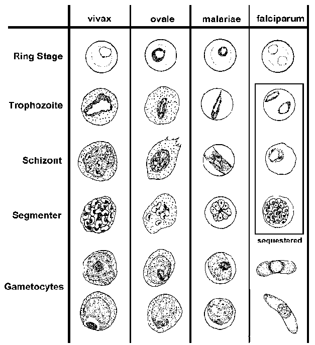
Until the use of direct detection of the antigen of the malaria species by PCR was introduced, the microscopic examination of blood slides for the presence of parasites was the accepted method of diagnosis of malaria.


  Falciparum                         Vivax                     Ovale                      Knowlesi
  • numerous rings
  • smaller rings
  • no trophozoites or schizonts
  • Cresent-shaped gametocytes
  • enlarged erythrocyt-e
  • Schüffner-'s dots
  • 'ameboid' trophozoit-e
  • similar to P. vivax
  • compact trophozoite
  • fewer merozoites in schizont
  • elongated erythrocyte
  • compact parasite
  • merozoites in rosette


Special features of falciparum malaria.

P. falciparum fever is also called malignant malaria and cerebral malaria. Worldwide, 2 million people are infected and ½ million die on yearly basis. The initial symptoms marked by shaking chills last only 30 min, followed by high fever, headaches, and confusion and children develop convulsions and coma rapidly. The fever returns every 48 hrs. Falciparum induces swelling of the RBCs, and the surface membrane protein of the parasite changes to knob like projections and attaches to the endothelium of capillaries. The circulation is compromised and endothelial cell hypoxia induces adhesive properties of the endothelial cells and locally released cytokines, cells facilitate platelet thrombi and the microcirculation of the tissue is cut off. Hypoxic injury of the brain occurs in the basal ganglia and hindbrain. Generalized swelling of the brain produces encephalopathy and various degrees of coma and convulsions.  With early medical intervention, cerebral changes are reversible. The mechanism of acute renal injury is multiple. Free hemoglobin in the circulation oxidizes glomerular capillaries, swelling, edema and piecemeal necrosis of uneven distribution of renal lesions leads to a marked reduction of GFR. The capillary circulation of the renal tubules is severely compromised and results in Acute Tubular Necrosis and renal failure.  Nearly 40 % of adults develop renal failure and cerebral malaria incidence is high in children and the mortality is high.

P. vivax.

Vivax malaria is less virulent than falciparum but deaths are still very high. Currently, southern Africa, Central & South American countries, the Indian continent, and Indonesia & Malaysia peninsular regions report most cases. Fever recurs every 48 hours, the temperature reaches 104F, and the febrile period is about 4-8 hours. Most of the symptoms are from released cytokines, TNF alpha and IL-6. Pulmonary involvement and enlarged spleen often rupture without trauma. Anemia is of chronic nature and secondary infections in endemic areas are not unusual. The parasite may remain dormant in the liver for years and one infection triggers six or more infections. In addition to renal failure, hypoglycemia, low platelet count and bleeding episode, loss of taste and occasionally lack of fever are some of the unusual features.

P. ovale.

Ovale malaria is the mildest form of human malaria.  It is seen in the Indian subcontinent. The fever recurs every 48 hours (tertian fever). Fever begins after 12 days following mosquito bites.

P. malariae. Massive splenomegaly from the abnormal immune response is a feature of P. malariae malaria. Fever recurs every 48 hours intervals. The incubation period is 16 to 60 days. Nephrotic syndrome is due to immune complex formation with malaria antigen and antibody and produces renal failure. 

P. Knowles. It is the malaria of Macaque monkeys; humans acquire malaria due to close contact with macaques. Fever recurs every day (quotidian fever). GI symptoms like hiccups and dysphagia are distinguishing symptoms.  Because of a few cases in the community, robust immunity does not develop. So, the fever continues and results in anemia and weakness.

Asymptomatic Carrier.

In endemic area, some people acquire immunity from repeated malaria infections. Unless their immunity is compromised by intercurrent infections or diseases, they remain malaria free. Once they move to other countries where malaria is rare, they can transmit the parasite to others by donating blood or organs.

Malaria continually develops resistance to drugs.

Plasmodium quickly develops gene mutations and renders drugs ineffective. For example, anti-folate medication becomes ineffective due to a point mutation of the Dihydrofolate dehydrogenase enzyme. Another point mutation involving two transporter proteins. By accruing mutations, the parasite increases the rate of expulsion of antimalarial drugs from the cytosol. Still, another way to develop drug resistance is by gene amplification of those genes mentioned above, making the mechanism work at a higher level, making unmatched therapeutic blood levels.

Defects in the DNA repair genes accelerate mutations, specially in P. falciparum.

Anti-malaria drugs.

Currently 5 groups of antimalarial drugs are available to treat malaria.

1. Endoperoxide – example Artemisinin.

2. 4-Aminoquinolines and related derivatives – Chloroquine, Mefloquine.

3. Napthaquinolies – atovaquone.

4. Antifolate – Pyrimethionine.

5. 8-Aminiquinols – Primaquine.

P. falciparum has developed resistance to every one of the above 5 groups of drugs.

Secondary drugs used in conjunction with the above 5 groups are -

Doxycyclines, Sulfadoxime, Lumefantrine.

New class of Drugs.

Monoclonal antibodies are used subcutaneously, prevent schizonts from attaching to RBCs and subsequently infect liver cells when used in combination with other standard antimalarials. Imatinib, a small molecule anti-leukemia drug, works by inhibiting the Kinase, has also been used recently and has the potential to rapidly clear malaria parasites from blood and liver. A drug, Alisporivir, used in hepatitis C, was found to be effective in Artemisinin-resistant P. falciparum infection. 

Several drugs are used in various combinations like chemotherapy for cancer. WHO's recommendations of drug therapy and chemoprophylaxis are essential to keep informed about the therapeutic challenges posed by rapid drug-resistant malaria spreading from one region to the other endemic area.

                           .

                                             wormwood

 Chinese sweet wormwood, source of artemisinin.

Artemisinin was introduced by Dr.Tu Youyou in the western world, at one time Artemisinin was the mainstay of drug therapy of P. falciparum and was used extensively alone or in many various combinations with other compounds. For his work, he too received the Nobel prize in 2015.

Malaria therapy for neurosyphilis.

Before Penicillin was available to treat syphilis, fever by P. vivax infection induced fever as the therapy for neurosyphilis by introduced by Julius Wagner-Jauregg. In Europe P. knowles was used for this malaria therapy till 1950. For this therapy, he was awarded the Nobel prize in medicine in 1972.

*****************************


Tuesday, November 8, 2022

Heat Intolerence


                                                       Heat Intolerance.

                                                   PKGhatak, MD


Heat intolerance is not a disease; it is a symptom. Some might say, “It is too hot in here,” and others might look at him in surprise because they feel comfortably cold in the same room.

The question is, who is right, or is there a medical explanation for this temperature perception difference between people.

Yes, there is a physiological reason for heat intolerance.

The Heat Center is located in the brain, and is connected with the skin and the internal organ temperature receptors., The Heat Center (HC) is located in the Hypothalamus of the brain, which is connected with the pituitary endocrine gland, which in turn is connected with the sympathetic and parasympathetic nervous systems.

By varying the output, the heat center of the body can maintain a constant core temperature throughout the winter and summer.

Source of body heat.

The human body derives heat from 5 separate sources:

1. Basal metabolism

 2. The specific dynamic action of food,

 3. Physical activities

 4. Shivering 

  5. Non-shivering thermogenesis.

Heat Center regulates temperature settings, but the metabolic process generates heat. It is like a computer; multiple programs are running in the background. The metabolic process continues at all times during the lifetime of the individual. The organ functions continue at a basal, and is called Basal metabolism. In humans, the Basal Metabolic Rate (BMR) is 76 kilocalories/Kg body weight /Hour. This is the main source of body heat.

2. Specific dynamic action of food (SDA). The process of digestion of food generates heat. Protein gives out the most heat, about 20 % of the calories contained in protein food. This is the reason we feel comfortably warm after eating a steak.

3. Physical activities. All forms of physical activities generate heat,

4. Shivering. Exposed to cold and wind, the body begins to shiver. Muscle contractions generate body heat.

5. Non-shivering thermogenesis: It takes place in Brown Fat and muscles. Adrenaline is released at the sympathetic nerve terminal of brown fat tissue and muscle. Adrenaline accelerates the conversion of fatty acids and glucose to heat, ketones, H2O, and CO2.


Excessive body heat and heat intolerance:

1. Menopause is due to falling sex hormone levels. Various symptoms, mostly due to the perception of excess heat produce the symptoms of menopause.

 2. Hyperactive thyroid gland.  Excessive secretion of Thyroxine (hormone of the thyroid gland) increases BMR, generates fine muscle tremors, and increases heart rate and blood pressure. Catabolism of fatty tissues and muscles produces excessive heat.

Drug abuse. Amphetamines, cocaine, antipsychotic, and antidepressant drug use generate a sense of excess body heat.

Anticholinergic drugs. Anticholinergic drugs decrease sweat production and prevent cooling from evaporation and loss of heat. The core body temperature rises.

Caffeine. It is a cardiovascular and neuronal stimulant. It produces small vessel constriction and decreases heat dissipation. A mild increase in temperature is usual in therapeutic doses. In posing, caffeine raises the core temperature. 

Heat stroke. When exposed to high heat in a humid environment, the body fails to dissipate heat by radiation and evaporation. The core temperature rises and derails the functioning of metabolic and enzyme systems. It turns into an emergency situation, and can permanently damage the brain and heart, and even death.

Malignant Hyperthermia. This is another life threatening medical condition. It is an inherited disease inherited as an autosomal dominant trait. exposed to volatile anesthetic agents during general anesthesia, large amounts of calcium leave the calcium stores and enter the skeletal muscle fibers. Increased intramuscular calcium produces sustained muscle contractions. An excessive amount of heat is generated. Blood vessels may contract for a long time and can cut off blood flow, which is called Rhabdomyolysis. The released myoglobin from the necrotic muscle can block the kidney filters, producing metabolic acidosis and hyperkalemia (high serum potassium). Followed by cardiovascular collapse and death even death. 

Multiple sclerosis.  In Multiple sclerosis, the demyelination may directly affect the hypothalamus and the autonomic control center. Excessive heat generation may also be accompanied by derailed heat dissipation because of spinal cord lesions in multiple sclerosis.

Diabetes mellitus can damage the autonomic nervous system, and excessive heat and fluctuation of body heat may result.

Multisystem atrophy of the nervous system. The autonomic center damage is the result of increased heat.

edited May 2025.

**************************************
Friday, November 4, 2022

How We See the Outside World

                                               How we see the outside world.

                                                   The science behind it.

                                              PKGhatak, MD


Looking straight ahead, we see a vast area, horizon to horizon. Using a visual field measurement chart and recording one eye at a time, we find each eye has a visual field of 155 degrees on the horizontal plane and 130 degrees on the vertical plane. By adding the visual fields of two eyes together, our visual field should be 310 degrees but in fact, the visual field is smaller because the nasal field of the two eyes overlaps.

That overlap gives us the dept of vision, meaning, we can judge the distance of an object in front. It is an essential quality for reading, writing, driving, painting, target shooting and other activities which require bringing objects in sharp focus.


The diagram above illustrates visual fields and where the images develop.

The visual field is divided into the Nasal field and the ear (Temporal field. To make a clear distinction between the left and right visual fields, the right-hand side of the visual field is colored red and the blue color for the left.

Note that the nerve fibers carrying the image from the nasal half of the right eye, represented by blue lines, are crossing the midline and going to the left side to join the nerve fibers carrying the image from the temporal half of the left eye. The same is true for the left eye, marked with red color.

It is interesting to note that crossed and uncrossed nerve fibers join and carry images representing the right and left visual fields and not the left or right eyes.

The second point to note is that the nerve fibers make a connection at a nerve center called Lateral Geniculate Body (LGB). The right visual field image goes to the left side and the left visual field image to the right side of the LGB. The stratification of nerve fibers is maintained in the LGB, meaning the temporal fibers remain outer and nasal fibers on the inner nuclei of the LGB.

The third point is that the same stratification is also maintained in the Visual Cortex. The visual cortex is located in the back of the cerebral hemisphere, one on each side in the Occipital Lobe.

Not shown in the diagram, there is upper and lower field stratification on the visual cortex. The lower half of the visual field is represented in the upper part of the opposite cortex, and the upper half in the lower visual cortex.

By knowing the detailed anatomy, the doctors are capable of detecting any segmental visual field loss and treating properly without wasting much time.

How two images merge into one.

This fusion of two images in the visual cortex and perceived as one depends on unison eye movement and convergent reflexes that make image generation on the corresponding points on each retina. When initial images are close but not exactly one, then a further adjustment is made by the eye muscles to focus properly.

Why does an object too close to the eyes appear blurred.

The eyes are situated 2 inches apart. The images of the two eyes are not precisely the same, the right eyes see a little more on the right-hand side and the left eyes see more on the left-hand side. The images fuse completely when the object is at a comfortable distance from the eyes. The disparity grows more and more when the object is brought closer and closer.

How images are formed on the retina.

Light rays travel in straight lines in all directions from an object. The pupil by changing its size brings light rays in focus on the retina. The photosensitive pigment of the retina converts the electromagnetic energy of light into electrical stimuli. Next, the rods and cones cells of the retina perceive the stimuli and then pass them to nerve cells which convert the electrical stimuli into nerve impulses and send out nerve impulses to the brain.

 How the color is represented in the brain.

In the daytime, sun rays are bright but colorless. But we know that daylight hides 7 colors from red to violet (color of the rainbow) in it. The color receptors of the eyes are cone shaped cells, called cones of the retina. Cones are plentiful in the central part of the retina and are only a few in the periphery. Cones are densely packed in the Fovea of the retina. The red, green and blue colors are detected by three different cone cells. The blue color generates maximum brightness of 2.56 to 2.76 eV and the red color is the least, generating 1.65 to 1.90 eV. These three cone cells make a unit known as the Trichrome unit. By various combinations of the impulse generation by the trichrome units, we are able to perceive the entire color spectrum. This is the basis of the printers' color cartridges we are so familiar with.  The pigment of the cone is called Opsin. It is a protein and it is most sensitive to light waves with a wavelength of 550 nm, blue responds to light waves with a wavelength of 450 to 485 nm and green to 500 to 565 nm wavelength.


White and Black color.

There is no black color receptor.  Instead, black, white and gray color perception depends on various shades of gray colors. The pigment of the gray scale is Rhodopsin, and the receptors are known as Rods. Rods are most plentiful at the periphery of the retina and are only sparsely distributed in the central part of the retina. Rhodopsin is most sensitive at light waves of 555 nm.

We see better in the corner of the eyes when light is very faint. There are no cones at the periphery of the retina, which is the reason we are unable to see color in the dark.

Why are black and white images not as clear as a color picture.

Each cone is connected with one neuron of the retina that sends out stimuli to the brain. 5 or 6 Rods are connected with one nerve cell, as a result, color picture has more pixels so to speak, and is sharper and clearer than black and white images.

Upside down and revered sidewise image.

This diagram illustrates why the upside down and left side on the right side image projected on the retina. 


This is a pinhole camera taking a picture of a tree. The same Laws of Optics work for the eyes.

How the Brain rights up images.

A newborn child grabs things with hand and tries to put them in his /her mouth. That is the beginning of the learning process of the brain and by the time the baby is 2yr old the proper interpretation of images by the brain is nearly complete but the learning process of the brain continues for lifelong.

Near and Distant vision.

Refractive errors of one or more components of the eye (cornea, anterior chamber, lens and vitreous) fail to bring light precisely on the retinal receptors and when the light rays fall short- then the condition is known as Near- Sightedness and when the light rays converge beyond the light receptors then it is called Far- Sightedness.

Double vision.

Double vision may be monocular or binocular. Mono-ocular double vision comes from the eyes, or contact lens injury, or foreign body injury of the cornea.

Binocular double vision is due to the failure of mages to merge into one. The defect may lie anywhere from the optic nerve to the visual cortex. Since eyes are extensions of the brain and when an eye is damaged it may not improve unless treatment starts immediately.

A quick way to determine mono-ocular or binocular double vision

The mono-ocular double vision disappears when the eye producing the double vision is closed and the person looks through the other eye. In binocular double vision persists by closing and opening one eye at a time.

In daylight, our world appears in full complements of spectrum colors - clear and beautiful and images up-righted and sides correctly positioned. In the dark the story is different. In fading light, the world appears in various shades of gray, fuzzy and best visible to us by the corner of the eye provided one is neither near-sighted nor far-sighted.


*******************************************************


*** 


Archive

  Archive. P.K.Ghatak, MD 2025  March. Laser Endoscpoe 2025 February. Use of Ultrasound in Medicine MRI 2025 January. Bacteriology and Rober...