Tuesday, January 17, 2023

Cornea and Cornea Transplant.

                                           Cornea and Cornea Transplant

                                         PKGhatak, MD


Eye to eye contact is an essential characteristic of humans. It is said one can see the soul of a person looking through the eyes of others. Medical science has not advanced to that level to know where the soul resides, but what a person sees looking directly into the eyes of the other is a colored iris within which is a central beautiful transparent zone, the dept of that remains elusive, probably the source of the idea of the location of the human soul. That central transparent zone is the pupil of the eyes, a part of the Cornea.

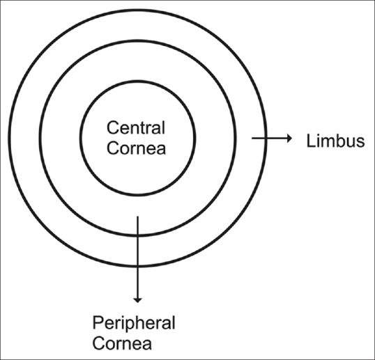
                                              Parts of the cornea.


Eyes are extensions of the central nervous system and develop at the same time as the brain is taking shape. The outermost layer of the eye is made up of tough collagen tissue, the white of the eyes is called the sclera and the transparent part is the cornea. The main function of the sclera is to protect the inner delicate eye structures and prevent infection. The function of the cornea is to bring objects in focus on the retina for vision. Cornea is one of a few immune privileged tissues, which are out of reach of immune surveillance cells and immune directed inflammation. Others such as tissue/organs are the placenta, fetus, sperm and articular cartilage.


Anatomy and the property of the cornea.

The cornea is 11 to 12 mm in diameter, the thickness varies from 0.5 mm in the center to 0.8 mm at the periphery. The cornea is devoid of blood vessels and lymphatic channels.

Histology of cornea.

                    Cornea from outside to inside.

Epithelium.

This layer of non-keratinized stratified epithelium consists of 6 layers of cells. The epithelium of the cornea is continuous with the conjunctiva of the eye. The cells of the basal layer of epithelium have fast regenerative capacity and quickly replace any damaged cells.

Bowman's membrane.

It is a tough tissue made up of collagen I fibrils, and the fibrils are tightly interwoven and adhere to each other. Bowman's capsule is about 14 micrometers in thickness. and devoid of any cells.

Stroma.

It is also called the substantia propria. This layer is made of regularly arranged collagen fibrils of type I collagen, arranged in thin sheets like pages of a book and has 200 layers. In this stoma, a few scattered but interconnected keratinocyte cells are present, these cells are responsible for the daily maintenance and repair of the stroma.

Descemet's membrane.

This layer is also called the posterior limiting membrane. It is 10 micrometers thick and made up of collagen IV fibrils. The thickness of Descemet's membrane increases with age and can be 20 micrometers in thickness. A tough layer of the innermost part of Descemet's membrane is known as the Dua layer, which is about 15 micrometers in thickness but can withstand 2 bars of pressure.

Endothelium.

Anatomically, the term endothelium is appropriate but these cells do not come in contact with blood. The endothelium is only one layer of cells, 5 micrometers in thickness, and the cells contain a large number of mitochondria. These cells are bathed in aqueous humor and regulate a proper fluid balance of the cornea. The endothelium cannot regenerate. When one cell dies, the adjoining cells stretch to cover the empty space. When a significant loss of cells happens, the cornea swells and becomes opaque and vision falls.

Nerve supply.

The cornea is innervated by sensory fibers of the ophthalmic division of the trigeminal nerve. Unmedullated fibers are very sensitive and carry pain sensation. The pain receptors of the cornea are 500 times greater than the skin and any corneal injury is excruciatingly painful.

The nerve terminals enter the cornea through three sites – at the episclera, sclera and conjunctiva. The nerve fibers form a network in three levels -midstromal, subbasal and epithelial and from these networks, all the structures of the eye are innervated. 

Optical property of cornea.

1. Refractive index. The cornea is highly transparent and allows 95% of daylight waves to penetrate inside. A radial colored diaphragm, the Iris of the eye, by varying its size of the pupil, regulates the amount of light to enter the eye. The size of the pupil can vary between 1.5 mm to 8 mm in diameter. As stated earlier, the cornea is composed of layers of cells of different types, the refractive index of the epithelium, the stromal anterior and posterior wall is 1.401. 1.38, and 1.373 respectively.

The refractive index of the cornea, as a whole, is n = 1.3765 +/- 0.0005. (see footnote)

Diopter of eye.

The property of refracting light in ophthalmology is expressed as Diopter (D). The eye contains two focusing lenses, the cornea and the crystalline lens. The cornea has 45 diopters and the lens 15 diopters for a combined 60 diopters of focusing power. The accommodation apparatus of the eye can add additional diopters required for near vision.

Cornea Transplant.

In December 1905, Edward Zirm, an Austrian ophthalmologist, was the first person to successfully perform a cornea transplant on a human. Cornea transplants are relatively problem free because the cornea is a privileged tissue and does not attack the transplanted cornea. Various modifications and advances in corneal transplant, like selected corneal layers rather than full-thickness cornea grafts, refined sutures and the use of the surgical microscope and eye banks, have resulted in high demand worldwide for cornea transplants. However, for every 1 successful transplant,  70 others are waiting because of the limited availability of donated cornea.

Indication of cornea transplant.

The opacity of the cornea from any conditions and even with associated other eye conditions amenable to medical treatment like glaucoma and cataract is considered for a cornea transplant.

Statistics.

About 10 million people in the world are blind due to corneal diseases.

In 2012, some 46,000 people in the USA had corneal transplants; 185,000 corneal transplants were performed in 116 countries of the world in the same year.

Eye diseases are treated by a cornea transplant.

1. Penetrating injury to the cornea.

2. Opacity from corneal ulcers or wounds

3. Keratoconus. In this condition, the cornea bulges forward due to structural weakness.

4. Fuchs dystrophy. It is an inherited condition acquired by dominant inheritance. The endothelial cells begin to die out slowly, as one cell dies, the adjoining cells stretch to cover the void. When many cells are gone, the remaining cells form little clumps. Fluid in the stromal layer accumulates and the opacity of the cornea becomes evident.

5. Thinning and tearing of cornea.


Types of transplants. The medical term for a corneal transplant is Keratoplasty.

 1. Full-thickness Keratoplasty.

In penetrating wounds of the cornea. a full-thickness graft is best suited. A circular portion of the damaged cornea is cut out and a graft, exactly matching the removed portion, is transplanted and kept in place by placing 16 sutures. In general, it takes 12 weeks for this type of graft to be fully functional and may still be dislodged in blunt trauma to the eye.

2. Endothelial transplant.

Descemet's stripping automated endothelial keratoplasty is the choice of operation for Fuchs endothelial dystrophy and bullous keratopathy.

3. Deep Anterior Lamellar Keratoplasty.

Conditions like keratoconus and corneal stromal scar are suitable for this operation.

4. Posterior Lamellar Keratoplasty.

Corneal opacity due to damage to the inner layers of the cornea is treated by this method. Only the damaged layers are replaced by the same layer of tissue of the donated cornea. Technically, this operation has an advantage over a full-thickness graft, only 2 sutures can hold the graft in place and in 2 weeks, the graft is fully functional and the refractive power matches with preoperative evaluation. The graft is not displaced in blunt injury to the eye.

5. Artificial Cornea Transplant.

The Boston K-Pro company made an artificial cornea by using medical-grade poly-methyl-meth-acrylate (PMMA). A cornea graft tissue is encased in two layers of PMMA. Dr. Francis Price performed the first artificial cornea transplant in Indiana, USA in 2004.

Indication for artificial cornea graft.

1. Artificial cornea grafts have been used successfully in cases of failed grafting on multiple previous attempts.

2. Steven Johnson syndrome. 

3.Ocular cicatricial pemphigoid.

 4. Systemic autoimmune disease produces corneal opacity.

5. Ocular burns.

6. Aniridia (absence of the iris) and other conditions.

_________________________________________________________________.


Footnote:

Refractive Index of Cornea.

The refractive index is the bending of light rays when entering another medium. The refractive index varies with the wavelength of light; the rainbow is a result of this phenomenon. The diagram below shows the angle of the entering rays at the contact surface and the angle the refracted light makes inside the other medium. The ratio of these two angles is known as the refractive index.

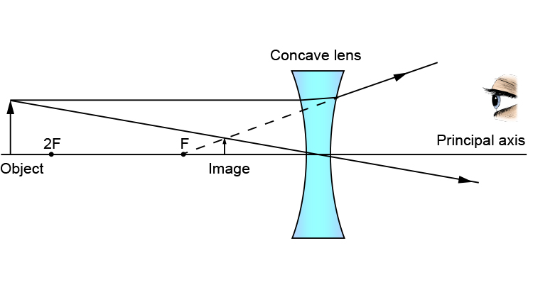
Focal Point of a lens.

The point is where parallel light rays converge into a point in the case of a convex lens.

Focal Length of a lens.

Focal length is the distance in mm from the distance from the lens to the focal point.

For a concave lens, these two definitions are the same, except in a concave lens the light rays diverge out from the parallel axis and the focal length is given in a negative value.

Diopter.


In medicine, the focal length of the cornea or the crystalline lens is expressed as Diopter (D). It is the reciprocal of the focal length of the lens expressed in meters. 1D is equal = 1m – 1. or, 1000/ focal length of the lens in mm.  If additional eyeglasses are required for clear vision, the prescription is written + D for correction of far vision, and - D for correction of near vision. The patient is said to have near vision when the image falls in front of the fovea centralis (used for reading) of the retina and a negative D lens will make the image on the fovea centralis.  For patients with far vision, the reverse is true and for correction, a positive value D lens is needed.

 The diagram shows the difference between convex and concave lenses.


      Converging lens (+D)  
                                                            Diverging lens (-D)

Additional graph representing a concave lens forming an image.


***************************************************************                                                                


Thursday, January 12, 2023

Nasal Septum


                                                         Nasal Septum

                                                PKGhatak, MD

Humans have one nose but two nasal openings called nasal passages. The two passages are separated by a central partition, made up of bony plates and one piece of cartilage. The diagram below shows pieces of bone and one cartilage forming a vertical nasal septum.


1. perpendicular plate of ethmoid bone (pink)

2. vomer bone. (green)

3. cartilage of the septum (khaki).

4. crest of the maxillary bone. (brown)

5. crest of the palatine bone. (blue)


A CT scan view of the nasal cavity and nasal septum.


How the nasal septum developed:

In the early embryonic stage of development, two nose buds (nasal placode) develop from the neural crest. Evolutionary embryology documented how the human nose transformed from an olfactory organ to dual functions of respiration and olfaction through various stages of transformations. In the end, several facial and skull bones participate to form the nose and the nasal septum. The cartilages fill in places where injuries and deformities are likely to take place. 

Nostril in other animals.

Sperm whales, dolphins, beluga whales and orcas have one nostril, also called the blowhole, the second nostril is modified as an echolocation organ.

Fish use gills for respiration and have 2 to 4 nostrils. One set is used to take in water to a chamber for detection of odor and the other two to discharge water.

Octopuses, crabs and butterflies have no nostrils. Snakes have no nostrils, an opening of the airway called the gullet is located just behind the tongue and is kept closed until the snake swallows air, then directs air to its lungs. Snakes can protrude the gullet to one side of the mouth while swallowing a large prey and at the same time breathe. The snake collects odor molecules from the air by flicking out its tongue repeatedly and when the tongue is retracted into the mouth, a special organ located on the palate generates the sense of smell.

Variation of size and shape of the nasal septum.

It is said that people in hot and humid tropical areas have flat, wide noses and a flatter nasal septum because for a given amount of air, the oxygen content is lower than what is present in a cold climate. As a result, a larger amount of air is necessary and a wide short nose is more suitable for such a purpose. In a cold climate, the air has to be warmed up to body temperature quickly. A higher nasal bridge and a larger nasal septum increase the surface area and warm up the air in a shorter time.

Embryology:

Nasal septum development is a part of the development of the face. It is difficult to summarize its embryology in a few short sentences. The link below can be used to access a more detailed description.

https://www.ncbi.nlm.nih.gov/pmc/articles/PMC7965203/figure/F4/

[Also see the footnote].

Head ectoderm, neural crest and prechordal plate by repeated fusion and separation from a common oral-nasal cavity. The midbrain neural crest migrates below the nasal placode (a plate like thickened epithelial layer) and develops as the lateral nasal process and the forebrain neural crest forms the medial nasal process. As these cavities begin to deepen, a middle ridge appears. This medial ridge develops as the vertical plate of the maxillary bone. Other bones and cartilage follow the same pattern and ultimately a nasal septum comes into shape.

Histology of Nasal Septum.


The nasal septum is about 2 mm thick. Both surfaces of the nasal septum are covered by mucous membrane and the submucous tissue contains blood vessels and nerve tissue. The surface of the mucus membrane is covered by epithelial cells, which have 3 different forms. See the diagram below. 


At the front end of the septum, the surface is covered by keratinized stratified squamous epithelium. The major portion of the septum is covered by respiratory epithelium which is the pseudo-stratified columnar ciliated epithelium. The uppermost part, below the cribriform plate, is covered by olfactory epithelium. These cells are tall columnar cells and support the olfactory cells.

Olfactory epithelium.

The olfactory epithelium contains Odor sensing neuronal cells and supporting cells, basal cells and brush cells.

Olfactory cells. These are bipolar cells, the top part facing the nasal cavity is supplied by hair cells (modified cilia). Its function is to trap odor molecules, helped by watery secretion from glands in the submucosa. From the base of the bipolar cells, the nerve fibrils emerge and the nerve fibers pass through the cribriform plate and make synaptic connections with the Mitral cells of the olfactory bulb.

Supporting cells. These are pseudo-stratified ciliated columnar cells. Functionally, cells are of two types. The sustentacular cells and microvillar cells. The sustentacular cell metabolically supports the respiratory epithelium. Microvillar cells are endowed with immune functions.

Basal cells divide actively and transform into other cell types, including bipolar nerve cells.

Brush cells. They are columnar cells and have microvilli. Brush cells are general somatic sensory receptor cells of the trigeminal nerve branch of the nose.

Cavernous tissue. Erectile tissue covers the turbinate (overhanging structures of the lateral wall of the nasal cavity), it is also present at the junction of bone with the cartilage. The blood in the cavernous tissues quickly warms air when the artery is dilated by parasympathetic signals.

Blood supply to Nasal Septum.

The nasal septum has a rich blood supply and several arterial branches of both the internal and external carotid arteries, which join together into an arterial plexus. From this plexus, the septum receives its arterial supply. The anterior inferior part of the vestibule (the place people put their fingertips) is known as Little's area and is the main area of the source of nose bleeds. The four arterial branches that join together to form the Kieselbach plexus are - the Sphenopalatine artery, a branch of the internal maxillary artery, which is a branch of the external carotid. The anterior ethmoidal artery, which is a branch of the ophthalmic artery, originates at the circle of Willis, comes from branches of the internal carotid artery. Septal branches of the superior labial artery, a branch of the facial artery. Posterior ethmoidal artery, a branch of the maxillary artery.

Venous drainage.

A submucosal venous plexus drains blood to the Sphenopalatine vein and ophthalmic vein. These veins make a connection with the cavernous sinus of the brain (a potential source of brain infection).

Nerve supply to Nasal Septum. The origin of the Olfactory nerve is stated earlier. The general somatic sensation (pain, pressure, heat & cold) is supplied by the sensory division of the 5th cranial nerve. The ophthalmology branch of the 5th cranial nerve supplies the upper anterior of the septum, the maxillary division of the 5th supplies the posterior and inferior part of the septum.

Parasympathetic secretory innervation of nasal glands originates in the parasympathetic nucleus of the 7th nerve, the preganglionic fibers synapse in the pterygopalatine ganglion and postganglionic fibers innervate the nasal glands. Stimulation of this nerve dilates blood vessels and increases nasal secretion.

The sympathetic nucleus is present in the Thoracic 1st segment of the spinal cord, preganglionic fibers travel up and make a synaptic connection in the Caudal sympathetic ganglion. Postganglionic fibers travel along blood vessels to reach the nasal glands, Stimulation of this division produces vasoconstriction and relieves congestion.

Lymph drainage of the nasal septum to the posterior deep cervical and submandibular lymph nodes.

Structural abnormalities of the nasal septum.

A common deformity comes from a direct injury to the nose resulting in a fracture of the bones of the septum but the cartilage withstands many less severe blows because of its flexibility and side to side mobility. Both congenital and acquired diseases can alter the shape and character of the septum.

Genetic abnormality.

Cleidocranial dystocia is an autosomal dominant trait. It is characterized by short stature, sloping shoulders, absent collar bones and deformed nasal septum.

Down syndrome babies have saddle shaped nose, a short neck, a protruding tongue, and exhibit mental developmental delays.

Congenital.

Congenital syphilis produces destruction of nasal cartilage and a saddle nose, frontal bossing, saber shins and many other abnormalities.

Acquired condition.

Relapsing Polychondritis.

It is an autoimmune disease that develops around midlife. Inflammatory destruction of respiratory cartilages, a saddle nose is the common finding.

Cocaine abuse. Cocaine snorting produces ischemia and as a result perforation of the nasal septum develops. Cocaine use causes frequent sinus infections.

Leprosy. Untreated leprosy can destroy cartilage and bones of the face and nose, and many other systemic symptoms.

Leishmaniasis. Mucocutaneous sores may erode nasal bones and soft tissue and other areas of the mouth and face, leading to many secondary infections.

-------------------------------------------------------------------------------------------

Footnote: -

Root. The uppermost portion of the nose is just below the eyebrows.

Bridge. The part that connects the root to the rest of the nose.

Apex. The tip of the nose.

Nostril. The opening of the nose is also called naris (pleural – nares).

Ala. The side wall of the nostril (pleural – alae).

Philtrum. The part connecting the tip of the nose to the upper lip. It is concave in shape.

**************************************

Sunday, January 8, 2023

Eardrum

                                                         Eardrum

                                            PKGhatak, MD


The eardrum is known as the Tympanic membrane (TM). The word Tympanum is a Greek word, it means to beat or strike. The Latin of the eardrum is Myringa and inflammation of the tympanic membrane is known as Myringitis.

The eardrum is about 1 cm in diameter, located at the end of the ear canal, positioned in a slant fashion, with the external surface facing downwards and forward, looking towards the face.

Structure. It is made of three layers. The outer surface. The middle fibrous portion and the inner surface.

A thin layer of skin tissue covers the outer surface, these cells are stratified squamous keratinized epithelial cells. The middle layer is a tough connective tissue made up mostly of type II and type III collagen and this layer contains blood vessels and nerve fibers. The inner surface is made of cuboidal epithelial cells, which are continuous with the lying cells of the middle ear.

Embryology.

In a developing embryo, an invagination of the first pharyngeal groove joins the first pharyngeal pouch. These two layers form the TM. The outer layer is Ectodermal in origin and the inner layer is derived from the Enteroderm. The middle layer is derived from the neural crest, a mesenchyme tissue.

Blood Supply. The TM has two different blood supplies. The blood supply of the outer surface is provided by the deep auricular branch of the Maxillary artery. The inner surface is supplied by the anterior auricular artery, which is a branch of the maxillary artery. The posterior part of the inner surface is supplied by the posterior tympanic artery, a branch of the posterior auricular artery.

Nerve supply. The inner and outer surface of the TM is supplied by separate nerves.

The sensory supply of the outer surface. The cranial nerves – the 3rd (Trigeminal), the 7th (Facial), the 10th (Vagus) and the 11th (Glossopharyngeal) supply different areas of the outer surface.  The inner surface is supplied by the cranial nerve 11th.

Attachment of TM to the bones.

The outer rim of the TM is thick and cartilaginous. That fits snugly with a groove of the mastoid bone; however, the ring is not complete, a segment of the top is devoid of the ring, as a result, this part is less taught and called pars flaccida.

Bone attachment to TM:

In the middle of the inner surface of TM, the manubrium of the Malleus bone is attached. It pulls the TM inwards and gives it a conical shape; the inner side takes a convex shape and the outer side assumes a concave appearance.

When the TM is observed under direct vision with a scope, the different areas are identified with specific names; a diagram is easier to point this out.


                                              The Tympanic membrane

Protective muscle of the middle ear.

Two small muscles, the tensor tympani, and stapedius muscles protect delicate hearing organs by reflex action. The motor fiber for the Tensor tympani comes from the Trigeminal nerve (motor division) and the Stapedius muscle is supplied by a branch of the facial nerve. Tensor tympani by contracting increases the tension of TM and reduces the amplitude of vibration. Stapedius can disengage the stapedius bone from the oval window of the inner ear in order to protect the delicate sound receptors and the hair cells.

                                       Tensor tympani & Stapedius muscles.

Function of TM.

TM is a physical barrier between the middle ear and the ear canal. It prevents water, dirt, dust, small insects,  etc. from getting inside the middle ear.

TM transmits sound waves to the 3 small bones directly, the bones in turn transmit the sound waves to an opening of the bony Cochlea, the Oval window and the sound is transferred to the endolymph of the membranous cochlea and thereby to the hair cells.

The surface area of TM is 64.3 mm square. The surface area of the Oval window is 1/120 of the surface area of TM. The sound waves are magnified 27 times by the time it reaches the endolymph of the cochlea.

Diseases of TM.

In adults, diseases of TM are not common. Injuries usually result from blast injuries in certain professions using dynamite and in warfare. Inflammation or infection from the ear canal can spread to the TM. Such infections usually develop in swimmers, due to moisture-loving bacteria like Pseudomonas or Atypical tubercular bacteria.

In children, TM injury or infection is common. Injuries result from improper use of Q-tips. Throat infections rather easily spread to the middle ear and then to the TM because the Eustachian tube in children is short and does not drain so easily as in adults.

A few special diseases of TM.

Bullous Myringitis.

Bacteria, Mycoplasma and Virus infections occasionally produce not only acute infection of the TM but also produce a fluid filled blister on the TM. Blisters may be hemorrhagic. This was found to be common in Mycoplasma infection, subsequently, bullous lesions are also observed in bacterial and viral infections. Blisters may be hemorrhagic. Myringitis is a painful febrile illness, that often results in perforation of the TM and a temporary decrease in hearing.

Swimmer's ear itches.

Cotton from the Q-tip gets dislodged in the ear canal, which blocks the drainage path. Stagnant water favors Pseudomonas aeruginosa and Staphylococcus aureus growth. Acute infection later becomes a chronic spreading infection from the TM to the entire external ear.

Swimming pool granuloma. Mycobacterium marinum can cause a chronic ear canal and TM infection, usually from minor wound infection. It produces granulation tissues and damages the tissue if not properly treated.

Ramsey Hans Syndrome. It is an uncommon illness characterized by paralysis of one side of the face and the appearance of a bunch of blisters,  including on the TM due to reactivation of the chicken pox virus, often called shingles. Pain in the ear, loss of hearing on the side of facial paralysis and difficulty in speech and eating are some of the main symptoms.

Cholesteatoma of the ear.

Retracted TM or perforated TM left untreated for a long time causes accumulation of dead skin in the inner side of TM and in the middle ear. Occasionally skin grows into a lump or cysts develop. The accumulated wax starts to eat away the bone and recurrent infections lead to the formation of osteomyelitis in the mastoid bone. Fowl smelling discharge, loss of hearing, sensation of fullness of ear and dizziness develop. 

Rupture and perforation of TM.

Ruptures are mostly accidental due to a sudden blast of air hitting the eardrum in milliseconds before the protective reflex action of the stapedius muscle can disengage from the Oval window.

Barotrauma. Air pressure of both sides of the TM is equal due to a reflex action of swallowing, yawning or clearing the throat. In a sudden change of air pressure, like an airplane taking a nose dive or loss of cabin pressure and many other situations, the decreased ear canal pressure causes TM to bulge out so suddenly that it actually ruptures.

Perforation of TM. Acute otitis media with large effusion or pus formation in the middle ear pushes the TM outward, and treatment is delayed, the TM ruptures and drains pus outside. Other causes of TM perforations are instrumentation, attempts to clean wax using unorthodox methods. Severe head trauma. Deep water diving or Caisson's disease.

Deliberate act. Bajau people of the Philippines, Indonesia, Malaysia,  engaged in deep water hunting, used to deliberately puncture their ear drums so to prevent accidental rupture during deep dive, and miss to earn a living wage for a few weeks.

Retracted ear drums. Retraction of TM is a common occurrence, in most cases pars flaccida is retracted and nearly all resolve spontaneously. Only a few instances, the whole TM is retracted and due primarily to pharyngotympanic tube blockage. It is one of the causes of cholesteatoma.

 Cancer of TM:

Cancer of the ear drum is very rare, because the outer surface is skin, the following cancers are possible: Basal cell carcinoma, Squamous cell carcinoma, and Melanoma.

The inner surface is the same as the lining membrane of the middle ear, if cancer develops, it would be adenocystic carcinoma and adenocarcinoma. 


************************************************


Thursday, December 22, 2022

Neurotransmitter

                                             Neurotransmitters

                                        PKGhatak, MD

Nerve cells communicate with other nerve cells, muscles, and glands, or internal organs by chemical molecules. Even though the nerve impulse travels along the nerve fibers as an electrical impulse, at the end of the nerve terminal, a tiny gap exists, that gap is bridged by a neurotransmitter,

Neurotransmitters (NT) are chemical messengers that carry forward electrical signal from neurons to postsynaptic neurons or cells of the target organ and produce the intended action. NT must meet the following criteria. NT must be synthesized by the neuron, found in the presynaptic nerve terminal, must produce depolarization and propagation of the nerve impulse. NT must be quickly removed either by enzymatic degradation, diffusion or reuptake by the nerve terminal.


From the time the first neurotransmitter (NT) Acetylcholine was discovered in 1921 by Otto Loewi of Germany, many more NTs have been added and today well over 100 NTs are known. Not all chemicals fulfill all the above criteria, however, all of them produce depolarization and transmission of nerve impulses. Chemically some of the NTs are simple gases like nitrous oxide (N0), carbon monoxide (CO) and complex protein molecules, like pituitary adenylate cyclades activating peptide, are included in the newer class of NT.

In this review, only those NTs abnormality that produces significant changes in the body are discussed. These are-

1. Acetylcholine, and Biological amines, 2. Epinephrine, 3. Noradrenaline, 4. DOPA and 5. Serotonin.

Inhibitory neurotransmitter –  6. Gamma-aminobutyric acid (GABA) and 7. Histamine.

1. Acetylcholine (ACh)

Acetylcholine is released at synaptic junctions by the neurons of the central nervous system, as shown in the diagram.


Acetylcholine is synthesized by the nerve cells and then stored in vesicles at the nerve terminals waiting to be rereleased at the synaptic cleft.  


Chemistry of ACh.

ACh is synthesized from Acetyl CoA and Choline. Acetyl CoA is the end product of glucose metabolism; choline is either locally produced by the nerve cells from another amino acid serine or taken up from the blood. Chemically, choline is a Quaternary ammonium compound, acts almost like a vitamin in humans, except that it can be obtained from another amino acid. Eggs, organ meat, fish, milk and beans are good dietary sources of choline. Excess choline can cause low BP, weakness, increased sweating, fishy body odor, liver disease and cardiac problems. A deficiency of choline causes non-alcoholic fatty liver, muscle damage and hyperhomocysteinemia (high Homocysteine blood level).

Neurons take up free choline via the 1.Na+/Choline transporter,2. a degradation product of ACh by the enzyme Acetylcholinesterase from the synaptic cleft and, 3. also from blood as shown in the diagram below.


Synthesis of ACh.

Acetyl CoA + Choline = Acetylcholine, by the enzymatic action of acetylcholine-transferase.

Degradation of ACh into Acetyl CoA and Choline by enzyme Acetylcholine esterase.

Action of ACh.

Autonomic nervous system.

(a). Parasympathetic division. It acts both on ganglia and postganglionic endings and its effects are -

Eyes - Constriction of pupils and fall of intraocular pressure.

Glands - Increases secretion of GI glands,

Bronchi- Constriction of bronchi, increased mucus secretion.

Smooth muscles of GI tract – increased motility, spasm and colicky pain, contraction of gall bladder and defecation.

Heart and arterioles. Slowing of the heart rate and vasodilation of arterioles.

Urinary bladder. Precipitate micturition.

(b). Sympathetic Division - It acts only on the sympathetic ganglia and the Adrenal medulla and causes high BP and increased heart rate.

Central Nervous system. It causes restlessness, insomnia, tremors, dysarthria and convulsions.

Voluntary muscles. Contraction of muscles and fasciculation.

ACh has Nicotinic and Muscarine effects. Nicotinic receptors stimulate the ganglia of both sympathetic and parasympathetic divisions and the adrenal medulla. It also stimulates parasympathetic postganglionic nerve terminals. Nicotinic receptors stimulate the Neuromuscular junction of the skeletal muscles.

Muscarinic receptors are present in the parasympathetic nervous system and signal secretion from glands and smooth muscle contractions. Muscarine has no action on the skeletal muscles and does not act on the brain cells.

Diseases, due to autoimmune disease and some unknown causes, decrease the functions of ACh: these conditions are -  Myasthenia gravis, Eaton Lambert syndrome, Guillain-Barre syndrome and Ascending paralysis in febrile children.

When used as a drug -   it temporarily produces muscle paralysis and is used every day in surgery to ensure a better surgical outcome. Patients on mechanical ventilators at times require muscle paralytic drugs to prevent patients from struggling to breathe. 

2. Biological amines:

Walter Bradford Cannon of Massachusetts, USA, in 1932 discussed the properties of adrenaline and used this phrase -  Flight or Flight response.

Epinephrine and  Noradrenaline.

In Europe, these two neurotransmitters are known as Adrenaline and Noradrenaline. Both Adrenaline and Noradrenaline are hormones. Norepinephrine (noradrenaline) is the neurotransmitter of the sympathetic division of the autonomic nervous system.

Biosynthesis of Norepinephrine.

L-Phenylalanine, an amino acid, is the source of Dopamine and Epinephrine, the intermediate steps and enzymes involved in this process are as follows.

L-Phenylalanine to  L-Tyrosine by enzyme Amino acid Hydroxylase.

L-Tyrosine to  L-Dopa. (1-3,4 -Dihydroxyphenylalanine) by the enzyme amino acid Hydroxylase. 

L- Dopamine to  Norepinephrine by enzyme Betahydroxylase.

Norepinephrine to  Epinephrine by the enzyme N-Methyltransferase.

Noradrenaline is the transmitter for all the neurons of the brain and spinal cord of the sympathetic division of CNS, and outside the CNS, for the sympathetic postganglionic neurons. Noradrenaline increases blood pressure vessels due to increased tone. It produces bronchodilatation in the airways of the lungs and relieves nasal congestion. The difference in action is based on alpha and beta receptors and their subtypes.

Origin of noradrenergic neuron.

These neurons originate from the locus coeruleus, tegmentum and dorsal medullary group.

Action of Noradrenaline and Epinephrine

Effect on

Norepinephrine

Epinephrine

Heart rate.

Slowed

increased

Force of cardiac contraction

No effect

increased

Cardiac output

No effect

increased

Cardiac irritability

increased

Much increased

Systolic BP

Rises

rises

Diastolic

Rises

falls

Vascular bed in muscles

constriction

dilatation

Vascular bed skin & viscera

contraction

contraction

Vascular resistance in the heart

increased

decreased

Glucose metabolism

unchanged

increased

Bronchial smooth muscles

No effect

relaxed

Intestinal muscles

relaxation

relaxation

Intestinal sphincters

constriction

relaxation

Pregnant uterus

Increased contractions

Contraction lessened

Capillary permeability

No effect

reduced


Epinephrine is a lifesaving drug in an acute allergic reaction and anaphylactic shock producing laryngeal edema and death due to airway obstruction. Adrenaline has multiple applications in everyday medical practice and is too numerous to list here.

3. Serotonin.

Serotonin chemically is 5-Hydroxytryptamine (5lHT). It is formed from the amino acid Tryptophan to   5-Hydroxytryptophan by enzyme Trytophanhydoxylase (adding “OH” group) and then to 5-Hydroxytryptophan (serotonin) by enzyme Aromatic amino acid decarboxylase and coenzyme pyridoxal phosphate (removing COOH group).  5-HT is present in large amounts in the GI tract and the brain neurons and platelets.

Serotonin acts on the areas of the brain and is shown in green color in the diagram below.


Serotonin is the neurotransmitter for appetite, sleep, memory, happiness, mood, vomiting center, sexual arousal and body temperature maintenance by its actions on the forebrain, brainstem and cerebellum. Lack of serotonin produces depression, anxiety and other psychological dysfunctions.

Serotonin receptor.

Serotonin (5-HT) receptors are designated by digits 1 to 7 and the receptors of 5-HT1 are assigned - A to F. The receptors of 5-HT 2 have A to C subtypes. The action of serotonin varies according to its binding with the type of receptor.

 5-HT1A is mostly related to mood, learning, memory and behavior.

 5-HT1B produces vasoconstriction. 5-HT2 signals via activation of phospholipase.

 5-HT2A stimulates urinary bladder contraction.

 5-HT2B increases pulmonary hypertension. and in general, produces inhibitory actions and opposes 5-HT1A effects.

 5-HT2C stimulates appetite.

 5-HT3 can produce nausea and vomiting. 

5-HT4 increases GI motility.

 5-HT5 consolidates memory.

 5-HT6 increases depressive mood.

 5-HT7 opposes 5-HT6 effects on mood.

Location of Serotonin receptors in the brain.

The Raphe nuclei B1 to B9 are serotonergic. These neurons are most abundant in the reticular formation, the axons of these neurons connect the brain and spinal cord nuclei.

Serotonin has a dominant role in understanding psychiatric disorders and depression. Therapeutic drugs are used extensively to increase the concentration or prolongation of the action of serotonin in the synaptic cleft.

4. Dopamine.

Dopamine is primarily an inhibitory neurotransmitter, which controls agitation and excessive motor actions.

Biosynthesis of L-DOPA (L-dihydroxyphenylalanine).

Biosynthesis is mentioned earlier. DOPA, norepinephrine and epinephrine are collectively known as Catecholamines. L-DOPA can enter the brain but L-Dopamine is blocked by the blood-brain barrier. In the brain, L-DOPA is converted to L-Dopamine by decarboxylate and Vitamin B6 as a cofactor. This actions take place in Sustantia Niagra. After dopamine is release at the nerve terminals, it is quickly broken down by catechol methyl-O- transferase.

Dopamine receptors and action.

Receptor subfamily

Location

Action

Therapeutic potential

Central

D1 and D2

substantia nigra and striatum

motor control

agonists - Parkinson's disease

D1 and D2

limbic cortex and associated structures

information processing

antagonists - schizophrenia

D2

anterior pituitary

inhibits prolactin release

agonists - hyper-prolactinemia

Peripheral

D1

blood vessels

vasodilatation

agonists - congestive

D1

proximal tubule cells

natriuresis

heart failure

D2

sympathetic nerve terminals

decreases release

hypertension

Dopamine has both CNS and outside CNS effects.

In the brain, dopamine edits signals that are going out to the skeletal muscle. In the limbic system, it organizes and forms emotional memory in associated mood elevation.

Outside the CNS, Dopamine is produced locally and acts locally (action is called paracrine effects). Dopamine increases heart rate and BP.  It is suspected that Primary Hypertension is due to an abnormal dopamine action on the kidneys. In the pancreas, it decreases insulin production, on the GI tract, decreases motility, acts to protect the GI mucosa and boosts local cellular immunity. It appears that dopamine acts as a natriuretic hormone in the kidneys.

Diseases involving movement disorders, like Parkinson's disease, Restless leg syndrome and minor strokes, are treated with DOPA.

5. Gamma Amino butyric acid (GABA).

GABA is derived from Glutamate. Both Glutamate and GABA are neurotransmitters of the central nervous system. Glutamate is a neuroexciter and is widely distributed in the brain, mostly in small sized neurons. GABA is an inhibitor NT. GABA neurons are present in the Limbic area of the brain, from these nuclei GABA connects many areas of the brain. A diagram below shows the areas of influence of GABA. 


Chemistry:


Brain neurons produce GABA from glutamine by decarboxylation by the enzyme decarboxylase. The reaction is a rate limiting reaction. GABA concentration in the brain is high.

Mechanism of action of GABA:

GABA has two isoforms, Ionotropic (GABA A) and metabotropic (GABA B) forms. Two separate genes on two chromosomes encode the production of isoforms. GABA has two metabolites, Homocarnosine and pyrrolidinone and both these chemicals are anticonvulsants. GABA prevents hyperpolarization and reduces incoming and outgoing signal strength. GABA A receptors are present all throughout the brain. GABA B receptors are formed by the fusion of two G-protein molecules.

Blood-brain barrier prevents GABA from entering the brain but Glutamine is free to enter. Food containing glutamine is soybean, brown rice, chestnut, mushroom, tomato, spinach, cabbage and cauliflower.

Fluctuation of the levels of GABA has been linked with Autism, Parkinson's disease and anxiety.

Therapeutic drugs having GABA-like action are used as pain control medication but the therapeutic action of these medicines does not match the theoretical possibilities it promises.


6. Histamine.

Histamine is well known as a mediator of allergic reactions. It is present in the mast cells of the skin and in the basophils of blood. It enhances inflammatory reactions by increasing vascular permeability and the production of mucus.

As an NT is not that well known. In the brain, it acts via H1, H2 and H3 receptors.

Action in the brain results in wakefulness, alertness and quickens reaction time.

Distribution of Histamine containing cells.

In CNS: Hypothalamic region of the brain.

Outside the brain: The nose, mouth, feet, skin, and feet have a good number of mast cells. In the stomach, chromaffin cells produce and release histamine.

Chemistry: Histamine is produced from the amino acid Histidine by the enzyme histidine decarboxylase.

Enzyme histidine N-methyltransferase and diamine oxidase degrade histamine.

Receptors. Histamine receptors are H1 to H4.

H1 receptors. Dendrons (short arms of neurons) of the Tuberomammillary nucleus terminate in the dorsal raphe and locus coeruleus. These cells regulate sleep-wakefulness cycles, body temperature, appetite, coordinate endocrine hormones and cognition.

H2 receptors. Basal ganglia, hippocampus and dentate nucleus have histamine 2 receptors. Modification of motor activities is the main function. In the stomach, H2 receptors on the parietal cells are activated and histamine increases HCl acid production.

H3 receptors. The precise location is not worked out. The action appears to counteract acetylcholine, serotonin and norepinephrine actions.

H4 receptors. H4 receptors in the brain have not been detected.

The realm of antihistamine drugs is increasing with new discoveries of histamine receptors and their role in the etiology of diseases.

 8. Other NTs.

Glycine.


Glycine is an amino acid, present in every living cell and besides performing metabolic functions, it also acts as a neurotransmitter for incoming somatic sensations like pain and touch, vision and auditory sensation. Glycine also acts on strychnine-sensitive receptors and produces inhibitory action and modulates excitatory neurotransmission of glutamine in glial cells.

The final word.

Neurotransmitters are chemical messengers synthesized by neurons and delivered at the synaptic cleft in order to ferry the nerve impulse to the next neuron or to target cells. In the cerebral cortex, the prime NT is acetylcholine. In the autonomic nervous system,  ACh is also the sole NT for both parasympathetic ganglia and postganglionic nerve terminals. In the sympathetic nervous system, ACh is NT for the sympathetic ganglia but epinephrine is the prime NT for postganglionic nerve terminals. In the Limbic nervous system, multiple NTs interact, modify, enhance, or inhibit nerve impulses.

NTs are also classified as excitatory or inhibitory. The same NT may act as both excitatory and inhibitory depending on the receptor it binds. The molecular structure of NT must fit with receptors like pieces of Logo fit with another - a Levo (l) or Dextro (d) from; an aliphatic chain or ring form, makes that difference. And finally, even minor NTs have major effects on the body.

************************************


Wednesday, December 14, 2022

Connective Tissue & Mixed Connective Tissue Disease

                             Connective Tissue and Mixed Connective Tissue Disease.

                                            PKGhatak, MD

Connective tissue is the most abundant tissue in the human body. It is present underneath the skin, in the lungs, in the mucus membranes of GI and GU tracts, and in the fatty tissue and in the coverings of the brain. The connective tissue fibers are made up of two proteins, Collagen and Elastin. Collagen is present in the tendons and elastin dominates in the lungs and skin. The connective tissue (CT) is a loose collection of cells in the middle of a water rich extracellular matrix. Blood, bones, cartilage, adipose and reticular tissues are special forms of connective tissues.

Embryology of CT.

CT is derived from the mesoderm. A sheet of cells in the developing fetus folds into two tubes, the ectoderm and the endoderm and in between the tubes, the remaining cells from the mesoderm. The embryonic mesoderm is known as Mesenchyme. From the mesenchyme, the connective tissue, muscles, bones, cartilage, blood vessels and lymph tissues develop. The mesoderm cells undergo various stages of development, from the initial flat sheet of cells to tube formation and back to sheet form. CT cells migrate to the ectoderm and develop into the subcutaneous tissue of the skin; cells migrate to the endoderm and develop into the submucous tissue in the GI tract and interstitial cells in the lungs. The developing mesoderm receives cells from the yolk sac, these cells become the Microglial cells of the brain. The end result is connective tissue containing blood and lymph vessels and nerve fibers.

This basic embryology is helpful to understand various manifestations of Connective Tissue Disease (CTD).

Connective tissue disease.

There are a number of Autoimmune CTDs that are simply identified as Connective Tissue Diseases. These diseases are Systemic lupus erythematosus (SLE), Scleroderma, Sjögren's syndrome, Polymyositis, Rheumatoid arthritis (RA) and Mixed connective disease.

The laboratory screening test for connective tissue disease is the ANA (anti nuclear antibody) test. The test is expressed as I:80 to 1: 640 dilutions. However, about 15% of the population have a positive test without ever having any disease, and these ANA tests are positive only in lower dilution (1:80).

The mixed connective disease (MCTD), as the name indicates, has some of the characteristics of other CT diseases and was previously named Overlap Syndrome.

MCTD (mixed connective tissue disease)

MCTD is a rare autoimmune disease; it is triggered by an unidentified agent. The antibodies generated are directed against collagen fibers wherever they are present. Those cells turn out excess collagen which causes the thickening of skin and fibrosis of internal organs. To describe a full spectrum of MCTD, a synopsis of common manifestations of the other CTDs is presented here.

1. Lupus Erythromatosus (SLE).

SLE is a systemic disease that affects many organs and is a steadily progressive illness. Deaths are due to renal or respiratory failure if treatment is not available. However, not every patient follows the progressive course, many just have skin or liver disease only, while others may have skin and lung involvement. The lesions produced by SLE on the individual organs are summarized below. -

Skin. The term erythematous of SLE is taken from a butterfly red colored skin rashes that develop over the bridge of the nose and on the cheeks. Other skin lesions are coin sized dry skin patches on sun-exposed skin, resembling psoriasis. And subcutaneous lesions with rolled up raised margins, localized lesions are seen on the neck, front of the chest and exposed areas of arms.

Joints. The finger joints of both hands and feet are commonly involved. The stiffness, swelling and joint pain are usual symptoms.

Kidneys. Glomerulonephritis is common. Nephritis is due to immune-complex induced damage to the capillaries of glomeruli. This leads to hypertension, renal insufficiency and renal failure.

Pulmonary. Pleurisy is the most likely to develop when the lungs are involved. Pulmonary fibrosis, pulmonary hypertension and respiratory insufficiency lead to respiratory failure. At times small vessel vasculitis produces pulmonary infiltrates and patchy areas of consolidation.

Liver: Lupus hepatitis is a known feature, often just the liver enzymes are elevated and in others, more liver damage and jaundice develop.

Heart. Sterile pericarditis, myocarditis, coronary artery disease and conduction abnormalities are usual findings. In addition, cardiac valve damage and sterile vegetative emboli to the brain may happen.

Brain. Encephalitis, small vessel strokes, transient ischemic attacks. delirium, psychosis, anxiety and seizures may develop.

Eye. Blurred vision, exudative macular detachment, cystoid macular edema, and ptosis may develop.

Hematology. Anemia, thrombocytopenia, leukopenia, lymphadenopathy and splenomegaly are seen.

Coagulation. Prolongation of coagulation due to lupus-anticoagulant can cause a serious problem in certain situations.

Confirmatory serology for SLE. In SLE the ANA test is invariably positive. Several antibody tests are also positive. But the confirmatory test is anti-Native double stranded DNA (dsDNA). This test is the gold standard diagnostic test of SLE.

2. Systemic Sclerosis (SS), and Scleroderma.

It is a rare disease, one in a million per year is seen in cold climate countries, still less prevalent in tropical countries.

SS is characterized by diffuse fibrosis of the skin and fibrosis of the internal organs. But not every patient has the same degree of involvement. All patients have positive ANA in high titers, and 70 % of them have positive Anti-SCL 70 antibody tests, this test is known as the Anti-topoisomerase I test. This test is positive in about 50 % of cases of diffuse SS.  When the lesions are confined to limited areas of the skin, the term Scleroderma is used and in 20% of cases, this test is positive.

 The following variants of SS are important.

a. CREST syndrome. CREST stands for calcinosis, Raynaud's phenomenon, esophageal dysfunction, sclerodactyly and telangiectasia. The skin lesions in CREST Syndrome are limited to the distal extremities, the face and the neck.

b. Diffuse disease. The thickening of the skin with areas of increased pigmentation and patchy areas of depigmentation is present all over the body, including the trunk. A test that measures antibodies against Topoisomerase I titer becomes positive in over 50 % of patients.

 In contrast, in diseases limited to the skin and few other places, a different antibody test - The Antocentromere antibody test becomes positive in over 50 cases. About 20% of patients with limited SS have anti-topoisomerase I positivity. In 14 % of cases of limited. The SS U1-RNP antibody test is positive; it indicates the presence of Pulmonary fibrosis.

c. Raynaud's phenomenon. Intense blanching of fingers and toes during exposure to cold is associated with pain. Anti U 11 and U12- RNP antibody tests in Raynaud's is associated with GI, Pulmonary involvement and higher mortality.

When SS produces fibrosis in the lungs, kidneys and GI tract, the disease produces pulmonary insufficiency, renal insufficiency, hypertension, reflux esophagitis and hypomotility of the GI tract, including the esophagus, respectively. In renal crisis due to uncontrollable hypertension and renal failure in systemic sclerosis, Anti-RNA polymerase III antibody test becomes positive.

 3. Sjögren syndrome. Dryness of mouth and eyes and their consequences are the presenting symptoms. The lachrymal glands and salivary glands typically show lymphoid foci with the destruction of glandular elements and fibrosis. ANA is positive in over 95% of cases and anti SS-A and SS-B positive in 65% of cases. SS is often associated with symptoms of RA, biliary ductal inflammation, Hashimoto thyroiditis, polyarteritis and pulmonary fibrosis.

4. Polymyositis and myopathy. Patients typically present with muscle weakness and muscle pain. Muscle enzymes- CPK and Aldolase levels are high indicating muscle necrosis and inflammation. Dermatomyositis is a variant of myositis. Dermatomyositis presents as bilateral "heliotropic rashes”. Another subvariant is Antisynthetase syndrome which is characterized by muscle disease associated with non-erosive arthritis, Raynaud's phenomenon, hyperkeratosis along the palmar and radial aspects of the fingers, pulmonary fibrosis and the presence of anti-Jo-1 antibodies.

5. Rheumatoid arthritis (RA). It is a common disease (about 1% of the population), in contrast with the previous illness mentioned under CTD. It is also different from the rest in having a negative ANA test in 80 % of cases. The blood tests for RA are Rheumatoid Factor (present in all cases), and anti-CCP antibodies (cyclic citrullinated peptides) present in 80 % of cases. Clinical features of RA are dominated by symmetrical polyarthritis of small joints of fingers and toes, The 1st (proximal interphalangeal) joint arthritis at the beginning of the illness is very characteristic. Swelling, pain and morning stiffness of hands and red discoloration of the overlying skin point to an early diagnosis. As the disease advances, other small joints like wrist joints, upper cervical spine, temporomandibular, and sternoclavicular joints are affected. Low grade fever, loss of weight and other clinical and laboratory tests follow the general features of CTD. X-rays of the hands show Erosion of bones at the joint faces and osteoporosis. These findings are also distinct features of RA. Extra articular features are subcutaneous nodules over the bony prominence, pleural effusion (small amount), pulmonary fibrosis, pericarditis, vasculitis and enlarged spleen.


                                  Mixed Connective Tissue Disease.

 Most patients with MCTD are young females between 25 and 30 yrs of age. In Native Americans of Minnesota, the incidence is 6 per100 000 and 2/100,000 in white females. The onset of the disease is insidious and begins with arthralgia of the fingers of both hands, which become white, numb and painful on exposure to cold. Later finger joints swell and become painful. Raynaud's phenomenon and myalgia are common symptoms. All MCTD patients carry HLA-DRB1 positive markers in their white blood cells (lymphocytes & monocytes). The blood levels of U-1 RNP (Ribonuclear Protein) antibodies in high titers (over 1:125) or over 25 U are seen in over 95% of patients with MCTD. MCTD is a slowly progressive disease but in some patients, the progression may be arrested for years, in others, the disease shows features of one or other connective tissue disease mentioned above. However, the initial phase of MCTD resembles RA but soon the disease takes a course resembling scleroderma and Raynaud's features dominate. Though dryness of the eyes is present, other typical features of Sjögren's syndrome are absent. Later, as GI, Pulmonary, and Renal symptoms develop, the diseases acquire SLE like illness, but the CNS is very rarely affected. Pulmonary fibrosis is the most devastating aspect of MCTD and death is generally from respiratory failure or pulmonary infection.

Treatment. 

The management of MCTD is based on individual patients because no two patients are having the same system involvement.

The general approach is to use well known immune suppression drugs and symptomatic therapy for individual organ system dysfunction and degree of disability. Disease modifying agents are added and watched for side effects and therapy is modified based on progress, and over adverse effects. 

Prognosis. Overall prognosis of MCTD is favorable over SLE and SS. A 10 year survival rate of MCTD is 80%. Those who develop Pulmonary fibrosis have the worst prognosis.

********************************************

Archive

  Archive. P.K.Ghatak, MD 2025  March. Laser Endoscpoe 2025 February. Use of Ultrasound in Medicine MRI 2025 January. Bacteriology and Rober...