Friday, November 4, 2022

How We See the Outside World

                                               How we see the outside world.

                                                   The science behind it.

                                              PKGhatak, MD


Looking straight ahead, we see a vast area, horizon to horizon. Using a visual field measurement chart and recording one eye at a time, we find each eye has a visual field of 155 degrees on the horizontal plane and 130 degrees on the vertical plane. By adding the visual fields of two eyes together, our visual field should be 310 degrees but in fact, the visual field is smaller because the nasal field of the two eyes overlaps.

That overlap gives us the dept of vision, meaning, we can judge the distance of an object in front. It is an essential quality for reading, writing, driving, painting, target shooting and other activities which require bringing objects in sharp focus.


The diagram above illustrates visual fields and where the images develop.

The visual field is divided into the Nasal field and the ear (Temporal field. To make a clear distinction between the left and right visual fields, the right-hand side of the visual field is colored red and the blue color for the left.

Note that the nerve fibers carrying the image from the nasal half of the right eye, represented by blue lines, are crossing the midline and going to the left side to join the nerve fibers carrying the image from the temporal half of the left eye. The same is true for the left eye, marked with red color.

It is interesting to note that crossed and uncrossed nerve fibers join and carry images representing the right and left visual fields and not the left or right eyes.

The second point to note is that the nerve fibers make a connection at a nerve center called Lateral Geniculate Body (LGB). The right visual field image goes to the left side and the left visual field image to the right side of the LGB. The stratification of nerve fibers is maintained in the LGB, meaning the temporal fibers remain outer and nasal fibers on the inner nuclei of the LGB.

The third point is that the same stratification is also maintained in the Visual Cortex. The visual cortex is located in the back of the cerebral hemisphere, one on each side in the Occipital Lobe.

Not shown in the diagram, there is upper and lower field stratification on the visual cortex. The lower half of the visual field is represented in the upper part of the opposite cortex, and the upper half in the lower visual cortex.

By knowing the detailed anatomy, the doctors are capable of detecting any segmental visual field loss and treating properly without wasting much time.

How two images merge into one.

This fusion of two images in the visual cortex and perceived as one depends on unison eye movement and convergent reflexes that make image generation on the corresponding points on each retina. When initial images are close but not exactly one, then a further adjustment is made by the eye muscles to focus properly.

Why does an object too close to the eyes appear blurred.

The eyes are situated 2 inches apart. The images of the two eyes are not precisely the same, the right eyes see a little more on the right-hand side and the left eyes see more on the left-hand side. The images fuse completely when the object is at a comfortable distance from the eyes. The disparity grows more and more when the object is brought closer and closer.

How images are formed on the retina.

Light rays travel in straight lines in all directions from an object. The pupil by changing its size brings light rays in focus on the retina. The photosensitive pigment of the retina converts the electromagnetic energy of light into electrical stimuli. Next, the rods and cones cells of the retina perceive the stimuli and then pass them to nerve cells which convert the electrical stimuli into nerve impulses and send out nerve impulses to the brain.

 How the color is represented in the brain.

In the daytime, sun rays are bright but colorless. But we know that daylight hides 7 colors from red to violet (color of the rainbow) in it. The color receptors of the eyes are cone shaped cells, called cones of the retina. Cones are plentiful in the central part of the retina and are only a few in the periphery. Cones are densely packed in the Fovea of the retina. The red, green and blue colors are detected by three different cone cells. The blue color generates maximum brightness of 2.56 to 2.76 eV and the red color is the least, generating 1.65 to 1.90 eV. These three cone cells make a unit known as the Trichrome unit. By various combinations of the impulse generation by the trichrome units, we are able to perceive the entire color spectrum. This is the basis of the printers' color cartridges we are so familiar with.  The pigment of the cone is called Opsin. It is a protein and it is most sensitive to light waves with a wavelength of 550 nm, blue responds to light waves with a wavelength of 450 to 485 nm and green to 500 to 565 nm wavelength.


White and Black color.

There is no black color receptor.  Instead, black, white and gray color perception depends on various shades of gray colors. The pigment of the gray scale is Rhodopsin, and the receptors are known as Rods. Rods are most plentiful at the periphery of the retina and are only sparsely distributed in the central part of the retina. Rhodopsin is most sensitive at light waves of 555 nm.

We see better in the corner of the eyes when light is very faint. There are no cones at the periphery of the retina, which is the reason we are unable to see color in the dark.

Why are black and white images not as clear as a color picture.

Each cone is connected with one neuron of the retina that sends out stimuli to the brain. 5 or 6 Rods are connected with one nerve cell, as a result, color picture has more pixels so to speak, and is sharper and clearer than black and white images.

Upside down and revered sidewise image.

This diagram illustrates why the upside down and left side on the right side image projected on the retina. 


This is a pinhole camera taking a picture of a tree. The same Laws of Optics work for the eyes.

How the Brain rights up images.

A newborn child grabs things with hand and tries to put them in his /her mouth. That is the beginning of the learning process of the brain and by the time the baby is 2yr old the proper interpretation of images by the brain is nearly complete but the learning process of the brain continues for lifelong.

Near and Distant vision.

Refractive errors of one or more components of the eye (cornea, anterior chamber, lens and vitreous) fail to bring light precisely on the retinal receptors and when the light rays fall short- then the condition is known as Near- Sightedness and when the light rays converge beyond the light receptors then it is called Far- Sightedness.

Double vision.

Double vision may be monocular or binocular. Mono-ocular double vision comes from the eyes, or contact lens injury, or foreign body injury of the cornea.

Binocular double vision is due to the failure of mages to merge into one. The defect may lie anywhere from the optic nerve to the visual cortex. Since eyes are extensions of the brain and when an eye is damaged it may not improve unless treatment starts immediately.

A quick way to determine mono-ocular or binocular double vision

The mono-ocular double vision disappears when the eye producing the double vision is closed and the person looks through the other eye. In binocular double vision persists by closing and opening one eye at a time.

In daylight, our world appears in full complements of spectrum colors - clear and beautiful and images up-righted and sides correctly positioned. In the dark the story is different. In fading light, the world appears in various shades of gray, fuzzy and best visible to us by the corner of the eye provided one is neither near-sighted nor far-sighted.


*******************************************************


*** 


Monday, October 31, 2022

Movement Disorder

                                                   Movement Disorder

                                              PKGhatak, MD


Muscles move joints and joints move the body. Movement does not only refer to ambulatory movements but movements of part of the body when the person is stationary. In any movement, two groups of muscles work in a coordinated manner, one group contacts the other group must stay in a relaxed state for the movement to occur. If both groups contact simultaneously, the joint is held in a fixed position and is called spasm.

Well known Movement disorders:

Parkinson's disease, Parkinson syndrome, tremors, ataxia, chorea, tics, myoclonus and spasticity.

Parkinson's Disease (PD) and Parkinson Syndrome:

Parkinson's disease is due to the sickness of Dopamine (neurotransmitter) producing cells located in the Substantia nigra in the hindbrain. Slowly the cells die and many neurological abnormalities develop, including dementia and psychosis.

Movement abnormality in PD.

Slow movement and lack of associated movements, like swinging of arms while walking, are striking features. Increased muscle rigidity of neck, head, limbs and back muscles produces a stooping forward posture; and stiffness of legs and thighs produces a shuffling gait. Tremors of hands at rest produce pill-rolling movement of fingers; later writing, feeding, and grooming become difficult. Loss of balance causes frequent falls. The cause of PD is unknown.

Parkinson Syndrome. This term is reserved for Parkinson like features in patients with known causes, for example, poisoning from heavy metals and pesticides, repeated cerebral concussion, and adverse effects of drugs used in Psychosis, Schizophrenia, Depression, etc.

Parkinson's disease (PD) Subtypes.

Experts classify the movement abnormalities in two catigories.1. Tremor predominant. 2. Postural instability - gait difficulties.

1. Tremor-predominant.

Clinically tremor-predominant PD is of three kinds: Mild, Progressive malignant and intermediate varieties.

a. Mild.

Tremors are mostly seen while trying to work with hands.

B. Progressive malignant PD tremors.

Tremors are a rapidly progressive type. The resting tremors are present during waking hours and in light sleep, but during deep sleep, tremors disappear. Tremors prevent patients from doing most routine work - like cooking, cleaning, or answering telephone calls.

C. Intermediate

The symptoms of this subtype fall between the above two subtypes.

2. Postural instability-gait difficulty.

Due to increased rigidity, the patients assume a stooping posture. There is a lack of spontaneous corrective adjustments of posture. Decreased two-way communication with the cerebellum produces unsteady balance and a slight bump against a chair or wall the patient falls forward on the ground.

Subtypes of Parkinson's disease.

A. Progressive Supranuclear Palsy (PSP).

The most distinctive feature of PSP is recurrent sudden backward fall on attempted walking, however, during and after the fall the patient remains fully conscious.  Pseudobulbar palsy leads to difficulty in chewing and swallowing. Intranuclear ophthalmoplegia produces abnormal eye conjugate movements when looking downwards. The patient is unable to look upwards and on attempts to look upward produce horizontal nystagmus. The upper eyelids are kept half-closed. PSP patients have no rigidity and tremors unless the disease is far advanced.

B. Restless Leg Syndrome (RLS).

Patients develop an uncontrollable urge to move. Symptoms may be intermittent, and the degree may vary. Restlessness may also be felt in the arms, neck and head. Sitting on a chair for a long period, driving an automobile and sleeping at night become difficult. Patients also describe a strange sensation of insects crawling under the skin, pins and needles, throbbing and aching pain deep in the legs and lower body.

The use of Dopaminergic drugs -  Pramipexole, Ropinirole and Rotigotin  produce some relief in the early phase of the disease. Later L-dopa may be helpful.

C. Lewy body dementia.

Abnormal muscle movements are generally periodic, bizarre, uncontrollable and purposeless. Apathy and profound loss of memory lead to total dependence on others for activities of daily living.

Other Neurological Conditions.

Neurological conditions produce muscle disorders, for example, Multiple System Atrophy (MSP), Cortico-Bulbar syndrome, Low pressure hydrocephalous, Huntington disease, Wilson disease, Tourette syndrome, Motor stereotypies and Multi System Atrophy.

Multi System Atrophy. 

The impairment of the autonomic nervous system produces a loss of spontaneous adjustment of BP with postural changes and with physical activities. Postural hypotension is common. Of the GI symptoms, constipation and difficulty in swallowing are common. Men may develop erectile dysfunction and difficulty in voiding urine. Olivopontocerebellar atrophy produces stiffness and incoordination of movements. loss of balance and slowness of movements.

Corticobulbar syndrome.

Degeneration of neurons in the motor and sensory cortex produces symptoms of poor muscle coordination, slowness of thought processing, language difficulty and speech abnormality. Increased and prolonged increased muscle tone produces stiffness of posture.

Huntington disease.

Huntington disease is an autosomal dominant inherited condition. Progressive severe dementia, obsessive compulsive disorder, mania, and bipolar disorder are major components. Writhing slow dance like movements, jerky movements of limbs, (called Chorea), language and speech abnormalities are present. Slow eye movements, muscle rigidity, impaired gait, balance and posture resemble PD. 

Wilson disease. It is a recessive mode of inherited disease involving excess copper accumulation in the brain and other vital organs. The muscle disorder includes tremors, difficulty in swallowing and speech and trouble in ambulation. Cirrhosis of the liver at an advanced stage is common. Copper accumulation at the periphery of the cornea of the eye produces a distinct diagnostic feature called the Kaiser Flacherie ring.

Tourette syndrome. It is an inherited disease, but the precise genetic abnormality is not known. Involuntary movements commonly involve the face and uncontrollable vocal sounds followed by the arms, legs, or trunk. Vocalizations of embarrassing words called Coprolalia often accompanied by obscene jesters, Copropraxia, make patients difficult to socialize with. The symptoms usually begin in childhood and spontaneously may disappear when older.

Motor stereotypes.

These are fixed repeated movements with no useful purpose, appear at the same predictable location and time. Movements last a minute or longer and occur several times a day. Typical motor stereotypes are thumb sucking, lip biting, hair twirling, teeth grinding and head banging. Symptoms start in childhood and rarely persist in adult life. Sometimes more complex motor dysfunctions involving hand and arm movements, like waving arms and wiggling fingers in front of the face and closing and opening hands.

Essential tremors. In this disorder, rhythmic involuntary fine shaking movements of hands generally appear in midlife. Tremors mostly occur when the patient is in the company of others and performing normal tasks like drinking a cup of coffee or buttoning a shirt. Tremors may also happen in the head, producing nodding of the head as yes-yes or no-no jesters.

Spasticity. Prolonged muscle contractions lead to increased tone of muscles that interfere with normal walking or performing activities of daily living. Spasticity is a feature of cerebral stroke, Multiple sclerosis, spinal cord injury, spina bifida and severe brain injury.

Dystonia. Dystonia is an involuntary muscle spasm producing twisting motion of the limbs or trunk producing abnormal posture and positions. An example is Writer's cramp. The onset of dystonia is gradual and always follows a specific muscle action. Muscles fail to relax in between contractions preventing smooth functioning.

Ataxia. Ataxia means uncoordinated muscle actions, mostly noticeable in arms and legs. Cerebellar ataxia is more common. In the younger age group, viral infections, tumors, cysts and vascular disruption in elderly people are common causes.

Tardive Dyskinesia (TD). Tardive means late onset. Dyskinesia is defined as difficulty in performing voluntary functions. Muscles of the face, tongue and upper extremity show repeated involuntary movements like grimacing, lip smacking, pursing of lips, eye blinking, and rapid involuntary arm, torso and finger movements. Long term use of anti-Schizophrenia, Anti choline and anti-depression drugs use are associated with TD. Discontinuation of drugs improves symptoms if done early.


*****************************


Monday, October 24, 2022

Epstein - Barr Virus Infection.


                                                          Epstein Barr Virus.

                                                   PKGhatak, MD


The Epstein Barr virus was initially called the Burkitt lymphoma virus because the virus was identified in the cells of the Burkitt lymphoma in an African patient in 1964. It is known today as Human Herpes Virus 4. This virus is the most common viral infection among all other herpes virus infections in humans. About 85 % of the world population have serology positive blood for Epstein Barr Virus (EBV), indicating past infection.

EBV virus characteristics.

EBV belongs to Gamma-Herpesviridae in the genus group of Lymphocryptoviral. The double stranded genome contains 85 proteins and 50 non-coding RNAs. The viral particle has an outer lipid envelop which fuses with the respiratory cell membrane at the time of infection. The DNA of the virus can turn out 9 different molecules of Entry Proteins, gP 1 – 9, and use them to infect many other cell types of the patient.

The EBV virus, once inside the respiratory cells, multiplies by redirecting the host cell to duplicate virus particles. The virus then leaves respiratory cells and infects B-lymphocytes. Inside the B-lymphocytes, the virus persists for life and goes through 3 cyclic phases, namely active phase, dormant phase and re-infective phase.

A person infected with EBE is unable to clear the virus from his body. A subsection of B-lymphocytes is memory cells. The memory cells continue to turn out immunoglobulins, the initial product is IgM and 2 to 4 weeks later all the products are IgG.

In addition, antibodies are produced against capsular proteins and nuclear proteins.


Why does the EBV virus live in perpetuity in humans.

Whenever antibodies are produced in adequate amounts, the virus goes into hibernation within B-lymphocytes and also in some cases in the respiratory epithelial cells. The NK cells, monocytes and macrophages are unable to find the EBV once inside the cells and antibodies are ineffective to neutralize the viral particles. This hide and seek game is played out throughout the life of the patient.

In certain circumstances, the EBV can also infect NK cells, monocytes and macrophages and produces more serious illnesses.

EBV human infection.

Children are the prime target of the virus and about 80 % of human infections take place in children under 2 years old. Most of the infected children remain free of symptoms or had minor symptoms. The only evidence of infection in these cases is positive serology tests.

The next venerable age is teenagers. Teenagers, however, become symptomatic and the illness is known as Mononucleosis or simply Mono and also commonly called a kissing disease. The most remarkable symptoms are the posterior cervical lymph node enlargement and enlarged spleen. Other significant symptoms are sore throat, fever and enlarged tonsils.

Persistent and recurrent activation of EBV.

The EBV can become active within the immune cells, whenever the immune systems are down. The repeated reactivation of the EBV within the immune cells produces Multiple sclerosis and rheumatoid arthritis.

Reactivation within the respiratory epithelial cell leads to Sjogren syndrome and Systemic Lupus Erythematosus (SLE).

The positive ANA, pANCA, cANCA and Rh factor tests are the results of back and forth infections switching cell lines over a time.

In patients with seriously handicapped immune systems, the recurrence of active and inactive stages of EBV leads to B cell Lymphoma and Nasopharyngeal Carcinoma.

Systemic Autoimmune Disease (SADs).

A group of autoimmune diseases has one common factor of connective tissue damage. There are overlapping symptoms between these entities and often some positive serological tests are also common. The diseases are Mixed Connective Tissue Disease (MCTD), Myositis, Rheumatoid arthritis (RA). Systemic Sclerosis (SS), Lupus erythematosus (LE).

Autoimmune vasculitis of various clinical entities is associated with EBV infection.

Serology tests for EBE viral illness.

Mono spot test. And Heterophile antibody test.

Mono spot is no longer recommended for routine use for the diagnosis of mononucleosis in children because of false negative and some false positive results. But the science behind it is solid.

EBV produces antibodies which cross react with RBC antigen of horses and cows. This property of antibodies leads to the name of this test as Heterophile antibody test.

The heterophile antibody test is modified as a Mono slide test, available as a test kit and commonly used in clinics and pediatricians' offices. It is a modified and improved Paul-Bunnell test.

Immunofluorescence tests.

Viral Capsid Antigen (VCA) IgM Test.

The test is performed using antibodies tagged with fluorescence dye mixed with the patient's serum. A positive test is seen in about a week after infection and disappears in 4 to 6 weeks.

VCA IgG Test.

The test becomes positive in 2 to 4 weeks and then declines slightly and remains positive for the rest of the life.

Early Antigen Test (EA) Test.

Anti EA IgG antibodies appear during the acute phase of the infection and disappear in 3 to 6 months. In 20 % the Anti EA IgG may remain positive for a very long time, otherwise, an Anti EAIgG positive test indicates a recent infection. 

EBV Nuclear Antigen Test.

It is also a fluorescence test against viral nuclear antigen that becomes positive in 2 to 4 months after infection and remains positive for the rest of life.

False Positive Heterophile Antibody Test.

False positive heterophile antibody results are seen in Viral hepatitis, Rubella, Toxoplasmosis, HIV/AIDS, Malaria, Lupus and Pancreatic Cancer.

A false negative Heterophile test is extremely rare in adults. Children do not readily produce Heterophile antibodies in the first two weeks following infection.

Blood Test.

The mononucleosis name is due to an increase in monocyte count in the peripheral blood. The total count increases only modestly in the range of 10,000 to 20,000. The lymphocyte count is generally over 50 %. And on the smear, Atypical lymphocytes appear.


*******************************************************

Sunday, October 23, 2022

Monkeypox


                                                             Monkeypox.

                                                  PKGhatak, MD


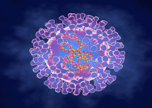
Monkeypox virus belongs to an enveloped double stranded DNA virus of the genus Orthopoxvirus in the family Poxviridae and belongs to a subfamily of Chordopoxvirinae. In the central and west African jungle, the monkeys are the natural host and this was also documented in squirrels, prairie dogs, rats, dormouse and primates.

                                                 Artist's version of a monkeypox virus.

The first human illness was recorded in the Democratic Republic of Congo in 1970, just two years after Smallpox was eliminated there. From time to time local outbreaks were reported by the WHO. Cases were seen in central and west African countries when children were bitten by monkeys.

There are two subclasses of this virus – one in the Congo Basin virus which is more virulent and has a higher rate of transmission, and the other is in West African. At present both varieties are infecting the people of Cameroon.

The first reported case in the USA was in 1980. In 2022, the USA declared monkeypox a public health emergency. Human to human transmission was seen in NYC in the homosexual community. Infection of humans takes place through the exchange of body fluids, respiratory droplets, prolonged close contact, and occasionally by the use of contaminated towels and bed sheets.

The incubation period is 5 to 21 days. The initial symptoms of malaise, body aches, fever and lymphadenopathy for 4 -5 days, then the rashes appear on the face and extremities, including on the palms and soles. In homosexual individuals, rashes are also present in the genital and anal areas. In contrast to other varieties of pox, monkeypox patients show striking lymphadenopathy.

Pox is known to appear on the lips and mouth, the conjunctiva of the eyes. The rashes go through the stages of macules, papules, vesicles and pustules. In three weeks, the scabs of pustules dry out and then drop off. Until all scabs fall, the patient remains a source of infection. The monkeypox illness is a mild disease but in immune deficient people, it can produce severe illness. In African countries as high as 10 % of the population is infected, and the fatality is generally below 3 %, and in children, the mortality has gone up to 6 %.

Diagnosis of monkeypox is done on samples obtained from the skin of the lesions or the fluid of vesicle/pustule by PCR test to confirm the monkeypox virus. Blood tests are not reliable and other varieties of pox produce the same serology test results.

Monkeypox vaccines.

Two vaccines are available in the US. In post exposure two doses of the Jynneos vaccine are given. Another vaccine ACAM2000 is less commonly given but available on a limited scale. In very high risk groups prior exposure vaccination is approved.

People vaccinated for smallpox have limited protection in high risk groups, for the general public provides 85 % protection against monkeypox.

Antiviral drugs.

In Europe, an antiviral drug, Tecovirimat, originally developed for the treatment of smallpox, is approved for monkeypox. In the USA, Tecovirimat is approved temporarily on an emergency basis and availability is limited.

*************************************

Cerebrospinal Fluid

                                              Cerebrospinal Fluid (CSF)

                                             PKGhatak, MD


The previously held view of the formation and circulation of the cerebrospinal fluid (CSF) has undergone modifications because current reproducible and non-interventional methods of investigation, like MRI and molecular and biological biology, discovered newer facts that contradicted the past theory.

To update the current thinking, a brief review of the histology of the Choroid plexus, Ventricles of the brain and Arachnoid granulations is necessary.


Choroid plexus.


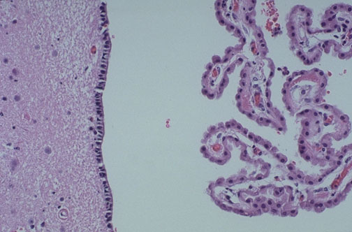
The Ependymal cells of the choroid plexus secrete the CSF by an active process. The central capillary of the choroid plexus is lined with one layer of fenestrated cuboidal cells. The albumin content of CSF is much lower than plasma. The transport of water across the ependymal cells of the carotid plexus is carried out by separate transport proteins, one for the ventricular side and the other for the capillary side. It is an active process that requires energy from ATP and likely also from K / Cl transporter. About 30 % of the total water volume of CSF comes from the inter-neuronal space, generated from the metabolic activities of the nerve cells, like Lymph formation in the other organs. There are to and fro moments of fluid between interstitial fluid and CSF.

Blood Brain Barrier.

This special characteristic of the blood vessels of the brain is due to the tight seal between the endothelial cells of the capillaries and supplemented by perivascular feet of astrocytes surround the basement membrane. In two locations this barrier is absent namely the choroid plexus and posterior pituitary gland.


Arachnoid Granulation:

Arachnoid granulations are collections of capillary networks kept suspended into the large veins of the subarachnoid space. The anatomical structure suggests arachnoid granulations are filters - a one way passage of CSF into the venous blood, getting rid of waste products and excess water. But that is not the case. Arachnoid granulation plays a minor role in this respect.

The perineural space along the cranial nerves and spinal nerves and the perivascular space known as Virchow Robin spaces are the main routes of drainage and filter of CSF into the general lymphatic system. This is specially evident in the Olfactory nerve as it travels through the cribriform plate of the nose.


Circulation of CSF:


The circulation of CSF in the brain and spinal cord.

The circulation of the CSF is an active process by the synchronous motion of cilia lining the surface of the ventricles and the central canal of the spinal cord. The CSF exits the brain through two openings of Luschka in the hindbrain and circulates in the subarachnoid space. The mechanism of a steady state of CSF formation, circulation and drainage has undergone many modifications. The circulation is pulsatile in nature due to and from movement of the CSF. The interstitial fluid exchange with CSF is a major component of circulation. A continuous bidirectional fluid exchange at the blood brain barrier produces flow rates. The general lymphatic system of the body is the main route of drainage of waste products of the brain and an entry point of the immune system in the brain.


******************************


Friday, October 21, 2022

What Cerebellum Does


                                           What Cerebellum Does

                                        PKGhatak, MD


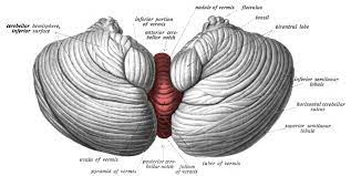
The human cerebellum occupies a small space at the back of the head, just above the neck, and it is a part of the hindbrain. Sensory information from joints, tendons and soft tissues from the limbs and trunk travel to the cerebellum. The motor stimuli coming from the cerebrum motor cortex, make connections with the nuclei of the cerebellum before reaching the lower motor nuclei of the spinal nerves and the cranial nerves. This interconnection of the motor cortex with the cerebellum results in a smooth sustained movement of the body, able to maintain the head position in space and in relation to the rest of the body and keep balance without falling. And that much knowledge people had till 1990.


Now the cerebellum is a subject of intense research for a variety of reasons.

Research in developmental biology documented three separate stages of growth of the cerebellum in humans. As the forebrain is added, the cerebral cortex, which has increased the mental faculties, knowledge gained about self, mind, higher state of consciousness, language, and development of culture has made humans as humans. But the cerebellum did not lag behind.

The growth of the cerebellum is more spectacular than the cerebral cortex. Cerebellum weighs 150 grams and contains 70 billion neurons, whereas the cerebral cortex weighs 1300 grams but contains only 16 billion neurons. The surface area of the cerebellar cortex is 1500 square centimeters, which is about 80 % of the surface area of the cerebral cortex. The cerebellar cortical folds are arranged like pages of a book - lots of surface area in a small space.


Compared with the cerebral cortex the layout of neurons and their connections in the cerebellum is much simpler and more uniform. Cerebellum receives input and sends out output to the same side of the body that is also a contrast with the cerebrum which controls the opposite side of the body. The central part of the cerebellum is old, but the lateral parts, the cerebellar hemispheres, kept pace with the growth of the cerebrum.

Cerebellum is the site of the formation and storage of working memory and the integration of automated sequences of thoughts and actions. This results in developing behavioral patterns, speech, dexterity of complex movements, a skill needed for technological advances and language development.

In association with the cerebrum, it forms kinetic memory, plans coordinated muscle movement patterns, retains reflex memory for self preservation and executes normal speech.

Cerebellum with two way communication with the thalamus and basal ganglia and receives direct input from the spinocerebellar tract and maintains balance and a stable head position both in states of body movements and at rest.

Vestibular sensory information is closely monitored by the cerebellum, that information is used to control conjugate movements of eyes, prevent double vision and reflex eye movements.

Rapid eye movement (REM) sleep includes Implicit memory involving perception and body movements. Implicit memory uses cerebellar retained memories.

*********************************


Tuesday, October 18, 2022

Science of Skin Color


                                            Science of Human skin color

                                           PKGhatak, MD


The skin is one of the body's large organs. The color of the skin prevents or minimizes the harmful effects of Ultra Violet (UV) light of the sun.

The darker the shade of color, the lesser amount of UV light penetrates the skin and decreases Vitamin D synthesis, the reverse is true for the lighter shades of color. It is said that women have lighter skin color because more vitamin D is needed for childbearing.

Not every part of the skin of an individual has the same shade of color and variability depends on the degree of exposure to sunlight. The sole of the feet and palms of the hands have different thicknesses of skin and a much lighter color, and that lighter shade is most noticeable in very dark colored people.

Theory of color variation in humans.

Scientific communities believe that the human race originated in equatorial Africa. Having dark skin gave humans an advantage over the other predators to hunt for a longer time each day. Because the black skin color developed as the coarse hairs were replaced by fine body hair. This combination allowed the body to avoid overheating. That advantage led to a population boom and subsequent migration of people to other lands. When people settled in the subtropical area, their dark skin turned to a disadvantage; the lack of calcium produced weaker siblings, and most did not survive beyond their early childhood. A genetic mutation made the color of the skin lighter shades which allowed more UV light to penetrate the skin and generate more vitamin D, and the population bounced back.

The so called white colored skin is the color of the pale white color of the subcutaneous tissue shining through the light-colored skin.

What are the color pigments of human skin:

Melanin is the primary color and minor color contributions come from hemoglobin, carotenoids and oxyhemoglobin.

Perception of color:

The combination of colors of melanin, oxyhemoglobin (proportional to blood flow) and carcinoids produce the final color perceived by the observer's retina.

Melanin:

Melanin is a group of chemicals very closely resemble each other in molecular structure. The skin Melanin in humans is Eumelanin and Pheomelanin. Neuromelanin is present in the brain.

Eumelanin and Pheomelanin are the same chemical but differ only in the number of polymer bonds in them.

Two colors of melanin:

Eumelanin is a dark pigment, has a range from brown to black, and Pheomelanin has a shade between yellow to pink color. The pink color of the lips is due to pheomelanin.


Eumelanin synthesis.

Amino acid Tyrosine is converted to Dihydroxyphenylalanine (DOPA) by the enzyme Tyrosine hydroxylase and in the presence of cofactor Tetrahydrobiopterin. DOPA is converted to Dopaquinone by Tyrosinase and then undergoes several steps of polymerization to form eumelanin or pheomelanin.

Site of melanin production:

Melanin is produced by the Melanocytes present in the basal layer of the skin. The pigment granules are nicely packed in an envelope and are called melanosomes. Melanosomes are carried along the long arm of the melanocyte and the pigment packets are deposited within the Granular cells of the keratinocyte layer. One melanocyte and 36 granular cells and one Langerhans cell make one keratinocyte unit. It is estimated one melanocyte can supply 500 granular cells with melanosomes in its lifetime.

Stimuli for melanin formation:

UV radiation stimulates the synthesis of melanin. A hormone, produced by the Pars Intermedia of the Pituitary gland, Pro-Opiomelanocortin, is turned into Alpha Menaocytotropic hormone (alpha MSH). UV light also increases ACTH (adrenocorticotropic hormone) secretion. MSH and ACTH increase melanin production. Androgen also influences melanin deposit of pigment in the form of male patterns.

Genes controlling Melanin.

In humans chromosome 11 is the primary chromosome and three additional chromosomes, 17, 6 and 13 have also influence melanin. The location of the genes on these chromosomes are 11q24.2, 17q23.2, 6q25.1 and 13q33.2.

Defective genes and abnormal skin color:

1. Albinism.  It is a Hereditary disease due to an inheritance of defective genes. Skin, hair, retina of the eyes are devoid of melanin and is known as Oculocutaneous albinism (OCA). It is inherited as an autosomal recessive trait. At present 7 varieties of OCA have been identified.

2. Hermansky-Pudlak Syndrome.  Hermansky-Pudlak patients lack skin pigment and in addition, have a tendency to bruise easily and bleed spontaneously. Patients have additional symptoms which may involve GI, Pulmonary and CNS systems. So far 11 gene mutations have been identified. This abnormality is also inherited as an autosomal recessive pattern.

3. Chediak-Higashi syndrome.  It is an autosomal recessive hereditary disorder consists of partial OCA, platelet disorder, immune deficiency due to abnormal Lysosomes and in addition to hematophagy lymphohistiocytosis syndrome

4. There are many other syndromes due to inherited defective genes. Symptoms involve various organ systems and skin color. Just to name a few – Acanthosis nigricans, Xeroderma pigmentosa, Treitz syndrome, Histiocytosis lymphadenopathy plus syndrome, Lengius syndrome, Cruzion syndrome, Incontigentia pigmenti, etc.

5. Loss of melanocytes.  Skin abrasions may peel off the basal layer of skin containing the Melanocytes, when skin lesion heals the area is devoid of skin color.

5. Autoimmune diseases. Some acquired autoimmune diseases may attack melanocytes producing pigment voided spaces.

6. Vitiligo. It is suspected to be an auto-immune disease. The color free areas are patchy and can be any spot on the skin. No genetic link has so far been documented.

7. Localized dark pigmentation on skin:

Freckles, moles, Mongolian spots on buttocks, Caffe au lait spots, and Birthmarks are examples of uneven distribution of melanin in keratinocytes. Some of these spots are transient, and others disappear and reappear later in life. Caffe au lait spots, if multiple, are associated with neurofibromatosis.

All these are benign conditions unless one starts to grow in size and becomes darker or bleeds.

 8. Melasma. Symmetrical dark patches on the face due to estrogen – progesterone concentration variation, as seen in pregnancy and menopause. Melasma has no health consequences and is important from the cosmetic point of view.

Carotenoids:

Carotenoids are Terpenoids having formula C40H56. Carotenoids are plant pigments and humans must depend on plant sources for carotenoid supply. Yellow, orange, or red vegetables and fruits contain carotenoids. They are fat soluble chemicals; animal fat is also a source like Salmon. Cooking shredded vegetables in oil increases the bioavailability of carotenoids. In humans, these molecules are antioxidants, boost immunity and provitamin A. Vitamin A is an essential vitamin for vision.

An essential organ:

The skin is an essential organ. The skin protects the body from the physical mechanical and thermal injury, prevents harmful agents from getting inside, maintains body temperature, synthesizes vitamin D, performs immune functions and the sensory receptors of pain, touch, temperature and pressure are located in the skin. Melanin in the skin protects tissues from the radiation damage of the sun.

All 8 billion people:

The color of the skin comes from one pigment - Melanin. All 8 billion people on earth have the same melanin. Color variation of the skin is due to the variation of melanin content in the skin cells.

People seeking advantage and try to divide human families into many color groups, like India did through the caste system. Businesses interested in selling their products claim about 110 tones of skin colors and have marketed matching colored products for sale.

Science has steadfastly maintained the scientific data which anyone can verify and as it stands today, all humans carry the same skin pigment melanin.

The difference in skin color is only in the view of the observer.

******************************

Sunday, October 16, 2022

Speech Disorder


                                                            Speech disorder

                                                    PKGhatak, MD


Speech is defined as the ability to express thoughts and feelings by articulated sounds. Language is in the same domain as speech but not the same. Language is the medium of communication with said words and symbols. 

1. Speech disorder in children is primarily developmental and congenital, after that comes others.

2. Developmental delay presents as uncoordinated brain function, manifest as Stuttering. 

3. The next group comprises Autism, dyslexia and cerebral palsy.

4. Birth defects such as tongue-tie, defects of the floor of the mouth, hare-lip and cleft palate and occiput occipito-facial abnormality. Many of them are surgically correctable.

 5. Apraxia and dysarthria are apparently similar defects but in apraxia, the brain fails to formulate a motor plan to supply nerve impulses to muscles of the larynx and vocal cords; in Dysarthria the muscles of the mouth and tongue fail to move properly due to mechanical and /or coordination abnormalities and fails to produce a normal speech.

 6. Congenital deafness or onset of early childhood deafness, leads to absent speech known as muteness. However, the mute babies did cry at the time of birth proving that absent speech in these babies comes from deafness.

Speech disorder in adults.

Speech problems in adults are always acquired after having normal speech for years.

There are three categories of this disorder - I. Transient and reversible. II. Secondary. III. Primary.

 I. Transient:

Alcohol intoxication is the most common cause of stuttering, fumbling for words and inappropriate answers. Opioid addiction and use of recreational drugs, tranquilizers, and sleeping pills are additional causes.

II. Secondary:

Multiple sclerosis (MS) produces various neurological deficits including speech. One special feature of MS is the exacerbation and remission of symptoms. ALS, Ascending paralysis, Myasthenia gravis, and myopathy of various types. and diseases involving the larynx, vocal cords and paralysis of the recurrent laryngeal nerve are further examples of speech defects.

III. Primary:

The brain is the center for speech formation (ideation), generation of motor signals and transmission of information to muscles involved in the execution and articulation of spoken words. Any disruption of the flow of information to the effector organ/tissues will adversely affect speech. Some of the pathological causes are interruption of blood supply, pressure from space occupying lesions, inflammation and infection of the brain, and traumatic injury to speech centers.

Ideation:   One can easily recall teachers telling “engage your brain before you blur out words”. That is the ideation – the speaker has instant knowledge of what he is going to say in a conversation or at the podium before a lecture. Ideation is the result of coordinated activities of many brain centers, including visual, auditory, and memory centers, which are located in the different areas of the cerebral cortex.

Motor function:   Cerebral motor center is located in the Pyramidal cortex of the brain. The axons from pyramidal neurons make synaptic connections to the lower motor neuron of the motor nuclei of cranial nerves and anterior horn cells of the spinal cord. Nerve impulses are carried to the muscles of the larynx, vocal cords, throat, lips, mouth, tongue, diaphragm and respiratory muscles of the chest wall transmitted by the axons of the lower motor neurons.

Monitoring and modification of speech are active processes and require communication via synaptic relays between various brain centers.

Localization of lesion:

At an earlier time, every speech abnormality was documented by neurological examination and then verified by autopsy. The medical practice of that time required memorizing named syndromes to identify each speech defect. Now neuroimaging by functional MRI and CT, with and without contrast, have made the diagnosis more refined and accurate at the same time names attached to lesions are not required to be remembered.

Types of speech abnormalities in adults.

 1. Broca's Aphasia.

Broca's aphasia is both a language and word disorder due to damage to Broca's area of the brain. Broca's area is a special speech center located in the left frontal lobe of the cerebral cortex. It is situated in the depth of the main horizontal fissure dividing the cortex into frontal and middle lobes and partially covered by the tip of the left temporal lobe. Anatomically, it is located in the inferior frontal gyrus. Blood supply of the Broca's area comes from the branch of the middle cerebral artery. Interruption of the blood supply produces a language and speech disorder known as Broca's Aphasia.

In Broca's Aphasia, the patient retains the capacity of Ideation but loses language due to partial loss of capacity to construct either the spoken or written language. Broca's aphasia has two components – Expressive aphasia and Receptive aphasia. In expressive aphasia, the patient is able to say important words but leaves out prepositions, pronouns and functional words. Speech becomes piecemeal, comes out in sprats and stops, says wrong words, missing the word orders. This type of speech is known as Telegraphic speech (at one time the telegram was the fastest mode of communication, but expensive and the charges were based on the number of words to be transmitted. That leads to cutting out words). In Broca's aphasia, the patient is cognizant of his disability and becomes visibly frustrated in his attempt to find his speech back.

In Receptive aphasia, the patient can speak in grammatical sentences but lacks semantic significance and exhibits trouble in comprehension.

Thrombosis or emboli obstruction of the Left middle cerebral artery produces Right sided paralysis including the right lower half of the face and loss of sensation from the right side of the body and loss of right visual field and Broca's aphasia.

 2. Wernicke's area. Werneck's area is located in the parietal cortex next to the lateral sulcus at the junction of the parietal and occipital lobes. In this area, the visual and auditory words are comprehended and an area for selecting words and phrases to construct a meaningful sentence. Patients with damaged Wernicke's area, from alcoholism and other diseases, produce articulated sentences but many lack any meaning. And some patients may speak fluently and are not aware they are uttering meaningless words. Alcoholism may lead to Korsakoff psychosis. During an episode of Korsakoff psychosis, the person speaks freely but confabulates and tells amazing stories.

3. Aphonia:

A lack of ability to produce any sound is known as aphonia. Direct injury to vocal cords, tracheostomy and bilateral recurrent laryngeal nerve damage can produce aphonia. And in addition, there are instances when a person is about to give an important talk and gets up to the podium and then is unable to make any sound. Some psychological or emotional neuronal activities overwhelm the natural order of speech making process.

4. Apraxia and Dysarthria are common in stroke and other lesions of the brain involving speech forming areas.

*************************

Thursday, October 13, 2022

Abnormal Red Cell Morphology and its Significance


                     Abnormal Red Blood Cell Morphology and its Significance

                                           PKGhatak, MD


Blood is known as the vital fluid and that characteristic belongs to the Red Blood Cells (RBCs). The hemoglobin, contained in the RBC, avidly combines with Carbon dioxide, a waste and toxic product of cell metabolic activities; and the RBC exchanges it for Oxygen in the lungs and carries oxygen to every living cell of the body (except the cornea).

RBC is routinely tested for - size, shape, color, volume, uniformity of distribution and for any structural anomalies; and all these are reported when a CBC (complete blood count) is ordered.

No other cell in the body has been studied that thoroughly.

A normal red cell (RBC) is a disc shaped and biconcave cell without a nucleus and inclusion bodies. The red color of the cell is due to the presence of oxyhemoglobin. RBCs appear brilliant ruby color when allowed to settle in a test tube filled with normal saline.

RBCs are 7 to 8 cu mm. in diameter, the central 1/3 is paler than the periphery. Normal RBC volume (MCV) is 80 -95 fL, and the normal number of RBCs is about 5 million per cu mm. The RBC shape is fairly inform - the RWD (red cell width distribution) is 11 to 15 %.

RBCs are formed in the bone marrow from the stem cells of the Myeloid line; Erythropoietin, a hormone produced in kidneys, stimulates RBC production. In circulation, RBCs survive for 100 – 120 days. Every part of the RBC is recycled by the spleen and other scavenger cells.

Size: Abnormal size is called Anisocytosis.

Normal size RBC is called Normocyte, Microcyte is < 7 cu mm (MCV<80 fL) and Macrocyte is over 8 cu mm (MCV >95).

When Anemia is detected, the finding is reported as either normocytic, microcytic or macrocytic anemia. Normocytic anemia is seen after a recent blood loss, like an accident, fracture of long bones, or surgery. Microcytic anemia is due to iron deficiency, thalassemia, chronic inflammation, slow GI blood loss due to cancers or bleeding duodenal/gastric ulcers and sideroblastic anemia. Macrocytic anemia is seen in Pernicious anemia, Folate and Vitamin B12 deficiencies, Steatorrhea, Myelodysplastic syndrome, Liver cirrhosis, Alcoholism and Drug- use of Hydroxyurea.

Shape: Abnormal shape is called Poikilocytosis. The abnormal shape could be Spiculated or Non-Spiculated.

Spiculated RBCs are called Burr cells, broken RBCs are known as Schistocytes resemble Napoleon hat), Helmet cells, Acanthotic cells and Dacrocytes (tear drop shaped) cells.

Non-spiculated RBC are Target cells, Ovalocytes and Stomatocytes, Pencil cells, Bite cells, Tear Drop deformities, Spherocytes and Elliptocytes.

Burr cells are seen in renal failure.

Ovalocytes and Elliptocytes are due to Hereditary causes; in addition, spherocytes are also seen in severe burns, ABO incomparable blood transfusion and acquired idiopathic hemolytic anemia.

Target cells are common in sickle cell hemoglobinopathies, thalassemia and iron deficiency anemia.

Bite cells are seen in Chronic Obstructive Pulmonary Diseases (COPD).

Stomatocytes (fish mouth like) are common in Liver disease, alcoholism, Rh-null disease and COPD.

Teardrop deformity cells are present in Myelofibrosis and pernicious anemia.

Helmet cells, Broken cells or Schistocytes in the peripheral blood are ominous signs. These cells in the blood indicate active hemolysis. Usually one of the following conditions is responsible – Decimated intravascular coagulation, TTP (thrombotic thrombocytopenic Purpura)and Thrombotic macroangiopathic anemia.

Color:

The red color of RBCs is proportional to oxyhemoglobin concentration in the cells. Based on the hemoglobin concentration per red cell, the cells are called Monochromic, Hypochromic or Hyperchromic. When color is not uniform, it is called Anisochromatic and when more than one color is present, it is called Polychromatic RBCs. Iron deficiency is the primary cause of hypochromia, and hyperchromatic is seen in sickle cell disease. Macrocytes in severe liver disease and Thalassemia, the RBCs can also be Hyperchromatic, this combination is called Leptocytes.

Increased turnover of RBC in the bone marrow results in the presence of immature RBC in the blood, these young cells retain some remnants of RNA and appear bluish when stained and laid out as a fine network called Reticulocytes. In normal conditions, the reticulocyte count is between 0.5 and 2 %. Reticulocytes over 2 % are seen in acute hemolytic anemia, massive hemorrhage, and in high altitude visitors.

When bone marrow is infiltrated, as in cancer metastasis and leukemia, Nucleated RBCs are visible in the blood and are called Erythroblasts. In times of stress,  the bone marrow also releases nucleated RBCs, e.g., hypoxemia, and removal of the spleen.

Inclusion Bodies:

Inclusion bodies are iron pigment, hemoglobin, remnants of DNA and RNA fragments in the red cell cytoplasm. Inclusion bodies are called Basophilic Striplings and Howell jolly bodies.

Basophilic stippling. These inclusion bodies represent defects in hemoglobin synthesis, appear as bluish stripes, widely distributed throughout the cell cytoplasm. Examples – Megaloblastic anemia, Post splenectomy and sideroblastic anemia.

Howell Jolly bodies. These bodies appear in the periphery of the cell as purple round dots when stained with Romanowsky stain due to the presence of fragments of DNA.

Siderotic granules. These granules are iron, which remains in hemoglobin and stains blue. Example: Sideroblastic anemia.

Parasites.

The malaria parasite belongs to the Plasmodium group. Several species of Plasmodium infect humans - mostly in people living in Sub-Saharan Africa and Southeastern Asia. As there are different species, several parasitic forms, like gametocytes, schizonts and trophozoites, are also seen in the blood of patients infected with malaria. Considerable experience is required to identify these intracellular malaria parasites.

Babesiosis. Babesia infection is seen in the New England States in summer months when tics are active. The parasites appear as ring forms, often in groups, and in heavy infestation rings in groups appear outside the red cells also.


Blood film in Babesiosis.

Important Points:

To a casual reader, this article may appear confusing and overlapping.

In clinical practice, however, the information is neatly and concisely presented. A few samples are presented.

Iron deficiency anemia.

Iron deficiency produces Hypochromic, Microcytic anemia. Besides a low hemoglobin, HCT (hematocrit), MCV (means corpuscular volume), MCH (mean corpuscular hemoglobin), Color index and Saturation Index are all low. RWD (red cell width distribution) is more than 15 %. This RWD finding is helpful to separate iron deficiency anemia from Thalassemia where RWD remains normal.

Macrocytic anemia.

A combination of low hemoglobin and low HCT in the presence of High MVH and MCHC (mean corpuscular hemoglobin concentration) is the rule.

Hemolytic anemia.

Intravascular hemolysis is a vast subject as such. Features of hemolytic anemia is the presence of normocytic and normochromic anemia with a normal RWD. If the hemolysis is less severe but recurrent then RWD rises above normal.

Sickle cell disease (SCD). In Benin, Senegal, and CAR (in sub-Saharan countries) death due to childhood malaria caused havoc. A mutation of the Beta Chain of the hemoglobin molecule at position 6 - substitution of amino acid glutamine to valine proved to be a lifesaver. That advantage led to a wide distribution of the mutated gene in the population. However, as people moved to the USA and Caribbean States from sub-Saharan Africa, that mutation has become a fatal mistake. In a low oxygen environment, the RBC of SCD patients changes from a disc to sickle form and obstructs blood flow in the capillaries and results in severe pain and loss of function. This results in death of cells, seen from the bone marrow to the brain.


                                Blood film in Sickle disease.

Thalassemia.

 A growing embryo in the mother's womb draws oxygen, due to the special characteristic of Fetal Hemoglobin. If fetal hemoglobin persists in postnatal life, due to a gene mutation, it is called Thalassemia. Thalassemic patients have anemia which has the following characteristics - Microcytic, hypochromic anemia with a high RBC count, normal MCV, normal RWD, high reticulocyte count, basophilic striplings and Target cells.

                                       Peripheral blood film in Thalassemia.

TTP (thrombotic thrombocytopenic purpura).

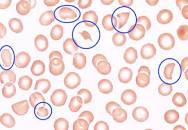
TTP is due to widespread platelet thrombosis in all organs producing an alarming medical situation. Hemolytic anemia due to mechanical break of RBC in their attempt to move forward in capillaries partially blocked by platelet thrombi, and a very low platelet count (10,000), the presence of schistocytes are characteristic features. Direct examination of blood film by hematologists can make a quick diagnosis. 


                             A blood slide in TTP, the helmet cells are marked by blue 
                                    circle.

Napoleon hat


*******************************

Monday, October 10, 2022

Eosinophil and Diseases associated with Eosinophils


                           Eosinophils and Diseases Associated with Eosinophils.

                                          PKGhatak, MD


Eosinophils are white blood cells that belong to granulocytes, other members of this group are Basophils and Neutrophils (also called polymorphs). The name, granulocyte, is derived from the presence of granules in the cell cytoplasm. The granules of eosinophils are coarse and turn red when stained with Eosin due to the presence of amino acids arginine and lysine in the protein, which reacts with Bromine in Eosin. The eosinophils are important Innate Immunocytes, involved in protecting the body from multicellular parasites, fungi, and some viruses, and are important determinants in certain diseases.

The easily identifiable feature of the eosinophils is their special characteristic, a Bilobed nucleus. 

Formation and distribution of Eosinophils.

The Hematopoietic stem cells of the bone marrow are the source of eosinophils; and in about 7 days the mature eosinophils leave the bone marrow. Most of them head for the thymus gland, spleen, lymph nodes, GI tract and the rest of the organs with the exception of the esophagus, ovaries, and uterus. Under normal health conditions, the eosinophils are not present in the Esophagus and Lungs and only a few eosinophils are present in the skin.

The Thymic Th2 cells initiate eosinophil production by secreting IL-5. (Interleukin-5).

Normal blood eosinophil count is 3 to 8 % and the total maximum count is 500 per microliter. In the peripheral blood, the eosinophils live about 1/2 days and in the tissues, they survive 8 days.

A total eosinophil count of 800 / microliter is abnormal and called hypereosinophilia, a count over 1,500 per microliter is called Hyper-eosinophilic syndrome.

There are very few conditions where the eosinophil blood counts are low. One is usually encountered when corticosteroid is administered. In Alcohol intoxication and Cushing's disease low eosinophil count are seen.

Normal functions of Eosinophil.

Eosinophils are immunocytes that act by releasing preformed substances, contained in the cytoplasmic granules. The pathophysiology of diseases is different from disease to disease. The substances carried in the granules can be grouped into – Oxygen radicals, Enzymes, Leukotrienes, Cytokines, Prostaglandins and Growth factors. In previous blogs, oxygen radicals, leukotrienes, prostaglandins and cytokines are discussed in detail and will not be repeated here.

Growth factors are TGF beta (tissue growth factor), VEGF beta (vascular endothelial growth factor), PDGE (platelet derived growth factor). In addition, the Eosinophils promote the development of post pubertal breasts, influence menstrual cycles and protect tissues from cancerous developments, prevent allograft rejection and help antigen presentation to T-Cells.

                               Hypereosinophilia

Herpesinophilia is seen in these conditions – Allergy, Asthma, Roundworm infestation of the gut, and Cutaneous larval migration like Filaria, Trichinosis, Cysticercosis. Eczema and Atrophic dermatitis, Chung-Strauss syndrome, Autoimmune diseases, Crohn's disease, Addison disease, Cancers of breasts, ovaries, and prostate. Leukemia and among the group of leukemias - Acute myelogenous leukemia (AML) and Eosinophilic leukemia are particularly striking. It is also seen in certain Fungal infections. Many prescribed medications produce allergic dermatitis and Penicillin leads the group; Penicillin may also produce Interstitial nephritis, which is characterized by the presence of eosinophils in the urine.

                                 Hypereosinophilic syndrome

When peripheral blood eosinophil count is persistently over 1,500 mcL for 6 months or longer and evidence of organ damage and allergy, parasitic infection and chromosomal abnormality are eliminated, the condition is called Hyper-eosinophilic syndrome (HES).

When the cause of high eosinophilia is known, the term Secondary Hypereosinophilic Syndrome is applied and many of the old HES are now classified under Secondary HES.

Chromosomal abnormality in hypereosinophilia.

The translocation of the gene between F1P1L1 and PDGFRA of the long arm of chromosome 4, at location 12 (4q12) produces a hybrid gene (F/P) which produces excess IL-5 causing rapid and excessive eosinophil production. Many other less prevalent Myeloid dysfunction due to chromosomal abnormalities are known, some examples are Chronic Eosinophilic Leukemia, Lymphatic HES, and Myeloproliferative HES.

Organs damaged by HES.

Esophagus, Stomach and intestine, Skin, Lungs and Heart. Multiple organs involved are the usual but only one organ or one system may be involved.

Clinical manifestation of HES.

Presentation and clinical features vary due to different organ /system involvement. Incidence in the USA is 0.3 to 6.0 per 100,000 population, including secondary EHS. General- Males are more affected by HES and ages between 20 and 50 are mostly affected. Systemic symptoms are fever, loss of weight, weakness. lethargy, pain in joints and muscles, and night sweats. Skin manifestations are itchy macular and nodular eruptions, angioedema and eczema. GI symptoms are heartburn, abdominal pain and diarrhea. Heart problems may mimic MI, endomyocardial necrosis, peripheral emboli and cardiomyopathy. Lung lesions resemble an attack of asthma, pulmonary infiltration and interstitial pneumonia and fibrosis. Unproductive cough. Hematological manifestations are many, chief among them are anemia, enlarged spleen and liver, and fibrosis of bone marrow. High serum B12 level. CNS manifestations are peripheral neuropathy, Transient ischemic attacks, strokes, and encephalopathy.

Diagnosis requires a complete blood count, serum B12 level, bone marrow biopsy and chromosome study.

Treatment of HES.

Intravenous corticosteroid is used to bring down the Eosinophil count rapidly, and Hydroxyurea is also used. Imatinib, a Tyrosine kinase inhibitor, is effective. Various other small molecules are available to suppress IL-5 production.

The prognosis is unfavorable without treatment. At present, with prompt treatment, the longevity on average is 80% in 5 years.

*************************************


Archive

  Archive. P.K.Ghatak, MD 2025  March. Laser Endoscpoe 2025 February. Use of Ultrasound in Medicine MRI 2025 January. Bacteriology and Rober...