Wednesday, April 5, 2023

Looking for Essentials Amino Acids.

                                  Searching for Essential Amino Acids.

                                     PKGhatak, MD


Essential amino acids for humans are 9 in number, namely: Histidine, Isoleucine, Leucine, Lysine, Methionine. Phenylalanine, Threonine, Tryptophan and Valine. Essential amino acids must be supplied in the food because our body can not synthesize them from other sources. Our body, however, can synthesize these non–essential amino acids, and common among them are Glycine, Alanine, Serine, Cysteine, Arginine, Aspartic acid, Glutamic acid, Arginine, Lysine, Tyrosine, Proline and Homocysteine. Amino acids are the main building blocks of protein molecules.

The human body is composed of 62 % water, 16% fat, 16 % proteins, 6 % minerals, 1 % carbohydrates and traces of vitamins and trace elements.

The function of Amino acids.

Both essential and nonessential amino acids are constituents of every cell and tissue of our body- from hair to the bone marrow cells and in between everything else. In addition, amino acids are required for growth, tissue repairs, immunity, hormone production and metabolic functions.

A few specific functions of individual amino acids are highlighted in the following paragraph.

Histidine - Blood cell formation.

Isoleucine - Increases growth hormone production. Skin and bone repairs.

Lysine - Calcium absorption, Collagen formation of tendons, cartilages, and skin.

Methionine - Antioxidant. Detoxify heavy metals.

Phenylalanine – Formation of the neurotransmitter, calcitonin and melanin.  and also the source of Tyrosine.

Threonine - Formation of elastin, the enamel of the tooth and mucin production from glands. Glucose metabolism.

Tryptophan - Formation of serotonin and melatonin, DNA repairs, Niacin    ( vitamin B3).

Valine – Muscle growth and health.

Are humans omnivorous.

No other living organism exists today that comes close to humans in choosing what to eat—from insects to the most poisonous puffer fish and snakes.

According to the WHO in the year 2021, a family of four in the USA consumed. 800 lbs of meat per month. 70 lbs of chicken per person per month was eaten during that time.

350 million tons of meat were consumed globally every year, of which pork was number one. China produced and consumed the most pork, and the USA came in third place. The USA is the largest consumer of beef and India came in 5th place; that must surprise some people. Fish consumption is highest per capita in Iceland, at about 200 lbs per year, the USA came 11th place by using 50 lbs per capita and still managed the second place by eating 290 eggs per person, only Japan consumed more, about 320 eggs per person.

The current trend in the USA.

In recent years, a vegetarian diet has gained popularity among the health and environment-conscious sections of the wealthier nations. Various degrees of vegetarianism exist. True vegetarians or vegans do not eat any meat or fish and avoid eggs or milk altogether. In India, where a major section of the population has been vegetarian for centuries, however, they consume milk from the cow, buffalo, goat and a few isolated tribes drink milk from camels. Some other Indians consider themselves vegetarian, but eat eggs and some also eat fish,  but not any animal meat.

Whatever form of vegetarian one may be, the main source of protein in their diet comes from plants. And most green parts of plants are not rich in proteins, the seeds of plants are. To meet the demands of plant seeds that are rich in proteins, more selective cultivation is required. It is becoming an important agricultural consideration for meeting this growing demand but must be met in sustainable and environmentally protective ways.

Protein requirement.

A growing child in utero needs 925 grams of protein and the mother supplies that amount during pregnancy. During lactation, an additional 1.3 grams of proteins per 100 ml of milk is needed.

Adults require 0.8 grams/Kg body weight ( stand height/weight). The elderly actually need less but defects in digestion and absorption are considered and the recommendation is like an adult - 0.8 g/Kg.

Obligatory Nitrogen Loss.

Protein turnover in the body is a continuous process of synthesis, breakdown and elimination of toxic nitrogenous waste products. When placed in a protein free but calorie sufficient diet, the body extracts the essential amino acids by breaking down proteins. This system is very efficient but the nitrogenous portion that is eliminated in urine and stool must be supplied in the food.

Effects of protein starvation.

Pictures of emaciated people ravaged by wars or severe famine need no further explanation. A short stature of a growing child, thin limbs, a pot belly, edema, fragile skin and orange hairs are the results of protein deficiency. Blood levels of albumin and hormones are low in these children. In nephrotic syndrome and severe liver cirrhosis, frequent infections and various complications are the results of derailed protein metabolic machinery of the body. Specific amino acid deficiency can occur due to inherited metabolic defects and the consequences of those can be found elsewhere.

Plant Proteins.

Many plant proteins are not available to humans due to the complex nature of the molecules, which are not easily digested or absorbed in the gut. Some plant proteins are poisonous, like in cassava.

Good source of vegetable proteins.

The high plant protein content is given here in descending order. It is the total amount of available protein but not in terms of essential amino content. The list of plant sources of proteins: Durham wheat, cashew nuts, quinoa seeds and ancient grains, dried seaweeds, rice, pumpkin seeds, beans, peas and raw soya beans.

Some individual plants with some good sources of essential amino acids are listed as -

Durham wheat supplies a fair amount of histidine, phenylalanine, tryptophan, valine and threonine.

Cashew nuts are a good source of phenylalanine, valine, leucine, lysine, Isoleucine and methionine.

Pumpkin seeds supply phenylalanine, tryptophan, Isoleucine, leucine and threonine.

Quinoa and ancient grains. Quinoa contains all 9 essential amino acids. Several other ancient grains are also good sources of proteins, chief among them are barley, farrow, buckwheat, millet, sorghum, kamut and teff. Each one has many other health benefits. Quinoa is an annual herb that belongs to the Chenopodium family. The grain contains 8 grams of protein per cup of cooked grain and is highly sought after because it supplies all nine essential amino acids. The protein of quinoa is 11s-globulin and contains no gluten. In addition, quinoa supplies unsaturated fatty acids, flavonoids, vitamins and minerals.

The rest of this group of grains contain more or less the same amount of proteins and are good sources of essential amino acids, but the amount varies from one to the other. All are good sources of micronutrients and antioxidants.

Soya beans have a good amount of leucine, lysine and phenylalanine.

Seaweeds are rich in histidine, leucine and lysine.

Peas and beans also supply fair amounts of phenylalanine, valine and threonine.

The world is always changing, and the rate of change has accelerated greatly in the last two or three centuries. The younger people of the present generation are much more concerned with animal sources of protein food along with other climate concerns. Eating proteins from nonanimal sources has come to attention lately from the processed food industries. This may be good or bad, only the future will tell.

Observing the effects of mass tourism causing accelerated degradation of the environment, like what has been done to Vienna and the Galapagos islands, should ring an alarm bell about this mass movement. No one can predict what that would do to the forested land in order to meet the demand for plant protein for human consumption.

*********************************************


Tuesday, March 28, 2023

Cholesterol and Lipoprotein

                                               Cholesterol and Lipoprotein.

                                              PKGhatak, MD

Cholesterol is a lipid. Lipids are a group of heterogeneous compounds of fatty acids and their derivatives which are not soluble in water and soluble in chloroform, ether and benzene. Oil, fat, wax and steroids belong to this group.

Protein when conjugated with lipids is called a lipoprotein.

Apo-lipoprotein:

Individual protein molecule contains a prosthetic group or a metal that gives it its special characteristic. When the prosthetic group or metal part is absent, then the protein is designated as Apo-protein. When a lipid molecule conjugates with an Apo-protein and then the conjugate combines with lipoprotein, the resulting molecule is called an Apolipoprotein.

Lipid molecules are wrapped around by proteins in order to make them soluble in the blood, making the transport of cholesterol between tissues possible. 

A diagram illustrates the makeup of lipoprotein molecules.


Chemistry of Cholesterol.

Cholesterol is designated as 3-hydroxy-5,6 cholestene.


The cholesterol molecule has a Sterol ring of 19 carbon structure and at position C-17 a side chain is attached, as illustrated in the above diagram.

 The rings A, B and C constitute a phenanthrene ring to which a cyclopentene ring D is attached. (this is cycleopenteno- perhydrophenerthrine ring or sterol ring) The double bond between position C5 and C6 gives the molecule its stability.

Source of cholesterol in humans.

Every living cell of the body can synthesize cholesterol, however, the liver is the primary organ where cholesterol is synthesized, in addition, the adrenal cortex, testis, ovaries, skin, aorta and intestine also produce cholesterol. Dietary sources supply only 15 % of cholesterol. Organ meat, egg yolk, brain and liver are good sources of cholesterol in food. The body recycles cholesterol and excess cholesterol is excreted by the liver in the bile as bile acids and neutral cholesterol. Some bile acids are reabsorbed with fat in the intestine and the rest is excreted in the stool.

The liver synthesizes about 1 gm of cholesterol a day and about 300 mg of cholesterol is obtained from food. Each 100 mg of cholesterol ingested will raise blood cholesterol by 5 mg/dL.

Synthesis of cholesterol.

The cholesterol is synthesized in the Endoplasmic Reticulum of the cytosol of the cell. Mitochondria of the cells generate energy via the Tricarboxylic acid cycle, and Acetyl CoA molecules generated in this process are available for cholesterol synthesis. Fatty acids, ketogenic amino acids and glucose are broken down to  2-carbon, Acetyl CoA molecules, and enter the tricarboxylic acid cycle.

All carbon atoms in a cholesterol molecule are obtained from Acetyl-CoA. Two molecules of Acetyl-CoA condense and form Acetoacetyl CoA by the action of the enzyme Thiolase. In the next stage, one molecule of acetoacetyl CoA condenses with another molecule of acetyl CoA forming one molecule of Beta-hydroxy-beta-methylglutaryl CoA. This synthesis is catalyzed by a rate-limiting enzyme HMG-CoA synthase (hydroxy-methylglutaryl Co-A synthase). Beta-hydroxy-beta-methylglutaryl is reduced by HGM-CoA reductase and NADPH acts as an H ion donor. In the next step, mevalonate is phosphorylated by ATP, and several steps later, condensation of several molecules of isopentyl takes place and Farnesyl pyrophosphate is generated.

Further downstream, one molecule of CO2 is lost and 5-C Isoprenoid units are formed. Six molecules of isoprenoid units condense to form Squalene. In the next step, the 19-C squalene assumes a ring form and is called Lanosterol or Steroid ring. There are more steps and at the end, a new cholesterol molecule emerges.

Regulation of cholesterol synthesis:

The rate-limiting HMG-CoA synthetase and HMG-CoA reductase enzymes are the main regulators of cholesterol synthesis. The rate of synthesis of HMG-CoA reductase- messenger- RNA is controlled by the steroid synthesis gene. Various hormones have roles in Cholesterol synthesis.

Cholesterol Esters.

Esters are formed by the conjugation of alcohol with cholesterol by the enzyme esterase reacting with the long chain fatty acid of the cholesterol linked to the hydroxyl group. Cholesterol esters are stored in the cytoplasm of the cells as oil drops. When energy is needed, esters are hydrolyzed and fatty acids are oxidized for energy production. The freed cholesterol is transported back to the liver by the Beta-lipoprotein in the form of HDL.

Cholesterol in the liver:

Cholesterol molecules go to form the cell membrane of liver cells. The rest of the cholesterol is free cholesterol, bile acids and cholesterol esters. Bile acids are Cholic acid, Chinodeoxycholic acid, deoxycholic acid and lithocholic acid. All are derived from cholesterol and each of them can combine with glycine and taurine and produce complex acids and salts.

2 gm of cholesterol is secreted by the liver into the bile. Daily fecal loss of cholesterol is 1.2 gm. Cholesterol does not supply any energy to the body and any excess cholesterol is eliminated from the body in the stool.

In clinical medicine, enzymatic analysis is used in measuring cholesterol. Gas chromatography is the gold standard of cholesterol measurement. Liquid chromatography and mass spectrometry are also used. Lipoproteins term is used for Apolipoproteins for convenience. There are four classes of Apolipoproteins based on their functions.

For the plasma membrane structural integrity: apo-B, E, A-I, A-II.

For Secretion: apo A-1, B-100, B-48.

For cofactors of enzymes: apo A-1, A-V, C-1, C-II, C-III.

For binding with receptors: apo A. apo B.

Apolipoprotein Apo-A: This is a large glycated protein of variable size.    Apo-A is a homolog of plasminogen.

Apolipoprotein B: It is perhaps more important that Apo A, is a greater atherogenic risk factor than LDL. The normal blood level of Apo B is less than 100 mg/dL. Apo B can bind with variable amounts of cholesterol. High levels of Apo B are seen in diabetes, pregnancy, and thyroid and kidney diseases. and kidney diseases. Low levels of Apo B can be associated with cirrhosis of the liver, acute hepatic necrosis, and congenital conditions.

Apolipoprotein E: Apo E is a major lipoprotein for cholesterol carriers. Genes controlling the synthesis of Apo E are responsible for the brain lipoprotein content. Alzheimer's disease and Alzheimer's disease carriers are due to carriers of the mutated gene. Apo E is also linked to cerebral angioid-angiopathy and age-related decline of cerebral functions.

Cholesterol- Esters- Transfer- Protein (CETP) facilitates the transfer of cholesterol esters and triglycerides and between LDL and HDL.

Each lipoprotein molecule is a spherical particle with a hydrophobic nucleus made with triglyceride and cholesterol ester and a peripheral envelope made by polar phospholipids, unesterified cholesterol, and one or more molecules of proteins. Proteins are bonded together with lipids in Non-Covalent bonds that make the molecule exchange its components easily.

It is customary in clinical medicine to classify lipoproteins according to their density and relative sizes. They are classified as chylomicron, very low-density lipoprotein (VLDL). Intermediate-density lipoprotein (IDLP). Low-density lipoprotein (LPL) and high-density lipoprotein (HDL)

LDL: LDL is derived from VLDL and IDL particles and carries 2/3 rd. of cholesterol in the blood. The liver turns out LDL to be transported to the muscles for energy generation and to the adipose tissue for storage of excess calories for future use. Because the LDL particles are small, they enter between endothelial cells and are deposited in the subendothelial space. Macrophages phagocytize LDL and turn themselves into ghost cells and become the site of atheroma formation. Atheroma can break and cause coagulation of blood and obstruct circulation and when this happens in the coronary arteries, a heart attack usually happens.

HDL: HDL is primarily produced in the liver and also produced in the intestine during fat digestion and absorption. In the peripheral tissue, cholesterol is released from cholesterol esters which are transported back to the liver combined with HDL. HDL plays an important role in reverse cholesterol transport from the peripheral tissue back to the liver. This property is considered Anti-atherogenesis. HDL also has anti-inflammatory, anti-oxidant, anti-thrombotic properties and inhibits macrophages from phagocytizing LDL molecules. In the liver, all extra cholesterol is secreted in bile as either free cholesterol and bile salts. 

Triglyceride:  Triglyceride is an old biochemical term. Structurally, triglyceride is Triacylglyceride. Glycerol is derived from glucose metabolism. It is esterified by fatty acids containing 16 carbon atoms or more. Triglycerides are the medical term for fat. The primary source of triglycerides is food, any extra calorie eaten is turned into triglycerides by the liver.

If the fatty acids have unsaturated bonds, the body can metabolize them rapidly. Most natural fats are mixtures of different triglycerides. The breakdown of glucose takes place in the cytosol and then the residue enters the mitochondria.

Looking at the table below, it is evident that triglyceride is a major component of Chylomicron and VLDL and IDL. It is now established that Triglyceride is an independent factor of atherosclerosis besides LDL. People with very high blood triglycerides suffer from Acute Pancreatitis. The normal blood level of triglyceride is 100 mg/dL or below in healthy adults.

Chylomicron: Triglycerides are the major component of chylomicron. After intestinal absorption, the chylomicron travels via lymphatics and then enters the portal circulation, and reaches the liver. After a high-fat meal, the plasma becomes milky opaque due to the presence of a high concentration of triglyceride. Lipoprotein lipase enzyme, present in endothelial cells of muscles, splits triglyceride and uses free fatty acids for energy. In 12 hours of fasting,  triglyceride returns to the normal level in the blood.

The table below summarizes important differences in various Lipoproteins.


Chylomicron

VLDL

IDL

LDL

HDL

Place of synthesis

Enterocytes of Intestine.

Liver cells

Liver

Endothelial cells of capillaries

Liver

Particle size

>75

25 -75

22-24

19 -23

10

Electrophoresis

None

Pro -beta

Slow- beta

Beta

Alpha

% of Triglyceride

90

54

20

4

3

% of Lipid

>75

70

24

23

10

% of Free cholesterol

1

7

9

11

5

% of cholesterol ester

2

12

35

45

30

Protein

2

10

12

20

50

Apolipoprotein

A I, II, IV,

B 48

C II, III

E


B 100

CII, III

E

B 100

C II, III

E

B 100

A I

C I, II, III

E

                             Taken from NIH publication

Cholesterol is an essential structural element of the cell wall of all living cells. Cholesterol provides stability to the cell membrane, which is primarily formed by phospholipids. All steroid hormones are derived from cholesterol, which is usually synthesized by the endocrine glands themselves. Many co-enzymes have cholesterol molecules in them. Bile salts are primary emulsifying agents for fat digestion in the intestine. The neurons of the brain have the highest concentration of lipids and cholesterol.

No discussion of cholesterol is complete without pointing out “Good and Bad cholesterol”. Cholesterol is neither bad nor good. Did anyone say glucose is bad because diabetes has high blood sugar? The word is HIGH or excess. All excesses in a biological system have a price to pay and cholesterol is no exception.
*************************************************************

Sunday, March 12, 2023

Anatomy of the Human Soul

                                                          Anatomy of the Human Soul

                                                                Pineal gland

                                                         PKGhatak, MD


                        Pineal gland.


A tiny endocrine organ in the brain that controls the circadian rhythm in humans is very much in the domain of Philosophers ever since the 17th century French nobleman Rene Descartes called the Pineal gland the site of the human soul. Today his concept of the soul residing in the pineal gland is dismissed, but the interest he generated still persists.

In the lower animal, the pineal gland acts as a light perceptive organ and is referred to as the third eye. But in the higher animals, light perception is the function of the retina. The pineal gland is also known as Conarium, Epiphysis cerebri, and Pineal body.


Descartes and the Pineal gland.

In the first book, Treatise of Man, Descartes describes a kind of conceptual model of man which consists of two ingredients, a body, and a soul. In the end, he, however, says nothing about the soul. The pineal gland plays an important role in Descartes' account. He believes body sensations, imagination and memory originate in the pineal gland and the body moves because the pineal gland directs them to do it. He sees animal spirits transformed into human bodily sensations and higher mental faculties when these animal senses reach the pineal gland via tubes, and threads, and are pressurized in ventricular cavities and directed to the pineal gland by these mechanical means.

The pineal gland, he believes, moves in three ways:

 1. By the force of the soul.

2. By the spirits randomly swirling about in the ventricles

3. As a result of the stimulation of these sense organs.

In his second book, The Passion of the Soul, published in 1649, he describes things other than the body's own parts, which are perceptually present within us, belong to the soul. The soul joins all body parts and so the soul belongs to the whole body. And the pineal gland is the only organ that joins the soul with the body with threads and spirits in the nerves. Descartes does not regard the soul as the principle of life but as the principle of thought. The ultimate and the most proximate cause of passion of the soul is simply the agitation by which the spirits move the little gland in the middle of the brain. [Please see the footnote]

Pine cone


Anatomy.

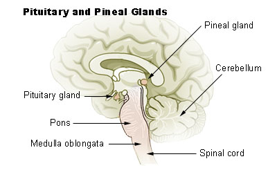
The pineal gland is located in the middle of the midbrain on the roof of the 3rd ventricle and situated below the tail end of the corpus callosum (the body of a major bundle of nerve fibers), in between the two Thalami. The pineal gland looks like a pinecone and so it was named the Pineal gland. The pineal gland is 0.8 mm in size and weighs 0.1 gm and is about the size of a rice grain. This endocrine gland is very vascular, second only to the kidneys (per each unit of mass). The blood-brain-barrier (BBB) is absent here and the hormone is secreted directly into the blood and also in the CSF. The cerebrospinal fluid bathes this gland through a small recess of the 3rd ventricle which continues within the stalk of the pineal gland.


Chemistry of Melatonin.

The pineal gland produces and releases melatonin. Melatonin is N-acetyl 5-methoxytryptamine. The amino acid Tryptophan is the source of melatonin.

Melatonin synthesis.

Tryptophan is converted to 5-hydroxytryptophan by hydroxylation. 5-hydroxytryptophan is decarboxylated to 5-hydroxytryptamine and this product is known as Serotonin.

Serotonin is converted to melatonin in two steps -

Step 1. A rate-limiting enzyme N-acetyltransferase transfers the Acetyl group from Acetyl CoA to 5-hydroxytryptamine and converts it to N-amino-5-hydroxytryptamine.

Step 2. N-amino-5-hydroxytryptamine undergoes methylation. The methyl donor is S-adenosyl methionine and the enzyme catalyzing this reaction is O-methyltransferase. And N-methyl-5-hydroxytryptamine is produced. This molecule is melatonin.

The reactions are shown as follows-

Serotonin + Acetyl CoA → N-Acetyl serotonin. This reaction is catalyzed by an enzyme N-acetyltransferase.

N-Acetyl serotonin + S- adenosylmethionine → N-acetyl 5-ydroxyserotionin.  This reaction is catalyzed by an enzyme O-methyltransferase.

Darkness induces Melatonin synthesis and release.

Darkness causes the release of Norepinephrine from the sympathetic nerve terminals of the pineal gland. The enzyme system is primed by norepinephrine and Cyclic AMP is generated. (cAMP). cAMP activates N-acetyltransferase and melatonin synthesis starts. As melatonin is forming, melatonin is secreted in the CSF and the blood. The pineal gland does not store melatonin in the gland.

If the artificial white light is of a certain strength, the effect of dark on the pineal gland ceases and no melatonin is produced or secreted. During international travel by airlines, the normal dark-light cycle is disrupted and resulting in sleep disturbances.

Breakdown of melatonin.

Melatonin is broken down in the liver by hydroxylation, then conjugated with sulfate and glucuronic acid and excreted in the urine.

Nerve supply of Pineal gland.

Somatic innervation. The 5th cranial nerve sensory nucleus, the Trigeminal ganglion, supplies nerve fibers to the stock and the gland. These fibers contain neuropeptide PACP which are vasoactive compounds. (PACP is pituitary adenylate cyclase acting polypeptide)

Autonomic innervation.

Sympathetic division nerve fibers come from the superior cervical ganglion. The parasympathetic fibers originate from the Otic and Pterygopalatine ganglia

Embryology of the Pineal gland.

In the 17th week of embryonic life, an invagination of the roof of the 3rd ventricle occurs. Initially, the pineal primordium contains Pax6 cells, arranged in a radial manner. After the neural tube fuses, the Pax6 cells rearrange into a rosette formation and then disperse in all directions. All pineal cells are derived from these progenitor Pax6 cells.

An adult pineal gland contains hormone secreting pinacocytes and microglia, astrocytes, and supporting cells. In the adult pineal gland, some progenitor cells remain. Calcium deposit in the pineal gland is common in the elderly, occasionally the entire gland may be calcified.

Role of the Photoendocrine system on the Pineal gland.

The retina of the eyes, supra-chiasmatic cells of the hypothalamus and noradrenergic sympathetic nerve fibers terminate in the pineal gland. Information about light exposure and circadian rhythmic variation is integrated into the pineal gland and regulated melatonin secretion.

Melatonin concentration in the CSF of the 3rd and 4th ventricles is higher than plasma and blood. What effect melatonin has on the neurons of the brain is not known.

Melatonin Receptors.

MT 1 and MT 2 are two types of melatonin receptors in humans. MT1 receptors are present in the suprachiasmatic cells of the hypothalamus, pituitary gland, retina and hypothalamus. When Melatonin binds with MT1, it produces inhibitory effects on the pituitary, and the release of hormones is inhibited and the blood level of Prolactin falls. Through the MT1 receptors, melatonin maintains the circadian rhythmic release of hormones of other endocrine glands.

MT2 receptors are present in the retina. When retinal receptors are stimulated, Dopamine release ceases. It also allows phase shifting of the internal circadian clock to the natural earth clock of the light and dark cycle. Other effects of MT2 receptor activation are increased phagocytosis and enhanced osteoclast activities and vasodilatation.

Hallucinogenic action.

DTM (dimethyltryptamine) is a hallucinogenic compound. Only a small amount of DMT is found in the Pineal gland. This fact might have started the notion that the pineal gland is a psychic center and controller of human behavior. 

Melatonin use.

Melatonin in the USA is an OTC drug (over the counter). It is available in 3 mg tablets, made solely in the laboratory. One melatonin compound. Ramelton is approved by the FDA for the treatment of insomnia, but the results are not consistent.

Indication of use.

Jet lag, Circadian rhythm disorder in the blind, Delayed sleep-wake phase sleep disorder in people who have delayed sleep and delayed wake time than required of them. In insomnia, melatonin reestablishes NON-RAM sleep. It is useful in shift workers and sleep disorders in children.

Adverse effects.

Melatonin is a safe supplement. However, it is a biological amine like Histamine and Dopamine. So, care should be taken when used with epileptic drugs, anti-platelet agents, BP medications, antidepressant drugs, immune modifying drugs, and anti-anxiety drugs.

____________________________________

Rene Descartes (1596 - 1650), a French mathematician, scientist and philosopher. He stated " Je pense, donc Je suis" (I think, therefore I am.)

Footnote: https://plato.stanford.edu/entries/pineal-gland/

********************************************************

Wednesday, March 8, 2023

C - Reactive Protein

                                            C-Reactive Protein (C-RP)

                                         PKGhatak, MD


In an article published in the Journal of Experimental Medicine in 1930, William S. Tillett and Thomas Francis Jr described a serological reaction with a molecule of pneumococcus. A clearer picture emerged from the authors, Abernathy and Avery, in 1941 from their work on the serology of pneumococcal infection describing a flocculation reaction with C-polysaccharide of pneumococcus and a plasma protein. That protein is now known as C-Reactive Protein.

Chemistry of C-RP.

C-RP is a member of the Pentraxin family of proteins. Pentraxin is a ring-shaped planer symmetry protein, with a hole in the center, like a doughnut. This structure gives a high degree of stability to the molecule and resists enzymatic attacks. The name pentraxin came from the Greek Penta Ragos meaning five berries. The molecular weight of C-RP is 115,135 Daltons and each molecule is composed of five identical non-glycosylated polypeptide subunits, each contains 206 amino acid residues. Not more than 15 sugar units are present in each C-RP molecule, but the glucose molecule is not one of them. The amino acids, asparagine, serine, threonine, hydroxylysine, and hydroxyproline, are the only 5 amino acids present, in repeats in a C-RP molecule. The glycosidic linkage between carbohydrate and protein occurs more frequently through oxygen rather than nitrogen.

Site of biosynthesis.

Hepatocytes synthesize C-RP when Interleukin 6 (IL-6) is released from the damaged cells into the blood and IL-6 is carried to the liver. IL-1 beta enhances IL-6 response in the synthesis of C-RP. The C-RP levels may go up 1000 times from a low normal blood level, 0 to 8 mg/L.  Protein molecules are synthesized elsewhere; an enzyme Glycosyltransferase, specific for each type of sugar molecule, catalyzes the attachment of the sugar to the protein moiety.

                                            Pentraxin molecule


Plasma level of C-RP.

Normal plasma levels of C-RP are between 0 to 8 mg/L. The half-life of C-RP is 19 hrs. and remains constant under all conditions of health and disease. The concentration in plasma is solely determined by the rate of synthesis by hepatocytes under IL-6 stimulus, which reflects the intensity of the pathological processes. Once the IL-6 levels fall, the CRP synthesis stops and plasma levels return to the base level in 24 -48 hrs. This property of C-RP makes it an ideal acute-phase protein for-

  1. Screening for physical illness.

  2. Monitoring the response to treatment

  3. Detecting any intercurrent infection in immunosuppressed patients

Highly sensitive C-RP (hs-CRP).

More than 30 years ago, researchers noticed lower than normal levels of CRP can be quantified and used in clinical medicine for cardiovascular risk assessment and prediction of future events. High-sensitive CRP determination is an immunoassay and is reported as mg /L. Interpretation of results of hs-CPR is as follows-

    1.hsCRP less than 1mg/L is not associated with any acute cardiovascular event and does not have increased incidence when followed for 20 years.

  1. hs-CPR 1 to 3 mg/L is a medium risk factor for Coronary events, Stokes and PAD ( peripheral arterial disease).

  2. hs-CRP over 3 mg/L results should be considered a risk factor for the diseases listed in No2., only if other causes of a more common condition like Rheumatoid arthritis, SBE (subacute bacterial endocarditis), or periodontal disease, etc., are eliminated by repeating hs-CRP a week later.

A few special features of C-RP.

Ligand binding molecules.

C-RP has the highest affinity for phosphocholine residue. It binds readily with small molecules of ribonuclear proteins. C-RP binds with histone, apoptotic cells (programmed cell death) and oxidized LDL (low density lipoprotein).

Complement Activation.

C-RP-ligand complex binds with the C1q complement of the classic pathway. In the process, complements C1, C3 and C4 are completely used up. This complex only minimally activates C3 complement and does not activate the complement of the alternate pathway C5 to C9. The outcome of activities is the initial innate response to tissue injury or infection and promotes the opsonization of cellular debris from the inflammation site and stimulates healing.

Autoimmune disease.

The first reported evidence came from works on the blood of Rheumatoid Arthritis (RA) patients.

C-RP deposits are present in the nuclei of the cells of the synovial membrane. The intensity of RA correlates well with plasma C-RP levels and has replaced ESR (erythrocyte sedimentation rate) for monitoring of disease activities and follow up of patients under treatment. Ulcerative colitis shows a similar pattern. But SLE (systemic lupus erythematosus), Scleroderma and Polymyositis show no close relationship with plasma levels of C-RP and disease activity. This is explained based on the works of certain patients who are unusually susceptible to pneumococcal infection and have very low C-RP in plasma. They have a polymorphism of the gene which encodes Guanine and Thymine ( G &T) nucleotides in the Intron of the gene, which accounts for a low plasma C-RP level.

Kidney.

C-RP binds with the immune complex deposited on the basement membrane of the glomeruli in several varieties of glomerulonephritis. In an acute renal transplant rejection episode, C-RP binds with renal tubular epithelial cells and endothelial cells of the peritubular capillaries in the kidney interstitium.

A short summary of the C-RP function.

  1. C-RP is an acute phase protein, though not specific for any particular disease, nevertheless is a useful clinical tool for the determination of the disease activities and follow up.

  2. C-RP activates the classic complement pathway and has an inhibitory effect on the activation of the alternate complement pathway.

  3. hs-CRP is a predictor of the future coronary event. Prognostic indicator of Cardiovascular events, Strokes and PAD (peripheral arterial) disease.  

  4.                          

  5. Taken from NIH publication.
  6. **************************************

Saturday, March 4, 2023

Liver Cancer

                                                        Liver Cancer.

                                                     PKGhatak, MD


Liver Cancer is a deadly disease. Cancers of the liver are of two main categories, namely, Primary Liver Cancer and Metastatic or Secondary liver cancer.

Primary Liver Cancer:

The liver is made up of about 15 thousand individual lobules. Each lobe is a structural and functional unit of the liver. Liver cells, Hepatocytes, when they turn cancerous, are called Hepatocellular cancer. The liver has a dual blood supply - the portal and systemic circulatory systems. A bile duct network drains the bile from the liver. 

The endothelial cells of the blood vessels and bile duct cells can turn cancerous. Cancers of different cell lines differ from each other in many aspects. In addition, the liver is richly supplied with immune cells and malignant tumors may develop from immune cells also.


 A. Hepatocellular cancer.

Hepatocellular cancer is by far the most common cancer of the liver. The annual incidence of hepatocellular cancer in the USA is 35,000. Native Americans have the highest incidence among all ethnic groups. Older Americans have more cancers than the younger age group. The incidence of hepatocellular cancer in East Asian countries is the highest in the world. A very rare form of cancer is occasionally seen in very young children in the hepatic stem E-MB  and MEM-HB cells.

Risk factors.

Hepatitis B and hepatitis C are viral infections and proven causes of liver cancer. A fungal toxin, Aflatoxin, is carcinogenic. Alcoholic liver cirrhosis and Non-alcoholic fatty liver disease (NASH) can turn cancerous. Cigarette smoking and alcohol are risk factors. Muscle-building androgenic steroids and oral contraceptive pills are additional risk factors. Liver cysts and cysts associated with polycystic disease of the kidney are also risk factors. Very rarely, cancerous cysts are seen in the use of anabolic steroids and birth control pills.

In addition, hereditary metabolic diseases like Hemochromatosis (iron), Wilson disease (copper), alpha 1 antitrypsin deficiency ( enzyme), porphyria cutaneous tarda, and glycogen storage disease are known to cause Hepatocellular cancer.

Symptoms.

A small and early liver cancer does not produce any symptoms. A patient with growing cancer develops loss of weight, loss of appetite, upper abdominal fullness, and abdominal pain. At this stage, the diagnosis of liver cancer leads to a better outcome. But most patients come to the doctors when they develop jaundice and dark-colored urine; at this stage, the disease has progressed too far for a good outcome.

Diagnostic tests.

Alpha fetoprotein in serum is elevated, and Liver function tests show elevated ALT over AST and high bilirubin. Ultrasound is a very useful tool to detect tumors. CT scans and needle biopsies under ultrasound guidance give a definite diagnosis.

Treatment.

In most instances, a complete resolution of liver cancer is not possible because of the advanced stage of cancer at the time of diagnosis. In suitable cases, local or regional resection of the liver is possible. The liver is a remarkable organ in its capacity to grow back to full size if it is free. In selected cases, a lobectomy or total hepatectomy followed by a liver transplant is done. Immunotherapy, chemotherapy, and several ablative therapy methods are available.

Prognosis:

In general, this is most disheartening. 5-year survival is less than 20%, and in advanced cases, it is less than 3 %.

 Carcinoma of the Bile Ducts.

The medical term for cancer of the bile ducts is Cholangiocarcinoma (CLC). CLC is less common than hepatocellular carcinoma. However, people who have habits of eating raw fish have an alarmingly high rate of CCL. 


The diagram above shows bile ducts inside the liver and also outside. The outside bile duct for this discussion is dealt with into two separate categories, namely Hilar and  Common bile duct cancers.

The life cycle of a Liver Fluke.

CLC (cholangiocarcinoma) is the second most common liver cancer in the world, but the incidence in the USA is much less than hepatocellular carcinoma. But overall, CLC is increasing in all countries, including the USA. In the USA, the Hispanic ethnic group has a higher CLC. In the world, Thailand has the highest rate of  CCL, about 40 in 100,000 people, followed by China, Japan, and other East Asian countries, with a clear association with Liver fluke infestation of the biliary system. In the Mekong River basin countries, Liver fluke infestation is between 30% to 70 % of the population, and the death rate from CCL is 3% of all deaths.

Risk factor.

Primary sclerosing cholangitis is a precancerous condition; a congenital bile duct disease called Choledochal cyst disease has a high rate of CLC. Ulcerative colitis and Crohn's disease have a higher incidence of CLC. Infestation of the liver fluke is a risk, as mentioned above. Both liver cells and the bile duct system developed from the same progenitor cell in the embryonic stage of development. As a result, the non-specific risk factors mentioned under hepatocellular cancer (alcoholic liver cirrhosis, NASH, diabetes, etc.) are also risk factors for CLC.

 A. Intrahepatic CLC.

Intrahepatic CLC is the least common among the three CLCs. Initially, CLC  is not distinguishable from hepatocellular carcinoma; the diagnosis is made only after a liver biopsy. Patients remain asymptomatic in the early stage. Occasionally diagnosed by chance when ultrasound or abdominal CT scans are done for other reasons. The prognosis at this stage is very good with surgery and chemotherapy. However, the majority of patients seek medical attention because of the development of jaundice. Diagnosis is relatively easy by ultrasound and fine needle biopsy. A standard care protocol in the USA hospitals uses a multidisciplinary approach and provides partial or complete removal of the liver and liver transplantations.

B. Hilar CLC.

Hilar CLC is the most common of CLC, accounting for about 70% of CLC. Pathologically, these are adenocarcinomas. Because of their location, jaundice develops earlier than in other liver cancers. The patient seeks medical attention because of worsening jaundice, dark urine and light stool, loss of appetite, and weight loss. Laboratory tests confirm obstructive jaundice. CEA and alpha-fetoprotein are positive. Diagnosis requires a fine needle biopsy. Curative surgery is not possible in the majority of cases. Initial therapy is draining bile by inserting a stent in the common bile duct, draining into the duodenum, done during an ERCP examination, however, a transcutaneous bile duct stent can also be done. 

C. Common bile duct CLC.

The risk factors are the same as the above group and an additional risk factor is an abnormal opening of the pancreatic ducts.

Abnormal Pancreatic Duct.

Anatomical variations of pancreatic ducts are common. Occasionally, one or both pancreatic ducts may join the common bile duct, rather than opening into the ampulla. In this circumstance, chemical inflammation of the duct from pancreatic enzymes leads to fibrosis and stricture, and also carcinoma

Obstructive jaundice is the presenting symptom of common bile duct CLC. In clinical practice, obstructive jaundice is common. The causes of obstructive jaundice are as follows.


                                                   Taken from NIH publication.

Among the benign causes, gallstone and bile duct inflammation are most common, followed by common bile duct stricture, and Mirizzi syndrome. Gallbladder cancer leads the malignant causes followed by cancer of the periampullary region. 12 % of cases are due to cholangiocarcinoma.

                              Mirizzi syndrome in a diagram.

Diagnosis and treatment. 

Initial diagnostic workup is no different from other types of CLC. Initial treatment is a stent placement during ERCP or can be placed transcutaneously. The definitive treatment is the Whipple procedure or a modified Whipple operation. The prognosis is encouraging with hepatic transplants and immunotherapy.

 [ See Chronic pancreatitis blog, dated April 9, 2022]. 

Childhood Liver Cancer, Hepatoblastoma, is seen in children less than 3 years of age. It is more common in premature and underweight newborns. The primary cause of it is unknown. Several inherited conditions are associated with hepatoblastoma. Resection of the liver when performed early produces the best outcome.


edited: May 2025.

************************************************


Sunday, February 26, 2023

Food Poisoning


                                            Food Poisoning

                                          PKGhatak, MD


Food and drinks can be contaminated with microorganisms if not handled properly. Certain bacteria grow in the leftover food kept outside the refrigerator and produce toxins. When people eat leftover food, these toxins produce an illness, which is called food poisoning.

In general, such a definition of food poisoning is not universally followed. Viral gastroenteritis, cholera, amebiasis, typhoid fever and others are also included in food poisoning. It is better to address those illnesses as food borne illnesses rather than food poisoning.


Bacterial Toxin.

Bacteria can grow over a wide range of temperatures, from 4 degrees C to 60 degrees C,  provided moisture and nutrients are present. A growing bacterial colony produces toxins.

Toxins produced by bacteria are in two groups-

A. Exotoxin. Endotoxins are secreted by bacteria in the food. Exotoxins can be heat resistant or destroyed by heating.

B. Endotoxin. Endotoxins are present in the bacterial bodies and released in food when the bacteria die or are killed by antibiotics.

Preformed toxins in food produce symptoms in 1 to 6 hrs. after ingestion, whereas, bacterial infection and toxin generation take over 8 hrs. and then symptoms develop. Bacteria commonly responsible for food poisoning are Staphylococcus, Bacillus, Clostridium, E. coli and Vibrio species.


Staphylococcus aureus food poisoning.

S. aureus readily grows when food is left on the kitchen countertop. Symptoms start within 1 to 6 hrs. after eating food. Reheating food before eating does not destroy the bacterial toxin. Nausea and vomiting are common symptoms and start abruptly. Campy abdominal pain and diarrhea follow. The illness is self limited and patients recover in 24 hrs.


Bacillus cereus.

Reheated fried rice is often the source of poisoning. Intense nausea and vomiting start within 1 to 8 hours after eating food. The toxin can be detected in stool and leftover food.

Symptoms last for 12 hrs. It is also a self limited illness.


Clostridium botulinum.

Clostridium botulinum is a soil bacterium. Canned vegetables not properly washed and low oxygen environment in a sealed can are ideal for the production of toxins. Another source is natural honey. The ingested toxin produces nerve paralysis and the illness is known as Botulism. Though the symptoms start days after ingestion of the poison, botulism is due to the toxin present in food and not a bacterial infection.

Botulism. The sudden onset of fluctuant and intermittent but severe muscle paralysis in a healthy person should alert the physicians of botulism. Botulism toxin prevents the release of a neurotransmitter, Acetylcholine, at the neuromuscular junctions.

Symptoms generally develop 3 days after ingestion of the toxin and continue to progress further for another 3- 4 days. Paralysis of eye muscles produces double vision, and eyelid paralysis causes drooping eyelids. Muscles of swallowing and speech muscles are also paralyzed, producing difficulty in eating and drinking, and a nasal voice. In severe cases, the muscles of the limbs may be paralyzed. Identifying the toxin in unused portions of food and serum helps in the diagnosis. An electroencephalogram(EEG) is also useful.

Antitoxins are available to reverse the effects of toxins. Hospitalization and close monitoring of respiration and blood oxygen and CO2 are essential parts of the management of botulism. Public health authorities must be informed.


Clostridium perfringens.

This spore-forming Clostridium is found in soil and feces. Uncooked beef, poorly cooked poultry and fish are the source of toxins. Between 6 to 24 hours after eating, symptoms of severe diarrhea and abdominal cramps develop. The illness can last one to several days. The initial onset may be from preformed toxin, but subsequently, the bacteria multiply in the gut epithelium and continue to generate enterotoxin and produce symptoms.

Enterotoxin producing E. coli.

This entity falls in between preformed toxin and bacterial infection and perhaps, both are operative.

Animals and humans harbor E. coli in their large guts. When food and drinks are contaminated with feces, severe watery diarrhea and abdominal cramps develop within a day or two after ingestion. It is a common cause of food poisoning among international travelers. Antibiotic fluoroquinolones are effective therapy.

Vibrio parahaemolyticus.

This vibrio usually contaminates shrimp, crab and shellfish. Eating raw oysters or lightly cooked seafood produces a sudden onset of abdominal pain and diarrhea within 2 to 48 hrs. Nausea, vomiting and diarrhea last for 2 to 5 days.

Proper hydration is required to prevent dehydration.


************************


Thursday, February 16, 2023

Peptic ulcer

                                                     Peptic ulcer disease.

                                              PKGhatak, MD


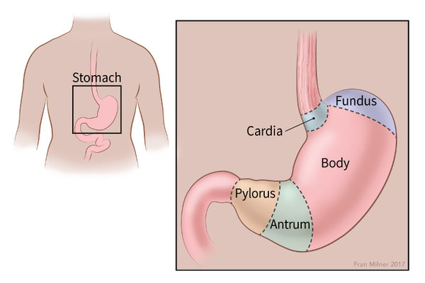
When ulcers develop in areas of the stomach normally bathed by acid and pepsin, those ulcers are called peptic ulcers. All sections of the stomach and the first part of the duodenum come in contact with acid and pepsin. And occasionally in an incompetent lower esophageal sphincter, the lower end of the esophagus develops ulcers due to gastric reflux. 


Typically, peptic ulcers are found in the first part of the duodenum and the Pylorus.

It is estimated that about 4 million new cases are detected annually worldwide. In the USA, about 5 to 8 % of the population have peptic ulcer disease. In the past several decades, the incidence of peptic ulcer fell but in recent times, the incidence has remained steady.

Gastritis and gastric erosion are included in Gastric ulcer diseases because the mechanism of development of these is the same as ulcers.

Why do people develop Ulcers of the stomach.

The immediate cause of peptic ulcer is excess acid and protein digestive enzyme pepsin production by the cells of the mucous membrane of the stomach.

Risk factors of peptic ulcer disease.

1. Infection of Helicobacter pylori (H. pylori).

2. Cigarette smoking.

3. Alcohol abuse. 

4. Chronic use of aspirin or non-steroidal anti-inflammatory drugs (NSAID).

5. Anxiety and stress.

6. Use of systemic steroids.

Peptic ulcer.

The common practice today is to detect peptic ulcers by endoscopy. Any area of the stomach and duodenum that appears abnormal under endoscope is usually biopsied and the histology of the tissue is examined. Gastritis and ulcers due to H. pylori can be detected and if any doubt exists, a biopsy should be enough to exclude cancer.

Peptic ulcers are small, less than 1 cm in diameter, have clear margins and a clean base. Cancers are usually located in the cardia and fundus of the stomach, usually large, and the margins are heaped up, the base is necrotic.


Panel A: are benign lesions. Panel B: is a cancer of the stomach.


A brief review of the characteristics of the gastric mucosa.

The inner lining of the stomach is called the mucous membrane. The diagram below shows the relative thickness of the stomach wall and a cutout section shows the different cells in the mucous membrane.


The function of the cells of the mucous membrane.

1. Parietal cells. These cells produce a strong inorganic acid - hydrochloric acid. The pH of the stomach is 1 to 2. Parietal cells also produce the Hematinic principle, a glycoprotein, which is essential for Vitamin B12 absorption.

2. Chief cells. Chief cells secrete an inactive form of protein digestive enzyme, Pepsinogen. Pepsinogen at pH 1 or 2 is converted to an active enzyme, Pepsin. And pepsin remains active in pH 1 to 8.

                                location of endocrine cells, stained brown.

3. Endocrine cells. The endocrine cells are G-cells, ECL-cells, and D-cells.

G-cells secrete Gastrin directly into the bloodstream. (that is the nature of all endocrine glands and cells). Gastrin is brought back to the mucosa by circulation. Gastrin stimulates the growth and development of parietal cells and enterochromaffin-like cells (ECL) cells.

ECL-cells produce and store Histamine and secrete histamine. Histamine is a potent stimulant for acid secretion. In addition, ECL cells secrete chromograninA derived peptides, pancreastatin and other peptides.

D-cells. D-cells secrete Somatostatin. It is an inhibitory hormone. It decreases the release of gastrin and decreases the motility of the stomach and intestine. Also delays the release of other peptides from ECL cells.

5. Goblet cells and mucus secreting glands. Goblet cells secrete mucus. The mucus has a very significant function. The mucus is of two layers, one of which is adherent to the cell layer and the free layer on the top of the adherent mucus layer. Both layers are about 1 mm thick and chemically are glycosylated mucin peptides. It contains a high concentration of bicarbonate ion [HCO3-]. HCO3 is immediately available to neutralize any HCl (hydrochloric acid) that comes in contact with the mucus layer.

The mucus barrier acts as a cushion on which gastric juice floats. It is like an atom or a proton kept separated from the copper tube and confined to a narrow zone by powerful electromagnets in a cyclotron (atom smashers).

Acid production in the stomach.

The parietal cell contains the Carbonic Anhydrase enzyme. This enzyme greatly accelerates the reaction between H20 +CO2 -->H2CO3, taking place within the cell cytoplasm. H2CO3 then dissociates into H ion [H+] and HCO3 ion [HCO3-]. Energy dependent [Na+]/[H+]  pump  (commonly called Proton pump)     expels  [ H+] into the stomach lumen and in exchange [K+] enters the cells. [HCO3-] is carried from the cells into the blood by a cation exchanger transport protein and it brings back [Cl-] inside the cells. [Cl-] enters the gastric lumen via Cl channels. [H+] unites with [HCO3-] because opposing charges attract each other and form HCl.

Factors accelerate Acid production.

The Vagus nerve is the secretory motor nerve for the GI tract. As soon as food enters the stomach, vagal stimulation produces Acetylcholine. Acetylcholine directly stimulates gastric acid secretion and indirectly by increasing gastrin release.

G-cell.

Stimulate acid production via gastrin.

Histamine.

Histamine is a potent stimulant for vasodilatation, increased glandular secretion, and other properties. In the stomach, the Histamine is produced and released by ECL- cells, nerve cells and mast cells. Histamine 2 receptors are present on the surface of the Parietal cells. Once histamine combines with the H2 receptors, acid production increases. Histamine is the main regulator of acid secretion influenced by local conditions.

D- cell.

D-cell produces Somatostatin. It inhibits acid production and decreases gastric motility and other inhibitors control beyond the stomach.

Conditions favor peptic ulcer.

Smoking.

Cigarette smoke contains Nicotine. Nicotine decreases Prostaglandin generation and makes gastric mucosa vulnerable to ulcer formation.

Alcohol.

Alcohol is a poison to cells when it comes in direct contact with cells. Alcohol destroys the mucus coat of the stomach, making it easier for alcohol to come in contact with the mucosa. An excessive amount of alcohol produces gastritis and chronic use produces ulcers. Once peptic ulcers are formed, even a small amount of alcohol damages cells and prevents ulcer healing and causes pain.

Aspirin.

Aspirin on ingestion turns into salicylate. Salicylate is toxic to cells and produces erosion of surface cells, disrupts the H+/K+ pump and loss of K+ from the cells. Hemorrhagic gastritis and gastric ulcers are common adverse effects of aspirin.

Since aspirin inhibits the enzyme cyclooxygenase permanently for the life of affected cells, adverse effects persist even after aspirin is withdrawn.

NSAID.

Non-steroidal anti-inflammatory drugs are commonly prescribed (e.g. Advil) for joint and bone injuries, post orthopedic surgery, and all varieties of arthritis and are also available as OTC (over the counter drugs). Peptic ulceration and gastritis have increased in recent years, going against the trend seen in several past decades.

NSAID inhibits the COX-1 enzyme. COX-1 is another enzyme needed for Prostaglandin synthesis. The effect of NSAID is however temporary, once the drug is stopped, the cells can recover.


Helicobacter pylori (H.pylori).


H.pylori is a gram negative, highly motile, unipolar-multi flagellated, microaerophilic organism. The body of H. pylori is spiral and so it is called Helicobacter. Scientists believe H. pylori infected the stomach of humans 2,5000 years ago and is still infecting humans. More than 50 % of the world's population is already infected, and 100 % of people in Africa and Asia have H. pylori chronic gastritis, although 80% are asymptomatic. Most people are infected in early childhood by the oral-oral route and the fecal-oral route. Once infected, 80 % of the bacteria are present in the mucus of the gastric lumen and 20% in the mucus secreting cells of gastric epithelium but absent in the parietal cells, endocrine cells and the chief cells of the epithelium.

The bacterium has developed a unique mechanism of evading the immune defense of the victims and continually inventing ways to resist antibiotics. The bacterial wall has several layers of envelopes of different proteins and in addition, has several adhesion proteins and these proteins keep the bacteria firmly attached to the cells. 

Detection.

Breath test.

The human body does not have urea splitting enzyme urease, but H. pylori contains urease that breaks down Urea into CO2 and ammonia. A tablet containing 13C  urea and citric acid is taken by mouth and the expired air is collected and analyzed. Presence of C13- CO2 in expired breath indicates H. pylori infection.

Biopsy.

Patients present with symptoms of peptic ulcer usually undergo endoscopic examinations. Biopsy from the suspected areas is taken. H.pylori is easily detected, if present.

Treatment.

Because antibiotic resistance is common, the antibiotic regimen is often changed. A commonly used regimen is mentioned and also a reference to the 2022 recommendation is provided.

Antibiotics:

Amoxicillin and one of the following two antibiotics: Macrolides and Metronidazole (an anaerobic bacterial antibiotic). Antibiotics are combined with Bismuth subnitrate. Bismuth forms a complex with the bacterial wall, inhibits multiple bacterial enzymes resulting in decreased synthesis of adherence protein molecules and deprives H. pylori source of nutrition.

Acid suppression.

Pantoprazole. It blocks the proton pump and acid secretion comes to a total stop.

Histamine receptor blockers. It acts by competitive binding with H2 receptors of the parietal cells and blocks the action of histamine in producing acid.

Antacids. Commonly used aluminum oxide and calcium carbonate as acid neutralizers are not therapeutically sufficient in the treatment of peptic ulcers.

____________________________________________________

https://www.gastroenterologyandhepatology.net/archives/june-2022/helicobacter-pylori-treatment-regimens-a-us-perspective/

(Copy & paste on your browser.)

*******************************************
Archive

  Archive. P.K.Ghatak, MD ghatak3@gmail.com 2025  March. Laser Endoscpoe 2025 February. Use of Ultrasound in Medicine MRI 2025 January. Bact...