Thursday, October 10, 2024

Syndrome in Clinical Medicine

                                               Syndrome in Clinical Medicine

                                                  P.K.Ghatak,MD

                                                  ghatak3@gmail.com


In a NIH publication in 2003, Dr. Franz Calvo et al, defined syndrome as a recognizable complex of symptoms and physical findings for which a direct cause is not necessarily understood. Once the medical science identifies a causative agent or process with a high degree of certainty, physicians may then refer the process as a disease and not a syndrome.

Some well known syndromes are now called diseases and to illustrate, an example is Cushing's disease and Cushing's syndrome. In 1912, Dr. Cushing reported a new disease he called polyglandular syndrome due to malfunction of the Anterior Pituitary gland. He published this entity in 1932 as basophil adenomas of the Pituitary body and named it Pituitary basophilism. Symptoms of the disease - gained excessive weight, mostly around the back of the neck, face and abdomen. Also developed high blood pressure, diabetes, acne and facial hair. In subsequent years, the condition was known as Cushing's syndrome. Further studies identified the cause was a tumor of the anterior pituitary producing an excess amount of ACTH hormone, which in turn put out excess corticosteroids from the Suprarenal glands. Similar changes were also detected when patients took steroids for a prolonged time, as one of the anti-rejection drugs following kidney transplants. And also seen in certain cancers of the lungs secreting an excess amount of polypeptide having similar hormone like actions of the suprarenal gland. Now Cushing's Disease term is reserved for symptom complex arising from tumor of the anterior pituitary and Cushing's syndrome is applied to all other causes which can produce some features of Cushing's disease.

If one looks up Syndrome in the Wikipedia, one will be surprised to find more than 1600 syndromes. In the index of any textbook of medicine, where every entry is listed in alphabetical order, one finds only 3 entries under syndrome, the rest of the syndromes are under the first letter of a name attached to the person who discovered the illness. Not all the listed syndromes, however, follow this rule. Some names are retained because the name refers to well understood symptoms by the public, like - Milk - Alkali syndrome and Irritable Bowel Syndrome.

Recent rapid advances in genetics, MRI imaging, sonography, immunology and biochemistry have solved many obscure causes of syndromes. In the lifetime experience of any well rounded physician, they might have not seen more than 30 syndromes. Certain specialties like Pediatrics are likely to see more syndromes.

A few syndromes will be presented, one or two from each group,  to give the readers a chance to the readers the mystery about the syndrome.

Down syndrome.

Down syndrome arises from abnormalities of chromosome 21. It occurs in 75% of cases as inherited and 25% as spontaneous. Each cell of the body contains an additional one full or partial copy of chromosome 21. This generally happens as non-splitting or translocation between chromosome 21 and 14 or 21 and 21 or 21 and 22. In 2 to 3 % cases it also occurs due to Mosaicism.

Some of the common features of Down syndromes are mental retardation, flat cranium and flat nose bridge, a short neck, large tongue compared with the floor of the mouth, prominent epicanthal fold, structural heart defects, truncal obesity, poor muscle tone an delayed mile stones of development.

Polycystic Ovary Syndrome.

Originally, the Polycystic Ovary syndrome was called Stein-Leventhal syndrome because this pair of investigators were the first to report it in 1935. In 1990, NIH added two additional criteria - multifactorial genetic errors and insulin resistance, and finally in 2003, ultrasonographic detection of more than 20 cysts in the ovary was added and named it Polycystic ovary syndrome. The cause of this syndrome is unknown and symptoms vary widely.

A typical case has these features: Symptoms of irregular and scant menstrual flow or delayed menarche. Development of acne and facial and body hair of masculine type, obesity, hypertension and insulin resistance and diabetes mellitus. Most cases are due to excessive androgen activities, but androgen is normal in the minority of cases. In a few cases, the polycystic ovary syndrome is detected during investigation of failure to conceive. If not treated properly, there is a high likelihood of development of endometrial carcinoma later in life.

Klinefelter syndrome. This congenital condition affects only male children due to having an extra X chromosome. The condition is also known as 47 XXX. The genetic abnormality occurs randomly either in the development of the ovum or sperm. The condition is generally detected at puberty when the child failed to develop muscle mass and develop a feminine body type - gynecomastia, no facial hair and wide hips. They have small genitalia and testicles, scant or absent sperm production and have long legs and a short torso. Many have learning difficulties and lag behind in reading and writing skills.

Not all affected have all the above features and may pass as normal and only correctly diagnosed much later when one fails to father children. They carry the risk of breast and extragonadal germ cell cancers and also osteoporosis.

Turner's syndrome. Turner's syndrome is also known as 46 XO. This congenital condition is seen only in female offspring, arises due to missing a whole or part of the sex chromosome X; but whether that missing X chromosome is paternal or maternal is not known. The gene responsible for bone growth is the SHOX gene and it is missing in Turner's syndrome, resulting in the abnormalities of bones in Turner's syndrome. At birth the child may appear normal except for a web neck and a broad chest. As the child reaches age 9 or 10, the growth slows down, develops no secondary sexual characters and menstrual cycles. Other features are cubits vulgus, aortic/ pulmonary stenosis and high BP, diabetes mellitus, hypothyroidism and osteoporosis.

Fragile X syndrome: It is a X link cause of mental retardation. Males are more severely affected than females. Incidence is 1:4000 male births and 1:10,000 in females. Symptoms in female are learning difficulties, variable degrees mental retardation and early onset of menopause. In males, the physical characteristics are large ears, a prominent jaw bone, a pitched voice, Mitral valve prolapse and increased immobility of joints. This syndrome is associated with autism.

The tip of the long arm of chromosome X carries the mutated Fragile RNA gene, results from undue expansion of CCG repeats. More repeats are present more mental retardation is detected.

Marfan syndrome. Marfan syndrome is an autosomal dominant inherited disorder of the connective tissue due to mutations in the Fibrillin gene (FBNA1) on chromosome 15. This results in aberrant TGFB1 and TGFB2 activities. Clinical features of Marfan syndrome are tall stature, long arms and legs, scoliosis, pigeon chest wall deformity(Pectus excavatum), dislocated eye lenses, mitral valve prolapse, aortic root dilatation, aortic incompetence and aortic aneurysm and dissection.

Ehlers-Danlos syndrome. This is another connective tissue autosomal dominant inherited disorder. Mutation of many genes, the last count 20 genes, are implicated, and based on the severity of symptoms, 11 varieties are known. Those who have severe symptoms have a mutation of COL5A1 and COL5A2 genes, other mutated genes are TNXB, ADMTS2, PLOD1, FKBP14 and many others. Mutations result in the formation of collagen tissue in a haphazard fashion and functionally of poor quality specially of the skin, skeletal muscles, arteries, bones and internal organs. Major symptoms include fragile velvety skin, which peels off easily and forms extensive scars. Aneurysm of the artery and dissection and rupture of aneurysm and other changes of the eyes, heart and bones mentioned under Marfan syndrome.

Irritable bowel syndrome. Irritable bowel syndrome fits the classical definition of the syndrome. A group of symptoms originates in the colon and small intestine but the cause remains unknown. Common symptoms are - a sense of not being able to evacuate completely after a bowel movement, constipation and low grade abdominal cramps, flatulence and diarrhea, often hard and lumpy stool and diarrhea on the same day, and several bowel movements a day. Various theories are put forward, including stress, pressure of modern life, precooked meals, food additives, change of colon bacterial colonies and low tolerance to pain. Both sexes are affected and the age of onset - from teens to elderly. It is a common illness.

Milk-Alkali syndrome. It is a self made syndrome due to taking an excessive amount of Calcium supplement to prevent osteoporosis and the condition is accelerated and made worse by taking a mega dose of vitamin D3. In the previous generation, people suffered this complication from taking excessive amounts of Tums, Maalox and Mylanta for heartburn and peptic ulcer diseases. Excess alkali present in the form of carbonate in these compounds shifts the blood pH towards the alkaline side. High blood calcium results in an excess amount of calcium excreted in the urine. Kidney stones and urinary bladder stones may develop. Renal colic, chronic pain around the loin and back are usual symptoms of kidney stones. Left untreated, patients may end in renal failure.

Abdominal Compartment syndrome. This is a serious and often fatal condition that arises from several causes but is often seen in severe burn victims. Other causes are organ transplants, prolonged abdominal surgery specially repair of abdominal aorta rupture, bullet or knife penetrating injury with severe intra-abdominal hemorrhage. Sepsis, abdominal abscess, severe peritonitis and others. Fluids and inflammatory exudates accumulate in and around organs and raise intra abdominal pressure over 20 cm of Hg. This impairs arterial supply and blocks venous and lymph drainage, producing further damage to the abdominal organs. Immediate proper fluid management and abdominal surgery is needed to relieve the high intra abdominal pressure.

Sudden Infant Death Syndrome (SIDS). A healthy newborn child suddenly stops breathing and dies during sleep. In the majority of cases are due to suffocation due to putting children face down in a soft bed for sleep; which favors obstruction of the mouth and nose. About 20 % of cases remain unknown.

Thoracic outlet syndrome.
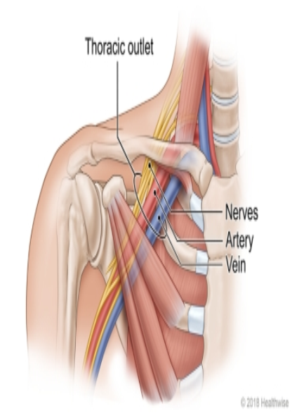
 Just below the clavicle at a point it turns towards the shoulder, the Subclavian artery the subclavian veins and all the nerves arise from the brachial plexus, enter the chest wall and course down the medial side of the arm. There is just enough room for the structure to pass. In an abnormal situation like fracture of the clavicle and hematoma formation, a cervical rib and osteoma of the first rib that space becomes too tight for these structures and pressure on artery produce pain in the arm and hand, pressure on the vein swelling of hand and forearm and bluish discoloration of fingers, Pain over the nerve produces tingling numbness of fingers and muscle weakness. The pressure must be relieved by surgery to prevent permanent damages to the hand.
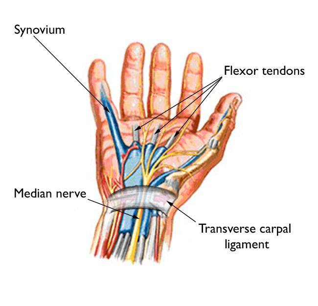
Carpal tunnel syndrome.

 This is another syndrome like the thoracic outlet syndrome due to anatomical peculiarity at the flexor surface of the wrist. The median nerve on the way into the palm of the hand has to negotiate through a narrow passage in between wrist bones- Pisiform laterally and Hamate and Capitate inferiorly and a strong transverse carpal ligament anteriorly. Pain, specially at night, numbness and tingling are the initial symptoms felt in the hand and fingers, at times the pain is felt in the forearm. Later weakness of fingers and thumb, muscle atrophy and ultimately claw hand develops. The causes of Carpal tunnel syndrome are several - Unaccustomed weight bearing, repeated flexion and extension of the wrist, pregnancy, fracture of wrist bones and malunion of fractures, synovitis of the flexor tendons, hypothyroidism and myxedema, rheumatoid arthritis, amyloidosis, sarcoidosis, acromegaly and leukemias.

Tarsal tunnel syndrome. Just like carpal tunnel syndrome, Tarsal tunnel syndrome may develop at the ankle joint. Symptoms and causes are practically the same except for the site.

Inappropriate ADH secretion. Blood volume and serum osmolarity are maintained by feedback loops on the control center located in the hypothalamus and posterior pituitary gland. In lower blood sodium concentration and excess water content of blood (low osmolarity), no Anti Diuretic Hormone (ADH) should be released from the posterior pituitary. But if ADH is still released in such a condition, the situation is called Inappropriate ADH secretion. The causes are many, however, Lung cancer, encephalitis, meningitis, subdural hematoma are main causes.

Syndrome X. Syndrome X is due to abnormal microcirculation of the coronary arteries or coronary vasospasm, which results in Angina pectoris.

Syndrome of apparent mineralocorticoid excess, This syndrome is due to 11 beta hydroxysteroid dehydrogenase deficiency inherited on an autosomal recessive mode. Symptoms are early onset of hypertension in childhood, low serum potassium and metabolic alkalosis.

Metabolic syndrome. Metabolic syndrome is a more recent entry in the syndrome category; previously the components of this syndrome were known as essential hypertension, hypercholesterolemia, obesity and diabetes mellitus. Under the new name, those previous entities are refined and redefined, instead of hypercholesterolemia, it is now low HDL cholesterol and high triglyceride. Obesity is now abdominal obesity, Diabetes is replaced by above impaired fasting blood glucose. People with Metabolic syndrome are at high risk of developing coronary arterial disease, diabetes mellitus and cerebrovascular accidents.

Stokes - Adam syndrome. This syndrome was well established a century ago, when physicians used their well cultivated skill of observation and taking pulse over several minutes. Main features of Stokes-Adam syndrome are sudden loss of consciousness, and fall to the ground and having epileptic seizures due to complete heart block ( third degree in new definition) and a pulse rate of 35 or below. Ventricular fibrillation and ventricular tachycardia also unconsciousness and seizures. At one time it was called Cardiac seizure and that name was better known than Stokes-Adam syndrome.

Long Q-T syndrome. This is an electrocardiogram finding. The time interval between the beginning of the q wave and the end of the T wave on the tracing of an ECG (EKG) is normally 0.43 millisecond when the heart rate is 72/ minute. In a slower heart rate, the Q-T interval increases predictably. In several cardiac diseases and quinine, procainamide, digitalis, over-the-counter cold medicines and many other medications can cause undue prolongation of Q-T interval. The longer the interval, the greater chance there is of Ventricular ectopic beats and precipitation of ventricular tachycardia and ventricular fibrillation. A particular form of multifocal and multidirectional ectopic ventricular beats called Torsades des pointes (a French word, meaning twisting around points) is a forerunner of Ventricular fibrillation and death.

Torsades des pointes

Sick sinus syndrome. This is another very significant ECG finding. The natural cardiac pacemaker is the sinus node. In oxygen deprivation from any reason, electrolyte imbalance and certain medications, the sinus node fails to generate the electrical pulse wave for the heart muscle to contract. This can manifest as slow heart rate over 3 seconds, 1st, 2nd, and 3rd degree heart block. These changes are variable and in some cases variable cardiac block can be intermittent. To detect the condition, a special cardiac monitor is used, which can trace every cardiac beat and record on a preset program. This allows closer examination of any abnormal heart beat and suitable treatment can be offered – often a pacemaker. Main symptom of sick sinus syndrome is missing a heart beat, irregular pulse, palpitation, fluttering sensation in chest, confusion and loss of balance and fall.

Goodpasture syndrome. Goodpasture syndrome is an example of how an antibody, generated to fight an unidentifiable infection, mistakes its own kidneys as foreign and attacks the collagen tissue present in the basement membrane of glomeruli and walls of alveoli of lungs, results in hemorrhage in the kidney and lungs. Main symptoms are bloody urine and coughing out of blood. Other significant symptoms are shortness of breath, chest pain, fatigue, anemia, high BP, fatigue, nausea and vomiting. It is a major illness and requires immediate medical attention.

Alport syndrome. Alport syndrome is a multisystem inherited disorder involving collagen IV tissue and results in renal failure, deafness and visual impairment. The defective gene is carried on the X chromosome. Male child develops glomerulonephritis at an early age and progresses rapidly and unless treated, dies by age 40, females are less frequently affected and the disease progresses slowly.

Nephrotic syndrome. Nephrotic syndrome is a stage in renal failure that arises from protein losing glomerulonephritis. Presence of a large amount of protein in the urine makes urine foamy, hypoalbuminemia produces puffy eyes, pale puffy face, ankle edema and scrotal edema. With the further progression of disease, ascites and plural effusion are seen. Anemia, fatigue, weight gain from accumulated water develop. Intercurrent infection is common.

Carcinoid syndrome. In a previous blog, carcinoid syndrome was discussed in detail. See footnote.

Carcinoid syndrome is due to the production of many polypeptides which have hormone like effects released from one variety of neuroendocrine tumor. Carcinoid tumors generally arise in the small intestine, stomach, colon, appendix, rectum and liver and pancreas, and also arise in the bronchial tree of the lung. Carcinoid secretes serotonin, histamine, kallikrein, tachykinins and prostaglandin. These are potent vasodilators and produce intense vasodilatation, hypotension, watery diarrhea, asthma like bronchospasm and intense flushing of the face and body. The tumors are small and can be benign or malignant, but pathologically can not be determined whether a tumor is benign or malignant. In malignant variety, the tumor metastasizes in the liver.

Dumping syndrome. Dumping syndrome is a number of gastrointestinal symptoms that occur a few minutes after a meal. Vagotomy and reducing stomach capacity by surgery are the principal causes of Dumping syndrome. Following any such stomach surgery, the food leaves the stomach prematurely and enters the duodenum in large amounts overstretching the duodenal wall, causing upper abdominal crampy pain, sense of abdominal fullness, nausea, diarrhea, weakness and lightheadedness. And later develops palpitation and rapid heart beats.

Short bowel syndrome. Certain medical illnesses require taking out a part of the small intestine. The small intestine becomes shorter. This changes the absorptive capacity of the small intestine and patients develop loss of weight and nutritional deficiency. Crohn's disease and ischemic bowel are most common causes.

Zollinger-Ellison syndrome (Z-E Syndrome). Z-E syndrome is due to Gastrin producing tumor - Gastrinoma of the duodenum or pancreas. Gastrin stimulates gastric acid production. The majority of Z-E syndrome cases are spontaneous in origin but 25 % are due to Multiple endocrine neoplasm of type 1(MEN1). Several complications generally develop and are pretty serious – gastric perforation and profuse Gastric bleeding, esophagitis and esophageal atresia.

Budd Chiari syndrome. This syndrome is due to hepatic vein thrombosis,  which produces liver enlargement, upper abdominal pain, hepatic necrosis, followed by centrilobular fibrosis and jaundice and liver failure. Patients may develop this condition due to a hypercoagulable state from inherited conditions like Factor V Leiden, Protein C and S deficiency or from acquired conditions like myeloproliferative disorders or Paroxysmal Nocturnal Hemoglobinuria.

Sjogren syndrome. This syndrome is named after a Swedish physician, Dr Henrik Sjögren, who reported cases of chronic arthritis associated with dry eyes and dry mouth. It is an autoimmune disease producing chronic lymphocytic inflammation of salivary and lacrimal glands. The microvascular and ductal blockage results from inflammation, which produces dryness. In addition, exocrine glands of the skin, GI tract, joints, lungs, CNS and kidneys are involved. Arthritis of the hand resembles rheumatoid arthritis. ANA blood test is positive over 1:256 dilution and in addition SS-A, SS-B and RP serology are positive. Sjögren's patients run a risk of B-cell lymphoma (non-Hodgkin lymphoma) and antiphospholipid antibody production results in vascular thrombosis.

Gillian Barry syndrome. This is an example of an Autoimmune nervous system disease. Antibodies attack the myelin sheath of the peripheral nerve and the damaged sheath eventually damages the core nerve fibers of axons. Peripheral neuritis causes weakness of muscles of the limbs, muscles of respiration and face. What triggers antibody production is not known but a viral infection is likely. From the onset of symptoms of muscle weakness and tingling, numbness and pain, the disability reaches its maximum in 3 weeks, and respiratory failure develops.

Pickiwian syndrome. Nearly 200 years ago, the Author Charles Dickens in his famous publication “The posthumous papers of the Pickwick club” described obese individuals who were awake but hypoventilate due to reduced sensitivity to hypercapnia of the respiratory center and frequently dozed off. Pickiwian syndrome is chosen to honor Dickens's astute observation of human nature. People with this syndrome have a BMI over 30 kg/square M. Fat accumulates around the neck, chest, and upper abdomen, and have a blunt respiration drive even in the presence of the strongest stimulus, respiratory acidosis and hypercapnia. Leptin insensitivity explaining the reason for obesity.

Premenstrual syndrome. Million of women suffers irritability, mood changes, weight gain and puffiness of face and hands and feet before the menstrual flow begins. These symptoms are due to hormonal changes necessary to induce shedding of extra growth of the endometrium, which developed in anticipation of fertilization of an ovum and the beginning of pregnancy.

Congenital Myasthenia syndrome. This syndrome is inherited by an autosomal recessive pattern, and also rarely by an autosomal dominant mode. Weakness of muscles of the eyelids, eye muscles that move the eyes, muscles of chewing and swallowing are commonly affected. Repeated movements make weakness worse. Mutation of CHRN gene is responsible for over 50 % of cases, the rest of the cases are due to mutation of RAPSN, CHT, COLQ, DBC7 genes. Children fails to suck breast milk. Swallowing and any normal physical activities are affected. Severity of disabilities varies depending upon the mutated genes. All these genes are responsible for providing the codes of protein synthesis required for the normal functioning of the transmission of nerve signal across the neuromuscular junction.

Eaton-Lambert syndrome. Eaton-Lambert syndrome is an autoimmune disease. Immune system produces antibodies which mistakenly attack the Calcium Channels on the nerve endings. This results in fewer functional pathways for nerves to deliver signals to the effector sites involving both somatic and autonomic nervous system. In contrast with Myasthenic syndrome, repeated action improves muscle functioning. Small cell carcinoma of the lung is the most commonly responsible for this syndrome, other malignancies also can produce this syndrome. The leg muscles are commonly affected; the effects of the autonomic nervous system can be severe in the cardiovascular system and the urogenital system.

Wernicki - Korsakoff syndrome. This syndrome is a combination of Wernicke encephalopathy and Korsakoff psychosis. Chronic alcohol abuse is the underlying cause of Vitamin B1 deficiency, which is the cause of this syndrome; however, other nutritional deficiencies are associated. The symptoms are - inability to develop memory of current events, confabulation and talkativeness, lethargy, confusion and coma. MRI shows atrophy of the thalamus, hippocampus, hypothalamus and enlarged ventricles. The symptoms are reversible if treatment can be started before Wernicki encephalopathy sets in.

Ramsey Hunt syndrome. This is a special case of Shingles. The one side of the face develops palsy of lower motor neuron type and is associated with vesicular rashes on the external auditory canal, pinna, mucous membrane of the oropharynx. This is due to reactivation of Varicella - zoster virus (chicken pox virus). The symptoms are pain in the ear, deafness, tinnitus, vertigo, balance and ambulatory difficulties.

Loeffler's syndrome. Loeffler's syndrome is due to a very high eosinophil count in the blood, associated with infiltrate of eosinophils in the lung tissue producing pneumonia like symptoms and bronchospasm. In early days, it was due to ascaris lumbricoides parasitic infestation when Ascaris larvae wandered around to find their final residing place in the small intestine. The allergic reaction generated an eosinophilic immune reaction. Now other parasites are known to produce similar reactions during their tissue migration.

Waterhouse-Feldman syndrome. Waterhouse-Feldman syndrome is a real emergency situation during a meningococcal meningitis with bilateral adrenal hemorrhage due to toxemia. It produces acute adrenal insufficiency manifested as shock, hypotension, vascular collapse and cerebral, renal and pulmonary inefficiencies. Many other bacterial and viral infections are also known to produce this syndrome. Immediate intervention to normalize blood volume and tissue oxygenation must be instituted and intravenous corticosteroids are administered along with antibiotics and other therapeutic agents.

Gilbert syndrome. This is an inherited benign liver condition involving high blood unconjugated bilirubin level causing jaundice. Other parameters of liver functions are all normal and no treatment is required.

Osler-Weber- Rendo syndrome. This is also known as Hereditary Hemorrhagic Telangiectasia. This is inherited in an autosomal dominant mode. It is due to the presence of multiple gene mutations. Arteriovenous malformation is present on the skin, mainly on the face and mucous membrane of the nose, mouth, stomach, GI tract, liver, lungs, spinal cord and brain. Bleeding from involved organs can produce a wide variety of symptoms, bleeding in the brain can be fatal.

Footnote: For further reading see https://humihealth.blogspot.com/Medical matters.

1. Carcinoid syndrome, - Carcinoid and Neuroendocrine tumors. Blog. Medical matters dated 13 June 2011.  2. Loeffler's syndrome. - Round worm Ascaris lumbricoides. Dec 6.2023 3. Korsakoff psychosis. - Speech disorder. Oct,16,202.    4. Sjögren syndrome. - Collagen & mixed connective Tissue diseases. Dcc14,2022.    5. Zollinger- Ellison syndrome. - Peptic ulcers. .Feb 16, 2023   .6. Polycystic Ovary syndrome. Insulin resistanceMarch10,2022.         7. Cushing's syndrome. Pituitary gland. August 20,2021. + Adrenal gland Oct22,2021.                                             8. Rumsey Hunt syndrome. Chicken pox Feb 16, 2023,    9. Myasthenic syndrome. Neuromusculat transmission, Dec22,2022.   10. Milk- Alkali syndrome. Kidney stones April 16, 2022.

revised; September 2025.

**********************************************************

Saturday, September 28, 2024

Cyst


                                                                        Cyst:

                                                               P.K. Ghatak, MD


What is a Cyst:

A cyst is a closed sac found anywhere in the body that may contain fluid or any other material - pus, blood, air/gas, or other substances. A cyst has a complete wall, and the wall may be composed of one or more layers. The fluid-filled cysts, in general, have an epithelial lining. Cyst, unable to drain its content because it had no duct.

Types of Cysts:

1. Congenital. 2. Infective. 3. Retention. 4. Malignant.

Congenital Cysts: Examples are Renal, Hepatic, Intestinal cysts,

How congenital cysts develop. - Those organs, like the liver and kidneys, have two sources of origin during the embryonic stage. In the end, two developed structures are joined together and the partition wall is absorbed, making it a functional organ. When one or more points of union between them fail to disappear, fluid accumulates. In the case of the liver, bile accumulates and urine in the case of simple renal cysts.

Infective Cysts: Example - Lung abscess.

Any infectious or Inflammatory process begins with the accumulation of WBCs, macrophages and platelets at the site of infection. To limit the spread of infection, the cellar layer acts as a barrier. This cell wall is gradually replaced by a fibrous wall. The enzymes released from the inflammatory cells dissolve the cells into a paste called Pus. This pus filled cystic structure is known as an abscess.

Retention Cysts: Example – Salivary cysts.

Inside the mouth, salivary glands of various sizes are present. One of the salivary gland ducts may be injured by dentures, a fish bone, or a chicken bone. An inflammation begins and the wound heals by fibrosis. Fibrosis may completely block the duct and saliva accumulates, resulting in a cyst formation

Malignant Cysts: Example – Cystadenoma of the nasal sinuses and lungs.

One of the mucous glands of the sinus or the lung may turn into a cancer. Some cancers retain glandular structures called cystadenoma, and other cancerous glands turn into anaplastic cancer.

Special cysts:

A few are discussed here.

  1. Sebaceous cysts.

Sebaceous cysts arise at the dermatomal junction due to accidental invagination of sebum secreting cells under the surface skin layer. The common sites are the temple, the head and the back.

These cysts are slow growing and painless. The wall is thick and fibrous and as a cyst grows, it raises the overlying skin. The cyst contains sebum. It looks like soft cheese and has an offensive odor. The cyst is attached by a long stalk to the deeper layer of the dermatome or to the suture lines of craniofacial bones.


Dermatomes junction where sebaceous cysts are seen


  1. Hemorrhagic cyst of the liver.

There are several reasons for the development of hemorrhage inside a hepatic cyst. Some hepatic cysts are estrogen dependent and bleed when women are on birth control pills. There is a sudden increase in the size of the cyst and the tense cyst wall causes sudden upper abdominal pain. When a cyst ruptures, the pain intensifies and is felt over a wider area of the abdomen. Bleeding may not be controlled by itself and patients may go into shock. Immediate surgical intervention may be necessary.

  1. Polycystic disease of the kidney.

This is an autosomal dominant mode of transmission of inherited disease. Patients are generally middle-aged individuals, who present with severe hypertension, which is not controlled by medications. A kidney transplant is necessary to control BP. Ultrasound study of the abdomen is diagnostic and for confirmation, a CT angiogram is generally performed. Polycystic liver is often associated with this condition.

  1. Cysts of the peritoneal cavity. Several types of Cysts are present in the peritoneal cavity. A cyst may originate from the mesentery, the peritoneum, or the GI tract. Besides sonography, other images are necessary, including MRI to diagnose cysts of the peritoneum. Based on locularity, wall thickness, partition walls, consistency of content and presence of calcification may narrow the preoperative diagnosis. But at laparotomy a definitive diagnosis is often made – such a cyst may be serous, chylolymphatic, or lymphangioma.

  1. Polycystic disease of the ovary.

If an ovary contains more than 20 cysts (follicles), it is termed polycystic ovary; if in addition, the woman has hirsutism and insulin resistance, an excess of androgen hormone activities and disruption of the anterior pituitary negative feedback loop, then the condition is called Polycystic Ovary syndrome. It is an autosomal dominant inherited disease. Patients have scant menstruation, infertility, facial and body hair of male pattern, diabetes mellitus 2, hypertension, obesity and more incidence of cardiovascular disease. If not properly treated, generally develop endometrial carcinoma.

6. Chocolate cysts of Ovary.

In endometriosis, the endometrial tissue may be deposited on the ovaries. The autoimplant tissues grow into cysts and bleed inside, coinciding with menstruation. These cysts are dark and are relatively large.

  1. Hydratic cysts. Echinococcus is a tapeworm that infects dogs, sheep and nearly all wild carnivorous mammals. In the dog's feces, infective cysts are present. When dogs' feces contaminate food and drinks and humans swallow these infected cysts, the larvae emerge in the stomach and spread out in various organs. Because humans are not definitive hosts, these larvae can not develop further and become cysts but continue to grow and produce daughter cysts and become multilocular cysts. The burden of the parasitic cysts is borne by the liver, lungs, muscles and brain. As these cysts grow, they produce obstruction of the flow of bile in the case of the liver and produce obstructive jaundice. In the brain, obstructed CSF (cerebrospinal fluid) circulation results in Hydrocephalus. Increased intracranial pressure becomes a serious medical problem. To cure, one or the other surgical procedure is required.

  1. Cysticercosis. Tenea solium is a tapeworm and it infects pigs. Infected animal muscles and tissue containing many infective cysts. When uncooked or lightly cooked meat humans ingest, these cysts hatch into larvae and the larvae are carried by blood to the liver, the brain and other tissues. In these organs, the larva develops into cysts. Like hydatid cysts, cysts are multilocular and increase intracranial pressure. Usually, in untreated cases, the walls of the cysts are calcified.

  1. Amoebic cystic abscess of liver and lungs. In tropical countries, humans are frequently infected by Entamoeba histolytica. Amoebic dysentery is the usual manifestation and amoeba burrows in the wall of the colon, producing colon ulcers and colitis. Occasionally amoeba are carried by blood to the liver and a liver abscess is formed. Adhesion develops between the upper surface of the right lobe of the liver and the right dome of the diaphragm. Amoeba enters the pleural cavity and produces empyema and also lung abscesses.

  1. Hydrocele. The covering of the testicle, which maintains fluid content. When fluid balance is disrupted and excess fluid accumulates in the testicular sac, it is called a hydrocele. Surgery is required to cure.

  1. Meningocele. Meningocele is a neural tube defect that develops during the early embryonic stage. Various degrees of severity, from simple meningocele with cerebrospinal fluid cysts associated with no neurological symptoms to herniation of the brain tissue with neurological complications, are seen. Treatment is surgical.

12. Myelomeningocele. The spinal neural arch fails to close completely and the condition is called spina bifida. It is often associated with herniation of spinal nerve tissues and CSF. The condition is called Myelomeningocele. Various degrees of neurologic defects and urinary symptoms are usual. Treatment is surgical but significant debilities remain after a successful surgery.

*********************************************

Tuesday, September 10, 2024

Ophthalmic Migraine

                                            Ophthalmic Migraine.

                                                    P.K.Ghatak, MD.


People, in general, understand Migraine; however, Ophthalmic Migraine may not be that familiar to the public. Retinal Migraine and Ophthalmic migraines are the same. Retinal migraine points to an abnormal condition of the retina that produces migraine. In contrast, a migraine with Ophthalmic aura consists of eye symptoms that are due to abnormal blood flow in one or other areas of the brain.


What are the symptoms of Retinal Migraine / Ophthalmic migraine:

Patients experience a sudden onset of blurred central vision in one eye, as if some smudge is on their eyeglasses or a film of mucus in one eye. As he/she tries to get rid of it, the blurred area enlarges, and lines appear with zigzag shapes. A bright, curved line or comma shaped figure appears, which simmers and the line has sharp edges like shattered glass.

The shimmering light does not disappear after closing the eyes. These symptoms last a few minutes to 15 minutes, then normal vision slowly returns and the shimmering line disappears. Some patients experience dull pain behind the affected eye.

Symptoms are recurrent and limited to one particular eye. Rarely, some patients have both eyes affected but always one eye at a time.


The gray area is the bind spot and the wavy zigzag line is the shimmering light.


Are there other causes of blurred vision and shimmering bright light.

The following conditions also produce some of these symptoms and generally last longer or permanently. The onset and progression of symptoms vary from one condition to another.

  1. Retinal tear

  2. Central retinal artery insufficiency or blockage.

  3. Central retinal vein thrombosis.

  4. Stroke and transient ischemic attacks.

  5. Migraine with visual aura, read a previous blog: Migraine - A Miserable Human Malady. date-May27,2019

  6. Macular degeneration

  7. Giant cell arteritis.


What causes Ophthalmic Migraine:

No identifiable cause is known. Various health conditions and several personal habits are linked to Ophthalmic migraine. Only genetic predisposition is perhaps acceptable.

It is also possible that irritation of some fibers of the Ophthalmic division of the trigeminal nerve triggers spasms of retinal arteries and initiates an attack of migraine.


Treatment:

Ophthalmic migraine is self limited and usually benign. It is very important, however, to see an eye specialist to confirm that the visual symptoms are due to Ophthalmic migraine and not due to the more serious conditions listed above.


General advice for prevention or shortening the duration of Ophthalmic migraine.

Limit time spent looking at the computer/ cellphone light. Limit exposure to cigarette smoke and any irritant spray or perfume. Control Blood pressure, blood sugar and cholesterol. Check vision and use eyeglasses if required.

*************************************************************


Sunday, July 14, 2024

Kennedey's Disease and Other Motor Neuron Diseases.


                           Kennedy's Disease and other Motor Neuron Diseases.

                                               P.K. Ghatak, M.D.


In 1966 Dr. William R. Kennedy reported a case from Minnesota of a 54 old man with progressive weakness of limb muscles and facial muscles. Working an the assumption that it was an inherited disease, he studied all the members of the entire family for evidence of similar cases. He concluded that it was inherited as an X-linked disease, a new type of spinal bulbar muscular atrophy. Subsequently, other investigators found abnormal sex hormone levels, gynecomastia, low sperm count, testicular atrophy, diabetes mellitus and some sensory impairments in these patients. This disease is known as Kennedy's disease.

Motor neuron disease(MND).

In 1933 Russell Brain of the UK coined the term motor neuron disease for a group of neurodegenerative diseases. Until then, these diseases were known by their neuroanatomical pathophysiological basis. MND is the third most common degenerative disease of the nervous system, after Alzheimer disease and Parkinson's disease.

The incidence of MND is 2 to 3 per 100,000 per year in the USA; 0.8 in the Far East, 1 to 2 in South Asia and 3 to 4 in Europe per 100,000 respectively.

Motor Neuron Disease (MND) includes - Amyotrophic Lateral Sclerosis, Progressive Bulbar Palsy, Primary Lateral Sclerosis, Spinal Muscular Atrophy, Post-polio Syndrome and Kennedy's Disease. However, many others consider all MNDs are variations of ALS; still others include Pseudobulbar palsy, Facial Skeletal Atrophy, Spinobulbar Atrophy and Hereditary Spastic Paralysis as distinct additional entities of MND.

The motor neuron cells of the cerebral cortex are called Upper motor Neurons (UMN). These cells make synaptic connections with motor neurons of the cranial nerves and the anterior horn cells of the spinal cord. UMNs do not directly carry impulses to muscle fibers. The motor neurons of the cranial nerves and the anterior horn cells of the spinal cord are called Lower Motor Neurons (LMN). They are the final common path of all motor actions – voluntary actions and involuntary reflex actions.

The common characteristic of Motor neuron disease is the progressive loss of either UMN or LMN or both UMN and LMN. The motor cranial nerves are 3rd,4th, 6th, 9th, 11th and 12th. The following cranial nerves are mainly sensory but also carry motor fibers – 5th, 7th and 10th.


Amyotrophic Lateral Sclerosis (ALS).

In ALS, both UMN and LMN are affected. In the USA, ALS is better known as Lou Gehrig's disease, named after a famous baseball player who developed ASL in 1939. The disease usually manifests in midlife as muscular twitching and stiffness, then progressively develops muscle weakness of limbs, larynx, throat and the rest of the body. There are wide variations of the site of origin of weakness, spread and rate of progression of the disease. In some cases, muscle weakness starts and stays limited to limbs and in others to muscles of swallowing and breathing. However, in most cases, ALS involves both groups of muscles and the disease progresses rapidly. The death is usually in 3 to 4 years from respiratory failure. [ amyotrophy = atrophy ]


Progressive Bulbar Palsy.

The muscles supplied by motor neurons of cranial nerves slowly degenerate, it is a Lower Motor  Disease. Health and survival of these muscles are vital for longevity, once symptoms related to muscle weakness begin, the prognosis becomes poor. In some patients, a full picture of ALS develops.


Primary Lateral Sclerosis (PLS).

In Primary Lateral Sclerosis, only the  Upper Motor Neurones are involved. The symptoms of PLS start between 40 and 60 years of age. The initial symptoms are muscle stiffness and unsteady balance. Weakness of skeletal muscles develops, but life is not cut short in many PLS patients, while others are. 

Spinal Muscular Atrophy (SMA).

The incidence of SpinalMotor Atrophy is less than ALS. It mainly affects the LowerMotor neurones. Limb and facial muscles become weak and atrophic, and spinal curvature develops. Four different types of SMA are known,

Type 1 is also known as Werdnig-Hoffman disease. The disease begins at 6 months of age, muscle tone is poor, and reflexes are slow and feeble. Milestone of development lags behind. Ultimately, the child develops difficulty in breathing and death is from respiratory failure.

Type 2. It is a milder form of SMA. The symptoms start at 6 to 12 months of age. The child shows difficulty in standing by himself and walking. But does not develop respiratory muscle weakness.

Type 3. It is also called Kugelburg-Welander disease. The symptoms start between 3 and 10 years of age, mainly in the form of joint and spinal column deformities due to short muscles and tendons.

Type 4. The symptoms appear at around the age of 30 years. The muscles of the arms and legs are weak and later become atrophic.


Post-polio syndrome.

Poliomyelitis virus affects the anterior horn cells of the spinal cord. Those cells survive the initial onslaught and recover, however, are susceptible to viral infection and affected by a variety of illnesses,  Patients do not tolerrate general anesthesia well. The most common organism that infect polio victims in post polio period is E-B virus. Patients develop flaccid paralysis of limbs or difficulty in swallowing, nasal intonation of speech and breathing difficulties. Recovery is the general rule with gamma globulin therapy and other measures incliding physical therapy, in order to to preserve and recover muscle functions.


Cause of Motor Neuron Diseases.

Most cases occur sporadically and are not inherited. Only about 5 to 10 % of cases are due to inhertated  mutations of one gene or multiple genes. As of today, many genetic mutations are mentioned but here only those mutations that are proven to be the cause.

Kennedy's Disease.

The proximal arm of chromosome X carries the DXYS gene which encodes CAG (cytosine, Adenine and Guanine) nucleotides. Mutation of the DXYS gene results in an expansion of  CAG in multiple times , usually 30 to 40 - 60 times. Overcrowding, which results from so many repeats, interferes with the functioning of the normal gene that encodes proteins for androgen receptors. Deficiency of androgen hormone results in testicular atrophy, oligospermia, and hypogonadism. 

ALS.

In ALS, the mutation is present in the SOD1(superoxide dismutase 1) gene and to a lesser extent in the SOD2 gene. Mutation of these two genes results in the accumulation of superoxide dismutase and other radicals. Accumulated radicals sicken and kill neurons. The chromosome 9 open reading frame 72 (C9orf72) encodes a protein that ferries nerve impulses across the synapses and to the muscle end plates. When muscles lose nerve connection, they undergo atrophy and degeneration. The speed of progression of ALS depends on the degree of mutation of these genes and is responsible for the wide variation of the clinical features of ALS.

The mode of inheritance is usually autosomal recessive, but autosomal dominant and X-linked recessive modes are also seen.

Spinal Muscular Atrophy (SMA).

 Motor neurons require a special protein, Survival Motor Neuron (SMN) protein. The SMA1 gene encodes SMA protein and the SMA2 gene also encodes SMA protein to a much lesser extent. SMA1 gene mutation varies greatly and some patients carry multiple copies of the mutated gene. The more mutated genes one carries, the earlier the disease manifests, and the clinical picture gets worse. A normal SMA 2 gene can have a beneficial influence, but if also mutated, then the outlook becomes dismal. The clinical types of SMA are due to this variation in the number of mutated SMA1 and SMA2 genes and their ratio.


***************************************************

edited. January 2026


Sunday, June 30, 2024

Jaundice


                                                              Jaundice

                                                       P.K.Ghatak, MD


Jaundice is a yellowish discoloration of the body, easily noticeable in the whites of the eyes, under the surface of the tongue and skin. It is a symptom and not a disease. It is the job of a physician to find the case of jaundice.

The immediate cause of jaundice is the presence of an excess amount of a pigment - Bilirubin, in the blood. Bilirubin is a waste product. Bilirubin appears in the blood from the breakdown of hemoglobin. A small amount of bilirubin is also obtained from the muscles and liver and from enzyme systems containing iron molecules,  as in Cytochrome. Bilirubin gives a straw color to the urine and a yellow-brown color to the stool. A normal bilirubin level in the serum is less than 1 mg/dL, and in jaundice, the bilirubin level reaches 2.5 mg/dL or higher.

The liver cells filter bilirubin out of the blood and excrete bilirubin in the bile. Bile reaches the small intestine intermittently from the gallbladder. The bilirubin is partly absorbed from the intestine and returned to the blood. In the kidneys, the bilirubin is filtered out and excreted in the urine, the part not absorbed in the gut, is eliminated in the stool.

The main causes of excess bilirubinin in the blood are  -  when an excess of Red Blood Cells are destroyed, in diseases of the liver cells, obstruction of the common bile duct, and gallbladder diseases.

Physiological Jaundice in all Newborns.

A fetus draws oxygen and nutrients from the mother's blood through the placenta. The oxygen level of the placental blood is lower  than mother's blood. To circumvent this problem, the fetus is provided with a special hemoglobin - Fetal hemoglobin, which is capable of drawing an adequate amount of oxygen in a low oxygen environment. After birth, the fetal hemoglobin is no longer needed by the newborn. The first cry the newborn makes the lungs are filled with air. This act switches the production of the adult type of hemoglobin and stop fetal hemoglobin production. The fetal hemoglobin , that remained in blood of the newborn, begins to break down and the bilirubin in the blood begins to rise. The normal range of bilirubin in the newborn is about 10 mg/dL on the 1st day and 15.18 and 20 mg on successive 2nd,3rd and 4th day. Then the bilirubin level declines rapidly.

Congenital enzyme deficiency diseases,:

 In Gilbert syndrome and Dubin-John syndrome. Both are inherited in an autosomal recessive pattern. Both are benign causes of jaundice and produce no ill effects. In Gilbert syndrome, the Glucuronosyltransferase enzyme is deficient due to a mutation of the UGT1A1 gene. In Dubin-Johnson syndrome, a transport protein is deficient, which produces accumulation of bilirubin in blood and in the liver.

Physiology of Bilirubin:

In normal adults, the bilirubin is present in two forms - conjugated bilirubin and unconjugated bilirubin.

The conjugated bilirubin is formed by the liver cells by reacting it with Glucuronic acid. In the laboratory, it is detected by a test called Direct Acting Bilirubin. The majority of the bilirubin in the blood under normal conditions is conjugated, direct acting bilirubin. A small amount of bilirubin in the blood is as unconjugated and is detected in the lab by adding alcohol, and the test, for this reason, is called indirect acting bilirubin.

Cause of jaundice:

1. Congenital causes

a. abnormal shape and size of RBC.

 In congenital abnormal RBCs, as in Hereditary Spherocytosis and Hereditary Elliptocytes, in the attempts to negotiate through the narrow capillaries, the membrane of the RBCs breaks down, releasing hemoglobin.

b. Congenital absence of RBC enzymes:   In G6PD (glucose 6 phosphate deficiency) and Pyruvate kinase deficiency, the RBCs are fragile and break down easily. In G6PD deficiency, the cell membrane fails to maintain integrity and the spleen breaks down damaged RBCs in Pyruvate-kinase deficiency, ATP level falls, leading to dehydration of RBC and deformed RBCs are similarly broken down.

c.  Abnormal hemoglobin:   In Sickle Cell Anemia, the sickle shaped RBCs are unable to negotiate through capillaries and break down. Similarly, in beta Thalassemia, the RBCs break down.

2. Other causes of Intravascular hemolysis and their mechanism, presented in the chart below:


1

ABO blood group mismatch


Presence of antibodies in the recipient attacking donor RBCs, on repeated transfusion, development of antibodies attacking the recipient's own RBCS

2

Mycoplasma infection and cold agglutinins

i-antigen initiates antibody production, which mistakenly attacks RBCs, the reaction takes place in cold. HIV, E-B virus and other viruses also produce cold agglutinins and complement is required for hemolysis.

3

Streptococcus infection

Hemolysin is generated by growing Streptococcus in the infected tissue and produces 3 types of hemolysis -alpha, beta and gamma

4

Staphylococcus infection

Alpha, beta, gamma, delta, and omega hemolysis generated by staph colonies. In addition, PLV toxin drill holes in the RBC membrane and hemoglobin leaks out in the serum.

5

Enterococcus

Most virulent of all coccal infections, generates alpha and beta hemolysins.

6

Clostridia perfringens

Produces gas gangrene. It produces Alpha toxin, which liquefies RBCs, muscles and soft tissues.

7

Autoimmune hemolytic anemia

Misdirected antibodies cause hemolysis, gand generally follow drug therapy. The common drugs are Cephalosporins, Penicillin, Levodopa, Levofloxacin, Nitrofurantoin, NASDs, Dapsone, Methyldopa, Quinidine and Pyridium.

8

Warm agglutinins

Warm antibodies are present in congenital syphilis, SLE and Scleroderma and bacterial infections

9

Malaria and Babesosis

Different morphological shapes and sizes of the stages of developing young forms and parasite load rupture the RBC membrane.

10

Complications of pregnancy

Toxemia of pregnancy, called Eclampsia and HELP syndrome

11

Non-immune hemolytic anemia

In paroxysmal cold agglutinins hemolysis, antibodies develop following viral or bacterial infection and unknown causes, hemolysis takes place at body temperature.

Paroxysmal Nocturnal Hemoglobinemia develops due to an acquired mutation of the PIGA gene, the main features and hemolysis, thrombosis and smooth muscle dystonia. Death occurs from bone marrow failure,

12

Poison and Toxins

RBCs are destroyed by the enzymatic action of snake poison and similarly by poison of other animals and plants.

13

Artificial mechanical heart,    valves and other medical devices

The plastic or metal surfaces of these devices, including the heart-lung machine, are noncompliant and as RBCs are thrown against them, the RBCs rupture.

14

Other illnesses, including hematological and solid organ malignancies.

Some hematological conditions produce abnormal shapes and sizes of RBCs, others produce hemolytic cytokines, and still others change coagulation factors and platelet function which causes capillary thrombosis, and intramuscular hemolysis.

16

Cirrhosis of the liver

Usually, chronic alcohol addiction and also in non-alcoholic steatosis.


2.Obstructive Jaundice.

Obstruction of the free flow of bile from the liver to the duodenum may be interruptrd in various conditions -  chief among them are gallstones, Cholangiohepatitis, and Liver flukes in East Asia countries, cancer of the head of the pancreas, metastatic cancer of the lymph nodes of the porta hepatis and lymphoma, and hepatocellular carcinoma.

The main feature of obstructive jaundice is progressive deepening of jaundice. Dark urine and light colored stool. Intense skin itching due to deposition of bile salt in the skin. Enlarged liver.

3.Viral Hepatitis.

Virus infection disrupts the cellular function of the liver cells and bilirubin is not excreted. The inflammatory edema  narrows intra lobular bile canaliculi, preventing the forward flow of bile through the duct system

The virus that causes the most serious form of hepatitis is Hepatitis B, followed by Hepatitis D and Hepatitis C. Hepatitis A virus and Hepatitis E. Other important hepatitis viruses are the Yellow Fever virus and the adenovirus. Lassa fever is a serious infection. Filovirus – Ebola and Marburg fever viruses. Herpes virus- Cytomegalovirus infection, Epstein-Barr virus and Human Herpes virus 6 are important in this group.

In summary the common causes of juandice are  -

1. Viral hepatitis. 2. Acute and chronic cholecystitis and gallstone obstruction of the bile duct. 3. Bacterial infections. 4.  Cirrhosis of the liver 5. Sickle cell anemia and Thalassemia. 6. Mismatched blood transfusion. 7. Cancer of the head of the pancreas and Hepatocellular carcinoma of the liver. 8. Lymphoma. 9. Therapeutic use of certain drugs. 10.Cholangiohepatitis. 11. Liver fluke infestation of the biliary system in certain South Asian countries. 12. Gilbert syndrome and Dubin-Johnson syndrome.

 In obstructive jaundice  a surgical consultation should be obtained. To relieve intense itching and painful enlarged liver, a transcutaneous bile duct stent can be placed to drain the bile in the small intestine, bypassing the obstruction.

edited November 2025

**************************************************

Tuesday, June 18, 2024

Situs Inversus


                                                   Situs Inversus.

                                               P.K.Ghatak, M.D.


Situs Inversus is an abnormal position of internal organs. When all the thoracic and abdominal organs are on the reversed side of the body, the condition is called Situs Inversus Totalis. Situs Inversus Totalis is rare, happening in 1 out of 10, 000 births, but it does not interfere with a normal life. This condition may be missed by a doctor when pretending examining a child, acting more like a TV doctor, pretending to be listening with a stethoscope but actually thinking something else.

Less complete reversed position of viscera in the body is known by these terms:-

  1. Dextrocardia. The heart is on the Right Side of the chest cavity, and other organs are in the normal side.

  2. Situs inversus with Levocardia. Only the heart is on the correct side, (Left) and the other organs on the wrong side of the body cavities.

  3. Situs Inversus Ambiguus. The organs are randomly placed in the body, some are on the correct side, others are on the reverse side. The Heart and the Spleen are often placed on the right side.

In this category, Reduplication of viscera and absence of viscera are also included.

     4. Malrotation of bowel. Mal-rotation and non-rotation of bowel are sub varieties of situs Inversus ambiguous.

In fetal life, the intestinal tract undergoes 3 stages of rotations. Normal rotation is 270 degrees. Rotation may be only to 180 degrees, called incomplete and 90 degrees called non-rotation.


In incomplete rotation the upper part of the mid-gut (Duodenum) is on the right side of the Superior Mesenteric Artery (SMA) and the lower part of the mid-gut (from distal part of the duodenum to 2 /3 of proximal colon) are in front and to the left of the superior mesenteric artery. The cecum, appendix and right half of the colon are unattached. In non-rotation, the gut does not rotate more than 90 degrees. The upper mid-gut is anterior to the right of the superior mesenteric artery, and the rest of the gut is anterior and left of the SMA.

[From the proximal midgut the small intestine develops and from the distal midgut the cecum and colon develop.]


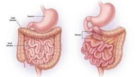
(On the left, a normal rotation on the right malrotated bowel).

Malrotation of gut is a serious health problem. Most patients experience intermittent incomplete bowel obstruction. Others develop volvulus and gangrene of the bowel. As soon as a diagnosis is made, surgical correction is needed, whether the patient has any symptoms or not.


Situs inversus is inherited as an autosomal recessive pattern, and at times it is linked with the X chromosome. Over 100 genes are implicated in this inherited disorder, but the NODAL gene mutation is best known for its clarity. In the very early development, the respiratory tract develops cilia. These cilia beat in unison in one direction - to the left. NODAL gene produces proteins which are components of ciliary axial structures. The directional movement of cilia creates a left to right orientation of the body. It directs the rotation of the gut and placement of viscera. Mutation of genes DNI 1 and DAH 5 result in Primary Ciliary Dyskinesia. Primary ciliary dyskinesia is associated with situs inversus, a well known condition is called Katagener's syndrome. Here patients develop chronic sinusitis, otitis and deafness, bronchiectasis, male infertility due to absence of ciliary movements in vas deferens, and later in life develop pulmonary insufficiency.

History of Situs Inversus.

It is reported that Aristotle noted the reversed position of viscera in animals. The first human case was reported by Fabricum in 1600, in a man with the liver on the left and the spleen on the right side of the abdomen. In 1663 Marco Severio reported dextrocardia and 1652 Rolan reported more cases of situs inversus.

Edited: November2025.

***************************************************
Wednesday, June 5, 2024

Changing Concept of Crohn's Disease


                                  Changing Concept of Crohn's Disease.

                                              P.K.Ghatak, MD


Crohn's disease is a chronic ulcerative granulomatous inflammation of the isolated segments of ileum, associated with some systemic manifestations. The cause of this inflammation has shifted several times -from syphilis, tuberculosis, autoimmune, genetics, rogue colon bacterial antigen derived immune reaction and unknown.

The Journal of the American Medical Association (JAMA) in October 1932 published a paper authored by Dr. B. B. Crohn describing a comprehensive summary of 14 cases of chronic diarrheal disease with histopathological findings. It was a new disease. He called the disease regional ileitis and speculated the etiology was a mycobacterial infection. The medical community named it Crohn's disease.

A review of literature showed Crohn was not the first person to publish this new disease. A short list of publications of the same illness appeared prior to the JAMA article is presented. Most of these are autopsy findings of patients with similar symptoms described by Dr. Crohn.

It is said that Alfred the Great of England and King of France Louis XIII died of complications of regional ileitis. Antonio Benivieni, in the fourteenth century, described autopsy findings of a chronic diarrheal disease as seen in regional ileitis. In 1769 Giovanni B. Morgagni described a similar case. In 1905 Wilmannis and 1907 Monyiham reported cases in BMJ. In 1909 Braun, 1911 Schmidt, 1912 Groto and 1924 Lawon published similar cases in German medical journals.

In 1912 Dr. Kennedy Dalziel of Glasgow, Scotland worked on regional ileitis.  It is now known that Dr Crohn worked based on Dalziel. Crohn, however, presented a comprehensive clinical and histopathological findings.

Characteristic features of Crohn's disease.

Men and women are equally affected, there is a genetic predisposition, but the precise nature of genetic mutation is unknown. Usual age of beginning of symptoms is late 20s. Exacerbation and remissions are features of Crohn's disease. Campy abdominal pain in the mid and lower abdomen and frequent bowel movements of blood mixed stool are initial symptoms. Other symptoms are a low grade fever, sclerites of the eyes, joint pains, and loss of weight.

Pathological findings- Skip lesions are typical (segments of the ilium are inflamed and in between sections of the ilium are completely normal). Granulomatous inflammation of all the layers of the ilium is a typical finding. The villi are atrophic and lymph follicular hypertrophy of the submucosal, muscular and serosal layers are thickened with granuloma formation. Fissures and sinus tracts are distinguishing features. Abscess formation, peritonitis and fistular communication with the urinary bladder and colon are complications.

Diagnosis of Crohn's disease is not difficult and usually made by endoscopy and biopsy of the lesions of the small intestine.

Another inflammatory bowel disease, Ulcerative colitis, should be excluded first because Crohn's disease can affect anywhere in the GI tract-from the mouth to the anus. Endoscopy in ulcerative colitis shows extensive inflammation of the entire colon and lesions are limited to the lining membrane of the colon only, sparing the other parts of the colon wall. And biopsy revealed acute necrotic lesion of epithelium of the colon, and no granulomas. Since granuloma is the distinguishing feature of Crohn's disease, a short review of granuloma is presented here.

Granuloma.

Macrophages and other immune cells appear at the site of invasion by infectious pathogens or the presence of a foreign body in the tissue. The macrophages encircle it and try to contain it from spreading. If macrophages fail to destroy it, more macrophages gather around it and summon other immune cells. The inflamed area takes the shape of a nodule. The center is formed by clusters of macrophages and is surrounded by layers of immune cells, chiefly by lymphocytes. In certain infections, the cells in the center of the nodule become necrotic, and in others no necrosis occurs. The first one is called Narcotizing granuloma, a typical finding in M.TB infection; the second one is called Non-narcotizing granuloma with no central necrosis, a typical finding in Sarcoidosis and Crohn's disease.

Causes of granuloma.

  1. Bacterial infection. Example- Listeria, Bacteriodes, Q fever.

  2. Mycobacteria tuberculosis are paratuberculosis (Non-TB mycobacteria).

  3. Virus infection. eg.- Epstein Barr virus, Cytomegalo virus, measles and mumps.

  4. Unknown antigen. -eg.- Scarcoidosis.

  5. Fugal infection. -eg.- Histoplasma capsulatum, Aspergillus species,

  6. Parasite. -eg.- Schistosoma sp.

  7. Foreign body. - Surgical sutures, talk, silicone and thorns.

  8. Cancer cells.

  9. Autoimmune diseases. -eg.- Lupus erythematosus, scleroderma.

  10. Special category,- Granulomatous angiitis, eg, Giant cell arteritis, Wegener's granulomatosis.

Etiology of Crohn's disease (CD).

The list of pathogens mentioned above is just a few in each category. After a diagnosis of CD is made, the first thing to do is to find the pathogen responsible for a granulomatous lesion in the GI tract. Microbiology and image studies are usually enough to exclude a known pathogen.

Heredity and gene mutation.

Mutations of several genes are suspected, but few are positively linked to CD disease. Mutation of the NOD2 gene (nucleotide binding oligomerization domain containing intra-cytoplasmic protein 2) which encodes a protein involved in apoptosis (cell death) is linked to CD.

ATGs (autophagy-related genes) encode many cytokines. When ATGs mutate proinflammatory activities proceed unchecked and result in inflammatory bowel disease.

A large section of each chromosome has no active gene, known as Junk genes. Newly discovered ETS2 gene, among the junk genes found to encode a protein which enhances Macrophage's activities.  Drugs that suppress the ETS2 gene help to heal the CD lesions in the bowel. It is now postulated that the mutated ETS2 gene is another inherited gene in the etiology of CD.

Mutation of these two genes and many other gene mutations are inherited by autosomal recessive and autosomal dominant modes.

Gut Bacteria.

Over 1000 different species of bacteria are present in the gut of any healthy adult. Among them, the dominant species are Firmicutes, Bacteroides, and Actinobacteria, and fewer are colonies of Protobacteria, Fusobacteria, Cyanobacteria, and Verrucomicrobacteria. Intestinal epithelium enters into a symbiotic relationship with these bacteria. Disruption of this relationship may happen due to antibiotic therapy and ingestion of other medications, certain illnesses, change in dietary habits, eating highly processed food and other environmental factors. Less abundant bacterial species multiply under this changed environment and break the intestinal barrier and invade the intestinal tissue. This results in activation of the adaptive immune responses and generates excess pro-inflammatory cytokines causing tissue necrosis and inflammation.

Bacterial antigen triggers antibodies by B-cells. Antibodies attack normal tissues instead of the pathogen due to an error of Th-2 cells (thymic 2) and the antigen complex may also trigger antibody production and tissue damage, a feature of autoimmune bowel disease.

Multiple factors.

Until a definitive etiology of CD is identified, a complex association of heredity susceptible gene mutation, disrupted intestinal microbiome, environmental factors, unidentified infective pathogen, autoimmunity and unknown factors are contributing to the development of CD.

***************************************************

Edited: November 2025.

Archive

  Archive. P.K.Ghatak, MD ghatak3@gmail.com 2025  March. Laser Endoscpoe 2025 February. Use of Ultrasound in Medicine MRI 2025 January. Bact...