Saturday, February 17, 2024

Prion


                                                        Prion

                                P.K.Ghatak,MD

Dr. Daniel Carleton Gajdusek in 1957, while studying a spongiform encephalitis called Kuru, in the local population of Papua New Guinea who were cannibals, proved that Kuru was an infectious disease when he successfully reproduced the disease by injecting the brain extract of Kuru victims in chimpanzees. He theorized Kuru was due to a slow growing unknown virus. It became evident that it was not a virus because Ultraviolet radiation did not kill the infective agent, on the other hand, was sterilized by a protein alerting chemical, bleach.

 In 1982, Stanley Prusiner of University of California at Sans Francisco called the infective agent "a proteinaceous particle and named it Prion.

Prion is simply a protein molecule made up of 259 amino acids. All living animals and plants, including yeast, have prions in the cell membrane and in the cytoplasm. Neurons have the highest concentration of normal Prion molecules.

Chemical compounds like toxins, snake poison, etc., are also compounds of proteins but Prions differ from toxins in that Prions multiply in huge numbers in the victim's body, whereas poison and toxins do not. The Medical community was naturally skeptical that an inert protein molecule could multiply in a victim without having Nucleic acids or DNA or RNA.

Dr. Prusiner proved that the infectious Prion (PrP sc), once introduced into the living cells it induces a change of configuration of normal similar protein molecules to adopt the shape and appearance of the PrPsc. The newly formed PrPsc, in turn, makes changes to more protein molecules and the chain reaction follows.

Normal protein molecule is alpha helical in their molecular folding, but abnormal Prion takes beta helical folding. Misfolded protein molecules prevent cells from carrying out normal cell functions, including elimination of metabolic wastes. Accumulated wastes clog the cells to death. The brain cells (neurons) have a high concentration of Prion molecules. As the misfolded cells die, they leave behind many small voids in the brain matter. Loss of vital functions of the brain cells results in progressive dementia, a movement disorder, and other symptoms, leading to premature end of life. The pathological process is called spongiform encephalopathy because of the resemblance with a sponge.

In the normal individual, the nature has provided a gene called the PRNP gene, that regulates the rate of conversion of misfolded proteins. When a mutation occurs in the PRNP gene, the mutation is passed to the next generation by an Autosomal Dominant fashion. Normal prion protein PRNP codon 129 also exists in polymorphic forms in association with variants Type 1 and Type 2 genes, which arise out of coding errors for amino acid methionine and valine respectively. The combination of these mutations results in 6 subtypes. This explains the variable penetration and low frequency of disease despite dominant inheritance.

Daniel Carleton Gajdusek and Stanley Prusiner received the Nobel Prize in medicine in 1976 and 1999 respectively.

These are the notations used when referring to a particular Prion and a prion disease.

Normal- PrP.  Scrapi - PrPsc.  Creutzfeldt-Jackob disease - CJD.  Variant CJD - vCJD, Familial - f CJD, Spontaneous -sp CJD.

*****************************************************


Diseases caused by prions in humans are

CJD Creutzfeldt-Jacob Disease.

vCJD (variant CJD).

Kuru.

Gerstmann Straussler -Scheinker syndrome (GSSS),

Fatal Familial insomnia,


In mammals prion causes Scrapie in sheep,

Mad-cow-disease in cattle.

Chronic wasting disease in deer, elk, moose, reindeer and caribou.

******************************************

Saturday, February 10, 2024

Pneumocystis Pneumonia

                                               Pneumocystis Pneumonia.

                                                  P. K. Ghatak, MD


In 1980, the medical community was challenged by an outbreak of a new disease that was rampant in homosexual communities in the major US cities. Within a few months, it was found to be a viral infection. The patients were the immunocompromised individuals and the virus was called HIV (human immunodeficiency virus). An interstitial pneumonia was almost constantly coexisted with the HIV infection. The organism was Pneumocystis jirovecii, a unicellular fungus, and the pneumonia was called PJP pneumonia.


The history of Pneumocystis is interesting.

In 1090 Dr. Chagas was in pursuit of the causative organisms of Trypanosomiasis of South America. He discovered a few tiny organisms in the respiratory tract of some of the victims. He thought those were immature Trypanosome trophozoites. In 1909, Antonio Carnii found 3 cysts in the lung of children which were similar to Chagas' trypanosome but he did not think they were trypanosomes. In 1910 Delano of the Pasture Institute discovered similar cysts in a rat's lung and found that the organism had many features of a protozoan and named it Pneumocystis carnii. In 1999, molecular analysis of mRNA and mitochondria of the Pneumocystis was conducted by Otto Jiroveci, a parasitologist from Czechoslovakia. He identified Galactosaminogalactan as a signature for a fungus. The organism now classified as a unicellular fungus. Pneumocystis exists in nature in three morphological stages – Protozoite, Sporozoite and Cysts (spores). Distinct genomic variability exists between host-specific members of the genera. For the humans, the pathogenic organism is called pneumocystis jirovecii. It was so named to honor Otto Jiroveci's work.


PJP pneumonia.

This fungus is ubiquitous, and exists as a parasite in the lungs of mammals and humans. The children acquire this fungus by 3 to 4 years of age. It spreads from person to person by being airborne. In good health, no adults or children show any ill effects of this parasitic fungus.

In severely immunocompromised individuals, with a CD4 cell count of less than 200 micro L, the Pneumocystis becomes an opportunist pathogen. In the lung, it produces an interstitial pneumonia. The infiltrates are a perihilar distribution, called a bat wing pattern. Like a viral interstitial pneumonia, the patients become severely hypoxic due to low oxygen diffusion capacity with a wide A-a gradient. Most severe infections produce acute respiratory syndrome. Various systemic symptoms develop due to tissue hypoxemia, specially cerebral manifestations. Other non-pulmonary features are – a. severe bone marrow depression producing pancytopenia, b. lymphadenopathy. c. cotton-wool exudates in the retina, d. thyroid gland enlargement and gastrointestinal symptoms.

Common symptoms are – fever, unproductive cough, chest pain, shortness of breath, cyanosis and tachycardia.

Diagnosis:

High LDH is a characteristic feature. Pneumocystis trophozoites and cysts are recovered by warm saline induced sputum and from bronchoalveolar lavage fluids.


Treatment:

Severely ill patients require tracheal intubation and mechanical ventilation.

PJP are very sensitive to Trimethoprim-Sulfamethoxazole (TMP-SMX). Usually given by IV in an acute situation. In tablet form, it is used as chemoprophylaxis. Alternative to TMP-SMX chemoprophylaxis are Dapsone, Atovaquone and aerosolized Pentamidine.


Prognosis:

Before Retroviral therapy, the mortality from PJP pneumonia was 80-90 %.  The mortality is 10 to 20%.at present.

********************************************


Friday, February 9, 2024

Amoebiasis


                                                             Amoebiasis

                                 Diseases caused by Entamoeba histolytica and other species.

                                                     P.K. Ghatak, MD


Amoeba: it is a unicellular organism. The cytoplasm of the amoeba is jelly like and contains mitochondria, other organelles, a large nucleus and various sized vacuoles. The cytoplasm is encased within a double layered membrane. The outer layer of the membrane is tough and elastic. Amoeba crawls peculiarly and is known as amoeboid movement. There are microvilli attached to the outer membrane, which prevent the amoeba from getting stuck to any solid surface, and the outpouring foot processes – pseudopods, help them float in water. Amoeba surrounds food particles by encircling it with pseudopods and incorporates it in its cytoplasm and after digesting food with various enzymes, the waste is eliminated and in the cytoplasm, this creates vacuoles of various sizes. Amoeba propagates by binary division. In rare occasions, it produces multiple identical daughters. The cellular form of the amoeba is called trophozoites, and it also exists in a cystic form. The cysts are the infectious form.

Three species of amoeba are pathogenic for humans.

Entamoeba histolytica. It is the primary amoeba causing intestinal diseases and its various other sequels.

Acanthamoeba. It causes keratitis of the eyes. People wearing contact lenses are susceptible to infection with this amoeba and if keratitis is not properly treated, it can produce a corneal ulcer and loss of vision.

Naegleria fowleri is responsible for a highly fatal disease, amoebic meningoencephalitis.


Entamoeba histolytica.

Humans are the primary host and in endemic areas the local people develop a partial immunity and 90 % of infections produce no symptoms because perhaps the people develop a kind of parasitic tolerance with the amoeba colonies in the colon mucosa. People who are immune depressed or on immune suppressed therapy, specially steroids, are vulnerable to severe illness. Visitors are susceptible to acute amoebic dysentery and often develop symptoms after returning home. The amoebic cysts are infectious and the mode of infection is the oral-fecal route. The incubation period is 2 to 4 weeks.

The cysts pass unchanged from the stomach to the terminal ileum, and here the cysts break open and release young trophozoites. Trophozoites release protein digestive enzymes and phagocytize the surface enterocytes. The amoeba is antigenic to humans. The body responds by both innate and acquired immune systems. This results in acute inflammation and formation of minute abscess in the colon. The abscess ruptures, forming ulcers. Ulcers cause a mucus dysentery, abdominal cramps and frequency of bowel movements. Low grade fever and other systemic symptoms also develop.

Direct extension of colon infection to the right lobe of the liver, through the right hemidiaphragm, produces liver abscess and then to right lower lobe consolidation of the lung, lung abscess and pleural effusion.


Complications and sequels.

The following complication may develop -

1. Colon perforation, toxic megacolon, fulminant colitis - specially in the immune compromised victims.

 2. Chronic non-dysentery colitis – specially common in the local population.

3. A localized mass formation in the colon called Ameboma. 

4. Anal and perianal fistula.

5. In some cases, the amoeba borrows through the colon wall and enters blood circulation, and distant organs are infected. This may result in peritonitis, ascites, pericarditis and pleurisy. Fallopian tube and uterine amoebic ulcers, left lobe of the liver abscess, abscess of the spleen and the brain.


Naegleria fowleri.

N.fowleri lives in warm water of lakes, ponds and untreated swimming pools. It enters the body through the nose. The amoeba penetrates the cribriform plate of the roof of the nose and enters the base of the brain, and a meningo-encephalitis follows. Because of the presence of a blood brain barrier, no amoeba killing drugs are effective in controlling the infection. This results in a catastrophe, and the fatality rate is over 90 %.


Diagnosis:

Examination of stool for amoeba cysts used to be the only reliable method. In the past, diagnosis of Entamoeba histolytica had happened due to another amoeba, Entamoeba dispar, which is far more common intestinal parasite, and it does not cause any human diseases, but the cysts are identical with the pathogenic amoeba species. The PCR can be used to detect amoebic systemic infections. At present, distant infections and the brain infections depend on this test.


Treatment:

In the past, Metronidazole was used indiscriminately and now in the endemic area, the amoeba is resistant to it. Other imidazole compounds also fell to the same fate. Emetine injections are discontinued because of cardiotoxicity. Chloroquine and other synthetic quinine compounds are in use in tissue invasive types of lesions like abscess in the liver, etc. Broad spectrum antibiotics are sometimes combined with Chloroquine.

A chat below, taken from the CDC showing the recommendation for Entamoeba histolytica infection:


Table. Recommended Treatment for Entamoeba histolytica Infection in Adults

Infection Type

Agent

Dosage 

Asymptomatic 

Paromomycin

25 to 30 mg/kg/d in 3 divided doses for 7 days

Invasive disease

Metronidazole

750 mg 3 times daily for 10 days or 2.4 g once daily for 2–3 days

Invasive disease 

Tinidazole

2 g once daily for 3 days

Amebic liver abscesses

Metronidazole

750 mg 3 times daily for 10 days

Amebic liver abscesses

Tinidazole

2 g once daily for 3–5 days

Amebic liver abscesses

Chloroquine* 

600 mg once daily for 2 days, then 300 mg once daily for 14 to 21 days 


************************************************************


Monday, February 5, 2024

Giardiasis


                                                              Giardiasis.

                                              Hill diarrhea, Beaver fever,  etc.

                                                        P.K. Ghatak, MD


Giardiasis consists of a number of gastrointestinal diseases caused by Giardia duodenalis. Giardia is a flagellated protozoan, present in the water of the lakes and small streams contaminated by human and certain animal feces.

Giardia exists in two forms - the trophozoite and cyst forms. The cysts have a tough outer shell which protects and makes them survive a long time. The cysts are the infectious form, and humans are infected when they eat and drink contaminated food and water. The outer shell of the cysts dissolves in the small intestine and each cyst releases two Giardia trophozoites. The trophozoites live in the lumen of the intestine by attaching to the surface cells and also live in the crypts of the intestinal villus processes. They extract food and nutrition from intestinal cells. Giardia multiply by binary fission.

The presence of giardia trophozoites inside the intestine provokes the body to react by activating CD4 cells and Interleukin-6 (IL-6) and other cytokines. The cytokines cause inflammation and giardia releases enzymes which break down proteins that bind surface cells of the intestine. This results in cell loss of enterocytes and destruction of villi. As a result of villus atrophy and a reduction of the surface area of the small intestine occurs.


[Protozoa means “ The first animal “. It is a unicellular organism, having a central nucleus but lacks a well defined cell wall and organized mitochondria in the cytoplasm. Protozoa exhibits free movement and predation behavior and lives independently as a free agent and/or as a parasite.]


Diseases produced by Giardia.

A. Acute gastroenteritis. B. Chronic diarrhea. C. Malabsorption syndrome. D. Irritable bowel syndrome, lactose intolerance and other sequels.


Acute gastroenteritis.

Within 2 weeks following infection, the patient develops acute explosive watery diarrhea of foul fishy smelling stool, several times a day. The stool contains flakes of denuded intestinal epithelium and undigested fat, which floats on the surface. Campy abdominal pain is common, a low grade fever is seen in some cases. Weakness and debility follow. Diarrhea may last for weeks and can turn to a chronic condition.


Chronic diarrhea.

Without treatment, diarrhea in general does not subside. Frequency of liquid stool lessens. Nausea and anorexia develop.


Malabsorption syndrome.

Reduction in number and height of villi resembles changes seen in tropical sprue and gluten enteropathy. Loss of nutrients, vitamins and minerals results in general debility, loss of weight and patients become susceptible to frequent infections.


Sequels:


Intestinal bacterial overgrowth, specially the pathogenic group, results in increased IgA production. Immune reactions manifest as arthralgia, muscle pain and weakness, irritation and headaches. Some patients have an aggravated immune disease like a flare-up of Crohn's disease.

Development of Irritable Bowel syndrome is thought to be an imbalanced growth of

beneficial/pathogenic bacterial colonies. Lactose intolerance is also common.


Diagnosis:

In acute diarrhea, the stool examination will detect Giardia trophozoites and a few cysts. In chronic diarrhea, the trophozoites have time to change into cysts. Consequently, diagnosis depends on recognition of cysts in the stool. Fluorescence antibody testing increases detection of giardia.

In malabsorption, endoscopic small intestinal biopsy detects typical atrophic intestinal villi. Lymphocyte infiltration of crypts and giardia trophozoites.


Treatment:

In any stage of infection, Metronidazole and analogs of Metronidazole used to be very effective. But overuse and misuse resulted in drug resistance. In such cases, some are using Albendazole. The WHO recommends Quinacrine and related synthetic quinine tablets.

In pregnant women, Paromomycin is used. FDA recommends Nitozoxanid for children.


Prevention:

Because sheep, cattle, mask rates. Beavers, dogs, and other rodents are infected by giardia; it is difficult, if not impossible, to protect lakes and small streams that flow down the hills into the valley. Chlorination of water does not kill the cysts of giardia. Boiling water and microwave radiation will destroy giardia cysts. Basically, it is the task of the Public health officials to educate people about waterborne diseases and how to protect themselves.

***********************************

Saturday, February 3, 2024

Whipworm infection


                                                                     Whipworm infection.

                                                              Trichuris trichiura infestation.

                                                                    P.K. Ghatak, MD


Whipworm is a roundworm and the sexes are separate. The worm is light pink in color and adult worms are about 2 inches long, females are longer than males. The mouth part of the body is thin and the bottom part of the female worms is thicker and rounded, whereas it is thin and coiled in males, resembling a whip that gave them the name Whipworm.

The worms live in the large bowel, from cecum to rectum, and can live up to a year. Within 3 months of ingestion of infected eggs, the female worm begins releasing 20,000 eggs in the stool daily. Using human waste as fertilizer and open air defecation made Whipworm infection more widespread. The WHO estimates about 1 billion people are currently infected with this round worm.

Mode of infection and people at risk:

By ingestion of matured eggs.

Poor people in poor countries, due to lack of education and access to clean water and having poor hygienic practices, are susceptible to infection and reinfections; children are specially so.

Chronology of symptoms:

Most infections or ingestion of eggs are asymptomatic. Eggs hatch and move to the small intestine. The newly released larvae penetrate the mucous membrane of the small intestine and mature into adult worms in 3 months. Some patients develop mild abdominal pain and diarrhea but most people do not feel any symptoms. The adult worms take up permanent residence in the large bowel wall. Heavy infection is the usual. Many adults and specially children develop a frequency of bowel movement and the stool is mixed with heavy mucus and some blood. Children can become very sick with rectal prolapse, nutritional deficiency, bloody dysentery and anemia.

Diagnosis:

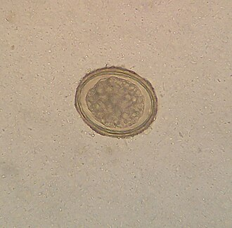
Stool examination shows typical eggs – barrel shaped brown colored eggs with distinct plug like ends


.

Treatment:

One dose of Albendazole kills all round worms. But reinfection is common.

Prevention:

Educating the public about sanitation and prevention of fecal-oral infection is an essential part of the eradication program of this common worm infection.

********************************************************


Wednesday, January 3, 2024

Tapeworm Disease

                                                        Tapeworm Disease.

                                                            Taeniases.

                                                   P.K. Ghatak, MD


Tapeworm infestation of humans was a universal disease in the old world, but improving the public health measures and general sanitary practices have eliminated this infestation from most of the countries of the world, except the tropical/subtropical poor nations.

Tapeworm belongs to the flatworm group. Adult tapeworms are usually 15 to 30 feet long. It has three distinct parts of the body – head, neck, and segmented body. The head, under the microscope, looks like an ugly monster with hooks and suckers. The body is segmented and each segment of the body is self-sufficient in reproductive units and it contains multiple eggs. The last few segments are shed in the feces and eliminated from the body during defecation.

Humans are infected with these species of tapeworms: Taenia solium (pork tapeworm), Taenia saginata (beef tapeworm), Diphylloborthium latum (fish tapeworm), Taenia asiatica (Asian tapeworm) and Hymenolepsis nana (dwarf tapeworm).

Tapeworms are responsible for two distinct illnesses in humans.

 A. Cysticercosis.

 B. Intestinal Tapeworm disease. Not all the worms listed above cause both diseases, but Taenia solium does.


Cysticercosis:


Cysticercosis is a serious illness due to the development of multiple cysts in the brain and eyes. Cysts may also develop in other organs but are called by different names, for example, echinococcosis is a cyst in the lungs.

Mode of infection.

This is primarily due to the contamination of food and drinks by human feces from bad sanitary practices by the individual, leading to self infection, or, from others like cooks or food handlers.

The ingested eggs in the intestine transform into spherules, penetrate the wall of the small intestine and enter the blood circulation. The circulation takes them to other organs. In the brain or eyes, the spherules develop a thick wall and secrete fluid within the cavity, and the cysts are known as Cyticerca. Most of the time, the cysts produce daughter cysts and cluster together. This causes an increase in intracranial pressure and the patient develops seizures. In the eyes, the cysts produce blindness.

Other neurological symptoms develop from damaged brain tissue by the cysts, and symptoms depend on the location of the brain cysts.

Diagnosis:

The cysticerci are easily diagnosed by MRI of the brain.

Treatment is anti-seizure medication for epilepsy followed by surgery. During surgery, care should be taken not to nick the cysts. The cystic fluid is very allergenic and can produce anaphylaxis, and daughter cysts will spread further.


Intestinal tapeworm disease.

This is the usual life cycle of the tapeworm.

When humans consume poorly cooked pork containing tapeworm cysts, the digestion of meat frees the cysts from the muscle. The cysts just open up and invaginate and attach themselves to the intestinal wall and begin to draw nutrition from the host and mature into adult worms. A tapeworm usually lives 7 to 10 years. And begins producing segments full of eggs within a short few months after entering the GI tract of the host.

Consequences of Tapeworm infestation of the intestine.

Most patients are not aware of the presence of the worms. However, children show signs of malnutrition and nutrient deficiencies. Occasionally the worm can enter the bile duct or pancreatic duct and cause Choledochocystitis or pancreatitis respectively. Similarly, appendicitis, bowel rupture and peritonitis also occasionally occur.

Diagnosis:

Stool examination detects the characteristic eggs of the tapeworms.

Treatment – Drug treatment is very effective and these 3 drugs are often used. Praziquantel, Albendazole and Nitazoxanide. Praziquantel is the preferred drug because it completely dislodges the parasite from the intestinal wall.

Prevention is essential and is very effective and costs practically nothing. Public education and proper handwashing with soap and water can wipe out intestinal tapeworm disease, and properly cooked meat and pork can control the spread of cysticercosis.

********************************************************

Monday, December 11, 2023

Blood Fluke


                                                          Blood flukes

                                                    P.K. Ghatak, M.D


Blood Fluke infection is the 3rd flatworm (Trematode) parasitic disease of humans discussed here, Liver fluke and Lung fluke were the other two mentioned in the earlier blogs.

Schistosoma hematobium is the parasite and the disease produced by the worm is called Schistosomiasis, also known as Bilharziasis and Snail fever. Schistosoma means a split - body. The shorter and stouter adult male has a longitudinal cleft along the length of the body and in that space, a slender, longer female worm is held in embrace by the male for its life. A German doctor, Theodor Maximilian Bilharzia, while serving in Egypt, discovered the flatworm parasite in 1851, and the disease is named after him.

Two distinct illnesses are produced by Schistosoma species.

  1. Urogenital Schistosomiasis. Produced by S. hematobium. The worms reside in the venules (small veins) of the urogenital organs.

  1. Gastrointestinal & Biliary Schistosomiasis. Several Schistosoma species are responsible and they are named according to the initial case originating country. The parasites invade the venules of the GI and biliary systems.

The life cycles of all the species of Schistosoma are identical and closely resemble those of the other trematodes.

Humans are the final host and act as the reservoir of the parasite. Other primates, ruminant farm animals and rodents are additional reservoirs.

Incidence of Bilharziasis.

The WHO estimates 200 million active cases are seen annually. Genitourinary bilharziasis is common in the Nile River Valley of Egypt and the adjoining northern African countries. Gastrointestinal/biliary bilharziasis occurs in China and countries surrounding the South China Sea. It is also prevalent in South America and the Caribbean islands. Indigenous bilharziasis does not happen in the USA.

Life Cycle of Schistosoma.

Human and animal feces and urine contaminate rivers, lakes and other bodies of water. Digging canals for irrigation has spread the risk of bilharziasis in much wider areas. Inundation during the rainy season is another hazard.

Schistosoma development stages are - Egg, Miracidia, Sporocyst, Cercaria and Adult.

Eggs: An embryonated egg has a spine, and the position of the spine distinguishes one species from the other. Each egg is elongated or oval, measuring about 175 X 45 micrometers. Inside an egg, one embryo is in the development stage. A hinged door at the head-end opens and lets a grown larva out in the water.

Embryos: Miracidia larva is 200 micrometers long and covered with cilia and is a free swimmer. Miracidia larva lives in the water only a few hours and must find the proper snails that live in sweet water to multiply and develop further into infective larvae.

Sporocytes: Miracidia after entering inside the snail, move to softer tissue and transform into cysts. Cyst develops many daughter cysts and the daughter cysts move to newer locations and continue to develop into Cercaria larvae.

Cercaria larva is 500 micrometers long, has a tapering head and a forked tail. Cercariae live only 3 days. It takes only a few minutes for cercariae to enter into the body of their victims by penetrating the skin. It drops its tail and moves inside the veins. Inside the blood vessels, it becomes a round ball and is carried by the blood to the heart, lungs, and finally to the liver. In the liver, cercariae develop in about 3 weeks into adult male and female worms.

Adult worms: Adult worms are 7 to 29 mm long, have a cylindrical body, colored gray-white, have two suckers and an alimentary canal but no anus and the body is filled with reproductive organs.


Subsequently, the life of Schistosoma depends on the species and victims.


Schistosoma species

Definitive host

Site of infection

Eggs discharged in

Endemic area

S. hematobium

Humans, other primates

Genitourinary system

Urine

Africa

S. japonicum

Humans, carnivores, ruminants

GI & biliary mesenteric veins

feces

South-East Asia

S. mansoni

Humans, rodents

As above

feces

Africa

S.mekongi

Humans, dogs

As above

feces

South-East Asia

S. intercalatum

Humans, rodents and cattle

As above

feces

South-East Asia

Symptoms: The initial infection produces no symptoms. Some people develop an itch at the skin penetration sites and it is called swimmers' itch. Some others develop eosinophilia and patchy pneumonia.

Acute symptoms. Eggs produce allergic reactions known as Katayama fever. It manifests as fever, weakness, fatigue, and lymphadenitis.

Abdominal pain, low grade fever and eosinophilia develop in others.

Chronic symptoms. Many eggs are carried away to different organs. The eggs get embedded in the tissues and generate immune reactions. The initial inflammatory reaction is followed by tissue necrosis, fibrosis and granuloma formation. Small granulomas coalesce together into polyps.  Polyps are seen in the urinary bladder and esophagus, stomach and intestine.

Genitourinary Bilharziasis: Hematurrhea, painful urination, urinary tract infection, glomerulonephritis, deformed external genitalia, calcified lesions surrounding the embedded eggs in tissues, specially in the urinary bladder, are common. Carcinoma of the bladder also develops. Fibrosis of the various components of the reproductive organs leads to difficulty in pregnancy and miscarriage. Blood in semen in a male patient is a striking feature.

Gastrointestinal and biliary bilharziasis:

Abdominal pain and diarrhea, dysphagia, bleeding varies in the esophagus and stomach producing anemia and malnutrition. Liver and spleen enlargement, anemia and leukocytopenia develop due to hypersplenism. Calcifications of blood vessels lead to various ischemic symptoms.

Distal organ involvement:

CNS. In the brain, the eggs are calcified and cause seizures, headaches and paralysis of limbs.

In the spinal cord: Transverse myelitis is a serious problem

Lungs: In the chronic stage, Pulmonary artery stenosis and calcifications produce pulmonary hypertension.

Heart: Myocarditis and heart failure occur.

Diagnosis: Stool examination detects eggs and the shape of eggs and the characteristics of the spine help diagnose the Schistosoma species.

Egg characteristics:


S.hematobium – Eggs are oval-shaped, the spine is long and sharp and attached to the terminal end.

S. japonicum - Eggs are round. The spine is rudimentary and appears like a hook and is attached to the lateral side.

S. mansoni –Eggs are elongated and the spine is attached at the posterior end of a side.

S. mecongi – Eggs are 30–45 micrometers long and have a short lateral spine.

S. intercalatum – Eggs are oval-shaped and the spine is attached to the terminal end; Its eggs resemble S. hematobium eggs.

In S. intercalatum infection, few eggs are excreted in the feces. The infection is in the lower colon and rectum. A biopsy of the rectum is required.

In CNS infection, more reliable tests are ELISA antibody test and parasite DNA identification by PCR test.

Treatment: Praziquantel is a very effective drug in killing adult worms but immature worms are not killed. A person is usually infected repeatedly, and both mature and immature adult worms are present at the same time. So Praziquantel is repeated weeks later. The Cure rate is 90 %.

Special features of Schistosoma. The adult worm does not produce inflammation or allergic reactions, only the eggs are allergenic.

The adult worm is classified as a flatworm, but it resembles round worms. The worms,  unlike other flukes, are not hermaphrodites, the male and female sexes are separate.

****************************


.


in other primates, ruminants, rodents and cattle.


Adult  The adult worms are 1–2 cm long with a cylindrical body that features two terminal suckers, a complex tegument, a blind digestive tract, and reproductive organs Schistosoma spp. [these species cause schistosomiasis/bilharzia in humans and ruminants]


Parasite morphology: Blood flukes form five different developmental stages: eggs, miracidia, sporocysts, cercariae and adult worms. Eggs are round to oval in shape, operculate (hinged at one end) and contain a developing embryonic larva (miracidium). Differences in egg morphology can be used to distinguish between Schistosoma species: S. mansoni producing oval eggs (115-175 x 45-7µm) with a sharp lateral spine, S. japonicum forming round eggs (70-100 x 50-70µm) with a rudimentary lateral spine; and S. haematobium producing oval eggs (110-170 x 40-70µm) with a sharp terminal spine. Miracidia are elliptical free-swimming larval stages (~200µm long) covered with cilia. Sporocysts appear as pleomorphic sac-like bodies which contain developing cercariae. Mature cercariae are elongate free-swimming larval stages (400-600µm long) consisting of a tapering head (with prominent penetration glands) and a forked tail (furcocercous). Adult flukes are elongate tubular worms (10-20mm long), with rudimentary oral and ventral suckers. Males are shorter and stouter than females, and they have a longitudinal cleft (gynecophoral canal or schist) in which the longer slender female lies folded.


Theodor Maximilian Bilharz 

Regarding Katayama syndrome (a possible clinical manifestation of schistosomiasis in naive patients characterized by fever, cough, myalgia, headache and abdominal tenderness [19]), descriptions fitting its clinical manifestations can be found in ancient books of traditional Chinese medicine referring to more than 2400 years ago

Wednesday, December 6, 2023

Ascaris lumbricoides


                                         Ascaris lumbricoides infestation

                               P.K. Ghatak, M.D.


Ascaris lumbricoides is a foot long intestinal parasite. The human infection has been known since ancient times. The first written record of human infection was given by Linnaeus in 1758.

Ascaris lumbricoides is a roundworm; the sexes are separate and the female worm is larger. Adult worms live in the small intestine of humans. Ascaris also infects pigs, monkeys and other animals. A female ascaris lives one to two years and can lay 200,000 eggs a day. The eggs are mostly fertile, but unfertilized eggs are also present in the stool of the victims. If a female worm does not find a male worm, it moves around, and crawls into the throat. If it enters the larynx, it causes respiratory distress and a bout of violent cough till it is dislodged. It may also come out of the nostrils.


People at risk:

The WHO says about 1 billion people are at risk of infection. The infective state of the worm is the fertilized 3rd stage eggs or embryonated eggs. Unlike any other parasite, ascaris eggs are well protected by their thick outer shell and can live for 3 years in the soil. People unknowingly swallow eggs carried on their fingers due to their unhygienic habits or ingestion of contaminated water and food. Humans and farm animals act as reservoirs of ascaris.


Life history of Ascaris:

The fertilized eggs are deposited in the soil, either by spraying the human waste on the agricultural land as fertilizer or by people defecating out in the open. The eggs undergo further development in the soil and become embryonated eggs in 15 -18 days.

Children playing in the dirt carry soil and eggs on their fingers or under their nails. The eggs enter the mouth when children eat with their fingers. The same way, adults are infected or, contaminated food and drinks by the food handlers.

Inside the duodenum, the larvae emerge. The larvae move to the lungs for further development. The larvae reach the lungs by way of the Portal vein and finally in the pulmonary circulation. In the lungs, the larvae become juvenile worms in about 15 days. The juvenile worms journey back to the small intestine by way of the trachea and are coughed up and swallowed by the victim. Back in the intestine, for the second time, they reach maturity and mate with the opposite sex and begin laying eggs in 3 months from the time of infection.

Researchers are not sure why ascariasis larvae must travel to the lungs. Several reasons are put forward but still remain unknown.


 Human disease.

During initial infection. Most of the victims are unaware of infection and remain symptom free. Some develop abdominal cramps and diarrhea.

During migration to the lungs. This period is generally asymptomatic.

The period of stay in the lungs. Symptoms are mostly due to the physical presence of foreign bodies in the airways and all related pulmonary complications, secondary infections and allergic asthma with eosinophilia.

During migration from the lungs to the small intestine. Cough and the horror of live worms being coughed up.

Small intestinal stay. Though the worms do not suck blood like hookworm, nevertheless,  they steal nutrients from the children and children become stunted and malnourished. Motile worms in the intestine can enter the bile duct, gall bladder, and appendix, causing acute obstruction and infection. A heavy parasitic load in the small intestine causes bowel obstruction.

During adult female searching for a male worm. When the worm reaches the pharynx and sometimes enters the larynx, cough and respiratory distress develop.


Diagnosis:


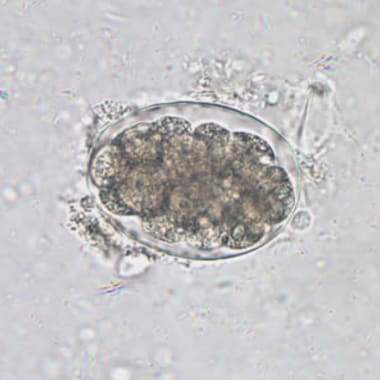
Fertilized ascaris eggs are diagnostic. Unfertilized eggs sink, while fertilized eggs float in water. Each egg is oval to round in shape, measuring 75 x 50 micrometers, and has a thick mamillated outer shell and is usually stained brown by bile.

Adult worms are also diagnostic. The worms are long, slender, cylindrical, unsegmented and colored light yellow. The mouth end is surrounded by 3 lips. The male worm is 30 cm long and 4 mm in diameter, and has a curved tail end. A female measures 40 cm (over a foot long) X 6 mm. The genital aperture is in the upper third of the body, and 2/3 of the body contains sacs containing 25 million eggs.


Treatment. Albendazole and Mebendazole are effective in killing the adult worms. A daily dose for 3 days is recommended for cure. Reinfections are common in endemic areas.

***************************************************


Monday, December 4, 2023

Hookworm


                                                      Hookworm.

                                                   PKGhatak, MD.

The name Hookworm is given to this roundworm because the worm has anchoring teeth or hard plates in the mouth to latch onto the interstitial wall of its victims. An adult worm is 8 mm long and the female is 11 mm. The worms are round in shape, have a long thin unsegmented body with tapering ends, and are pale creamy color. Each worm carries both sex organs. The outer wall is made of a tough cuticle

Human diseases produced by hookworm:

  1. Iron deficiency, anemia and protein malnutrition

  2. Allergic skin lesions and eosinophilia. Cutaneous larva migrants

  3. Eosinophilic enteritis.

The life cycle of hookworm.

Human excreta contaminates the soil, where hookworm infection is common. Children playing bare feet on the fields or farmers working on their fields, come in contact with the infective stage of hookworm larvae. It takes only 5 minutes for the larvae to find the tiny hair openings and or penetrate the skin. In the dermis, the larvae begin migrating to locate the capillaries or venules and enter. The blood carries the larvae to the right side of the heart and then into the pulmonary alveolar capillaries. This part of the migration takes 10 days. The larvae penetrate into the alveoli and enter the airways. Ultimately, the larvae enter into the major airways and are coughed up and swallowed. In the stomach, they are protected by the outer tough cuticle. Once inside the small intestine, the larva molts twice and develops a buccal capsule with teeth and a muscular esophagus. The larvae attach to the mucous membrane of the intestine from the lower duodenum and upper ileum. The larvae secrete tissue resolving enzymes which dissolve tissue and expose the blood vessels. The larvae bite and remain attached to the blood vessels of the intestine with their teeth or buccal plates. In humans, the parasites do not multiply.

The larvae quickly grow and attain maturity in a month and after mating, begin to release eggs. A female can release 30,000 eggs daily. Adult Nicator worm lives up to 5 years and Ancylostoma species for one year only.

In the soil.

The eggs release the immature Rhabdotiform larvae in a day or two. The larvae feed on the excreta and molt twice. In 7 days, rhabdotiform larvae become the infective Filariform larvae. The filariform larva has a short life outside and dies if unable to find a victim within 3 weeks.


;


Diagnosis:


The stool test for eggs is commonly done for the diagnosis of hookworm infection. The eggs with multiple larvae are diagnostic. Each egg measures 60 x 40 micrometers. The usual practice is to release the eggs from the fecal matter by placing a portion of the stool sample in a glass jar, diluting it with water and agitating it thoroughly. The eggs float on the top of the water and are collected by merely touching the top of the water with a glass slide.

PCR tests are available but are hardly utilized.

When the larvae are in migration and until the adult worm starts producing eggs, the stool tests will be negative. The PCR tests are needed for confirmation,

Treatment.

Albendazole is effective and requires a daily dose for 3 days or just one heavy dose.

Iron and vitamin supplements are also required.

Infection of hookworm does not provide protection from future infection. Vaccines are developed but not commercially available.

Epidemiology.

According to a 2010 WHO report, about 120 million people had hookworm infection. The countries where most cases are seen are poor - spanning from Sub-Saharan Africa to South Asia, East Asia and the Americas.

Species infective to humans.

Necator americanus infestation is the most common in humans, taking the world as a whole, followed by Ancylostoma duodenale.

Other hookworm species infective to humans are Ancylostoma ceylanicum,

Ancylostoma caninum

Ancylostoma braziliense.

A. barziliense commonly produce eosinophilia with creeping skin lesions called Cutaneous larva migrants.

Besides the common mode of infection, some species can infect humans via contaminated drinking water and breast milk in children. Ancylostoma craninum usually remains dormant in the skeletal muscles and produces eosinophilic dermatitis, bronchitis and asthma. Then move to the intestine to propagate.

Amount of blood loss and anemia.

It is reported that each adult Necator worm consumes 0.03 ml of blood and 0.2 ml of blood by Ancylostoma species. In addition, blood is also lost in the stool. On average, in a heavy infestation, humans lose 1 ml of blood per day per adult worm. In general, many dozens of worms live in the intestine at the same time. A heavy infestation is determined by the presence of over 4,000 eggs /gram of feces.

It does not take much time for the children to become severely anemic and the hemoglobin levels fall to 4 - 5 grams/dl.

An adult worm lives 1 to 5 years based on the species. An infestation of hookworm does not produce immunity against future infections.

Vaccines are known to protect against fresh infections in people,  but are not utilized and not produced commercially.

**************************************

Friday, December 1, 2023

Lung Flukes


                              Lung Fluke- Paragonimus westermani

                                          PKGhatak, MD


Humans are infected by Paragonimus westermani from eating uncooked or poorly cooked crab, crayfish and pork. Metacercariae stage of the parasite travel from the intestine to reach the lungs through the diaphragm. And within a month of reaching the lungs, the worms reach maturity and a pair of adults cross fertilize each other and begin laying eggs in the bronchus. The eggs are coughed up and spat out or swallowed. Eggs hatch in water, and newly released miracidia seek out snails. Within the muscles of the snails, the parasites multiply and develop into a motile form of cercariae.  Cercariae leave the snail. Circariae moves along the bottom of rivers or lakes and finds the 2nd intermediate hosts – crabs, shrimps and crayfish. Again, the parasites multiply and develop into infective metacercariae and are ready to infect humans or any mammals of the cat family.


In East Asia, lung fluke infections are mostly seen, however, the lung fluke infection is prevalent in a wide area of the world, wherever the people eat raw crab meat, or pickled crab and shrimps, and raw crayfish. It is estimated that 200 million people are at risk of lung fluke infection.


Symptoms and diseases produced by paragonimus parasites:

Following ingestion, the eggs hatch in the intestine and migrate to the lungs. For 2 days to 2 weeks, during migration of the parasites, the patients have symptoms of abdominal pain and diarrhea. These symptoms are followed by fever, chest pain, cough, rusty sputum production, blood eosinophilia and various forms of infiltrates in the lungs detected by chest x-rays. Occasionally, pneumothorax and a small pleural effusion develop.

Weeks later, the main symptoms become chronic cough, with a low grade fever, fatigue and sputum containing traces of blood, resembling pulmonary tuberculosis and or chronic bronchitis with bacterial infection.

In heavy parasite infection, in 25% of hospitalized patients, the parasites move into the cranial cavity and produce symptoms of meningitis and meningoencephalitis, and seizures. Occipital and temporal lobes are commonly infected. Diplopia to blindness develops. Characteristic soap bubble like pictures are seen within the lateral and occipital ventricles of the brain on CT or MRI of the brain. The CSF examination shows high eosinophil counts.

 Diagnosis.

Sputum examination detected P. westermani eggs, each one measures 80 -120 x 4 – 6 micrometers, and are diagnostic. Specific IgM and IgG ELISA tests are also used, specially in CNS infections.

 Treatment:

Praziqyental orally for 3 days is quite effective.

 Anatomy of P.westermani:


The four stages of the parasite are as follows: Eggs, Cercaria, Metacercaria and adult worm.

Eggs – An egg is 80 -120 x 40-60 micro M, brown in color, and resembles a coffee bean. 

Cercaria and Metacercaria are similar in size and appearance to any other Trematode worm,

Adult – the adult worm is 7 -12 mm long and 4 -6 mm wide. The outer wall is covered with scales like spines. It has two suckers like any other Trematodes, and each one has both the male and female sex organs. But unlike Liver flukes, Lung flukes meet as a pair and cross-fertilize each other. Adult worms produce fibrous cavities in the bronchi filled with their excreta and altered blood


Life Cycle of P. westermani.

In general, P. westermani needs a snail as an intermediate host, but unlike the liver fluke P. westermani, it requires another host – crabs and crayfish for the multiplication and development to an infective form. P. westermani uses any locally available snail species, unlike liver flukes.

************************************************************

.


Tuesday, November 28, 2023

Fasciola Hepatica


                                                    Fasciola hepatica

                                                    Liver fluke

                                 PKGhatak, MD


Fasciola hepatica is a flatworm, belongs to the Trematodes. It infects humans and other ruminant mammals.

Three other species of liver flukes, namely Chonorchis sinensis, Opisthorchis viverrini and Opisthorchis felineus have similar life cycles and modes of human infection.

In endemic areas, the liver fluke infects the hepatobiliary system and is an important risk factor for Cholangiocarcinoma (CCL). According to the WHO, people in areas of the Mekong River basin, the liver fluke infection is between 30 and 70 % of the population and the death rate from Cholangiocarcinoma is 3 %. CCL is the second most common cause of death in East Asian countries. Thailand has the highest rate of CCL, about 40 in 100,000 people, followed by China, Japan, and other East Asian countries.

The anatomy of Fasciola hepatica.


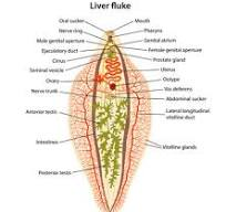
The adult worm is 3 to 8 cm in length and looks like a leaf. It has a tough outer membrane. The mouth functions as sucker and in addition has an anchoring sucker on its ventral side. It has no inner body cavity, it lives on the blood of its victim. Both male and female reproductive systems are present inside the body, and a worm usually lives for 9 years inside the bile duct of humans and mammals.

Life cycle:

Each fasciola releases 20,000 to 25,000 eggs daily in the bile. The eggs are plentiful in the stool. The eggs, when they come in contact with water, transform into embryonated eggs and seek out snails. Inside the snail, the embryos develop into elongated larvae with a long tail. This stage of the parasite is known as Miracidia. Miracidia leave snails and swim by undulating their tails to the nearby vegetation or fish, crabs and crayfish. In this second host, the larvae transform into cysts, called Metacercariae. Grazing animals or humans eat contaminated water cress, other vegetation and Crustacea and become infected.

In human victims.

The outer wall of the cysts prevents digestion in the stomach, and the cysts are propelled into the duodenum. Here the eggs are released. The larvae burrow through the intestinal wall and enter the peritoneal cavity and move to the liver. The worm grows rapidly in the liver and then enters the bile duct system and takes its final residence in the common bile duct or its branches. Within 90 days of entering the human body, the liver flukes become adult worms and begin releasing eggs in the bile.

Diseases produced by Fasciola.

Many people remain symptomatic in the early stage of infection. Others develop abdominal pain, nausea and mild malaise within 4 to 7 days when the larvae are migrating from the intestinal wall to the liver. An enlarged, tender liver and abnormal liver enzymes and eosinophilia are usually detected.

This period may last about 7 days and may last for months.

In the chronic stage. Months may pass without any symptoms. Then symptoms of biliary disease begin to occur.  The common symptoms are related to gallstones, Cholangio hepatitis, hepatitis, allergic skin lesions, and the blood eosinophil count is high, which are usual symptoms. In heavy infestation of the bile duct, obstructive jaundice may occur, but usually, a secondary infection is the usual cause of jaundice.

The most serious illness from the liver fluke infection is Cholangiocarcinoma. The bile wall mucosa is damaged by the worm, endothelial metaplasia and fibrosis become recurrent and chronic. This leads to cancer development. Patients seek medical help in a late stage when surgery is not feasible or if performed,  the results are not good.

Diagnosis:

Examination of stool is a simple test but diagnostic. In very early cases of suspected, asymptomatic infection, the saliva, urine and blood tests reveal the presence of Fasciola antigen. Antibody test in 3 weeks post infection becomes positive.

In advanced countries like Japan, Taiwan and others, endoscopy, ERCP, and MRI are available to detect parasites in the hepatic and biliary system.

Medical treatment.

Triclabendazole is very effective and requires only two oral doses.

People at risk:

A large population from Malaysia to Japan, China and Russia is at risk of infection of liver fluke infective cysts. Humans are accidental victims due to their lifestyles or lack of knowledge. The liver of sheep, goat and water buffalos when eaten raw or barely cooked is another source of infection besides drinking water contaminated and other food items already mentioned.

***************************************************


Archive

  Archive. P.K.Ghatak, MD 2025  March. Laser Endoscpoe 2025 February. Use of Ultrasound in Medicine MRI 2025 January. Bacteriology and Rober...