Saturday, April 29, 2023

Sugar and Diabetes mellitus

 11 Draft.


                                             Sugar and Diabetes mellitus.

                                              PKGhatak, MD

Sugar belongs to carbohydrates and is essential for humans. Sugar is classified as a monosaccharide and a disaccharide. Monosaccharides are Glucose, Fructose and Galactose. Disaccharides are composed of two molecules of monosaccharides and are Sucrose, Maltose and Lactose. When disaccharides break down in the gastrointestinal tract by specific enzymes, they yield the following monosaccharides.

Sucrose = Glucose + Fructose.

Maltose = Glucose + Glucose

Lactose = Glucose + Galacose.


All monosaccharides are six-carbon compounds. Fructose is the sugar of fruits and vegetables. The common source of fructose is cane sugar, table sugar, beet sugar, and soft drinks. Milk and milk products contain lactose. Maltose is a man-made disaccharide, obtained mostly from barley grain.

Glucose and fructose resemble each other both chemically; the only difference is that glucose is an aldehyde and fructose a ketone. Maltose is also an aldehyde like glucose and maltose is an Epimer of C-4 glucose. Epimer differs only in the position of the OH group of one C atom. All three monosaccharides are present as Isoforms (L & D isomers).

Carbohydrate metabolism:

The energy required for sustaining life comes from ingested carbohydrates, fat and proteins.

The breakdown of complex carbohydrates ( Cellulose, starch, and dextran) starts in the mouth by the enzyme - salivary amylase. The breakdown of carbohydrates continues in the intestine by the pancreatic amylase and various other enzymes, including Lactase, which splits Lactose into glucose and galactose. Many adults have Lactose intolerance due to either acquired lactase deficiency or congenital deficiency of lactase.

Glucose, fructose and galactose, once formed inside the lumen of the small intestine, follow their own path of metabolism. There is similarity and also a different paths of the three sugars.


                                 Glucose.

Glucose is ferried across the gut lumen into the intestinal enterocytes by the Sodium/glucose co-transporter 1 (SGLT1). SGLT1 is a member of the solute carrier family SLC 5 transporter. SGLT1 also selectively reabsorbs glucose completely from the glomerular filtrate in the proximal tubule, unless the amount of glucose in the filtrate exceeds the maximum reabsorptive capacity of SGLT, as that happens in diabetes mellitus.

Glucose utilization.

In the liver, glucose is converted into glycogen.

Glucose is taken up by all living cells by the action of Insulin. Insulin fuses with the cell membrane and creates an opening for glucose molecules to enter the cell cytoplasm and then glucose molecules are taken up by the mitochondria. Glucose is used up in the tricarboxylic cycle and produces energy, generates ATPs and the end products are CO2 and H2O.

In the muscles: Some glucose is reserved in the muscles as glycogen for future use. Most of the glucose is utilized as fuel.

Hexose monophosphate shunt (HMS):

HMS is also known by other names like pentose phosphate pathways. It takes two separate paths -

1. Aerobic. This path leads to the generation of energy and NADPH (nicotinamide adenine dinucleotide phosphate). NADPH is required for the biosynthesis of fatty acids, cholesterol, and neurotransmitters.

2. Anaerobic. This path leads to the production of pentose sugars. Pentose sugars are components of Nucleic acids and nucleotides. Also through the anaerobic path, Erytrose 4 phosphate is produced. Erytrose 4 phosphate is a component of aromatic amino acids.

                                      Fructose:

Important dietary sources of fructose are fruits, honey, high-fructose corn syrup (soft drinks), and table sugar (sucrose). The enzyme, Sucrase, breaks sucrose into glucose and fructose in the intestine.

Absorption.

GLT transporter is used for fructose absorption from the gut lumen by GLT 2 and from the enterocytes to the portal vein by GLT 5 transporter. There is no fructose in the peripheral blood or in the glomerular filtrate

Fructose utilization.

Fructose is taken up mainly by the liver and only a limited amount by the kidneys, adipose tissues, and muscles. In the liver, fructose is converted into glycogen and all excess fructose is used in the synthesis of Triglyceride. The majority of body cells are unable to utilize fructose directly, and all the reactions involved in fructose metabolism take place outside the mitochondria. And that too is limited to the liver, adipose tissue, gut and testes.

There are two paths for Fructose utilization:

Hexokinase.

In the muscles and fatty tissue, fructose is phosphorylated by fructose-6 phosphatase and then enters the glycolysis via the tricarboxylic acid cycle. Insulin is not required for fructose metabolism. The majority of cells are not able to metabolize fructose.

In the Liver:

The liver turns fructose to triglyceride and triglyceride is stored in the liver cells as fat. When excess fat accumulates in hepatocytes it causes non-alcoholic fatty liver disease. The liver delivers triglyceride to fat cells of the body and an excess amount of fatty tissue produces obesity.

Relation between Fructose and insulin.

Fructose does not stimulate Insulin secretion. Glucose on the other hand stimulates insulin secretion. Insulin, when it reaches the Hypothalamus, releases the Leptin hormone. Leptin suppresses hunger. Increasing fructose in the diet has no effect on hunger suppression and contributes to obesity.

The adverse effects of high triglyceride in the genesis of atherosclerosis and coronary artery disease are dealt with elsewhere in Humihealth. blogspot.com.

                                           Galactose:

Milk sugar is lactose. In newborns and young children, lactose supplies the majority of energy requirements. Lactase splits lactose into glucose and galactose. SGLT 1 transports galactose, like glucose, from the gut lumen to endothelial cells. Galactose along with glucose is taken up by the liver. Galactose is entirely utilized in the liver for the conversion into glucose by multiple enzymatic steps involving specific enzymes for those reactions. Intermediate compounds are galactose 1 phosphate and Uridine diphosphate glucose (UDP-glucose). The further enzymatic reaction produces UDP-galactose and glucose 1-phosphate and by epimerization forms UDP-glucose. Several other downstream metabolic paths are involved in galactose metabolism to produce other compounds.

Synthesis of Galactose and Lactose by lactating women.

The milk glands of the breast synthesize galactose from the 3-carbon Glycerol, an intermediate product of glucose utilization. The formation of lactose takes place within the gland by the reverse action of the enzyme lactase, and the glucose molecules are obtained from the blood.

                                 Blood sugar:

Blood sugar is Glucose, and glucose only.

It is customary to obtain blood chemistry at the annual physical examination for all adults and children in the USA. One of the routine tests is fasting blood sugar(FBS). If the FBS is 100 mg/dL or over, the result would be marked as abnormal. Based on the actual FBS, the attending physician informs the patient of the possibility of a prediabetic condition or diabetes mellitus.

FBS:

Although a fasting state is not difficult to understand and adhere to, it has many variables which may significantly alter the FBS results. When such a situation arises, the test is repeated under proper overnight fasting. That too is variable and controversial. Even then FBS is good only for that moment because blood glucose level varies from hour to hour based on food intake and physical activities.  Instant blood glucose is essential for the determination of insulin dose. But glucose levels for the long term determine the long-term prognosis of diabetes mellitus. This led to the introduction of the Hemoglobin A1C blood test (HbA1C).

                                    HbA1C test:

The reaction of Glucose with Hemoglobin:

The beta chain of the hemoglobin molecule has a terminal valine residue. Glucose molecules irreversibly bind with valine. The saturation of this N-terminal of valine with glucose is proportional to the average blood glucose level. Hemoglobin A1C is the percentage of glycated hemoglobin. Because the life of a RBC is about 120 days, the HbA1C is the sum of old RBCs and newly formed RBCs; the older RBCs have more time to combine with glucose whereas new RBCs hardly have time to be glycated. And HbC1 is a measure of the average blood glucose over 90 to 120 days. HbA1C is remarkably stable and reproducible in an individual.

False high HbA1C may result from excess hemoglobin, dehydration,  frequent blood transfusion, splenectomy, or polycythemia. A low HbA1C is due to low hemoglobin level which may result from iron deficiency anemia, beta thalassemia (absent beta chain of hemoglobin), hemolytic anemia, etc

The glycation of hemoglobin involves two stages. The initial reaction is the chemical binding, then the non-enzymatic rearrangement of the molecule to a stable ketamine ( keto-amine).

                 Classification of Diabetes and Diagnostic Tests:

Diabetes mellitus is a distinct entity, only the common link is elevated blood glucose levels.

Type I diabetes is due to a complete lack of insulin production from the beta cells of the pancreas. The onset of the disease is early, usually in teens and generally sudden, caused usually by a viral infection. Ketoacidosis and weight loss and debility are hallmarks of diabetes type I.

Type II diabetes is generally detected in middle age. It has an insidious onset and progresses slowly but steadily and produces microvascular changes in the retina, kidneys, brain and peripheral arteries. DM type II runs in the family. The blood level of insulin is usually high and insulin resistance in the tissue or structural abnormality of insulin is often demonstrated. Autoimmune diseases can manifest as diabetes by producing antibodies to insulin.

A group of diseases associated with high blood glucose levels due to various reasons other than lack of insulin or structurally abnormal insulin. These entities are Cushing's disease, chronic use of cortisone and other steroid products, pancreatitis, carcinoma of the pancreas, Liver disease, Cystic fibrosis, Polycystic disease of the ovary, stress, trauma and following severe burns, chronic use of epinephrine, use of Cyclosporine and Tacrolimus, hyperalimentation, obesity, pregnancy, poor diet habits, sedentary lifestyle and lack of physical activities.

People with high blood glucose are prone to infection, but non-diabetes hyperglycemic patients do not develop ketoacidosis or progressive microvascular change unless they are prediabetic, to begin with.

                              Pregnancy and Blood Glucose:

Pregnancy is a risk factor for the development of Diabetes mellitus (DM). To identify the pregnant individual at risk of DM, an Oral Glucose Tolerance Test (OGTT) is performed at 24 to 28 weeks of pregnancy on a routine basis.

OGTT:

One step OGTT. A fasting blood glucose is drawn. A concentrated glucose solution of 50 gm of glucose dissolved in 250 ml of water is ingested. The patient is asked to remain sitting or remain inactive during the test. Blood samples are collected at 60 and 120 mins following ingestion of glucose.

In a modified OGTT, the patient has been coached adequately, and fingertip blood sugar test kits and other supplies are given to the patient and asked to do the test at home; then mail back the blood samples by post.

In multi-step OGTT, in selected patients, the test period is 3 hrs. long and blood samples are collected at 30, 50, 90, 120. 150 and 180 minutes following oral glucose ingestion.

OGTT test is also done in very few selected Non-DM patients with an oral dose of glucose 1.75 gm / Kg body weight. Blood samples are taken at 60, 120 and 180 mins. The 3-hour OGTT is done where delayed gastric emptying or delayed pancreatic reactions to a glucose load are suspected.

Interpretation of OGTT.

Diagnostic Criteria of

Fasting Glucose in mg/dL

60 minutes post glucose dose in mg/dL

120 minutes post glucose dose in mg/dL

Normal Gestational

Less than 90

130 to 140

Less than 120

Gestational DM

Greater than 95

Greater than 140

Greater than 120

Normal Nonpregnant

60 to 100

Less than 200

Less than 140

Impaired Nonpregnant

100 to 125


140 to 200

DM nonpregnant

100 to 126


More than 200


Diagnostic Tests for Diabetes Mellitus:

Diabetes Type I.

In an infant, child, or teenager, low or high blood glucose should not be dismissed. A proper evaluation requires a repeat FBS or an OGT Test in selected cases. Blood insulin level is a preferred test when a young individual develops ketoacidosis or unusual loss of weight and exhibits low energy levels.

Diabetes Type II.

For adults suspected of having DM, an 8-hour fasting glucose is the initial test. If FBS is above 100 mg/dL, then the next test is the HbA1C test. Fasting is not necessary for the HbA1C test.

HbA1C criteria for normal, prediabetes and DM are as follows:

Normal or negative for DM –> HbA1c is 5.5 or less.

Prediabetic –> HbA1C is between 5.6 to 6.9

Diabetes mellitus-> HbA1C is 7 or over 7.

Care should be taken if patients have hemoglobinopathy or any red cell abnormalities. Iron deficiency anemia gives false lower values of HbA1C.

2-hour postprandial blood sugar, usually known as 2hr PP sugar, is a relic of the past and has no clinical importance in the present-day management of DM. A random sugar test is preferable instead.

Interpretation of Random blood glucose test.

Normal- Glucose less than 140 mg/dL or HbA1C 4.6% or less.

Prediabetic- Glucose between 140 to 200 mg /dL or HbA1C 4.7 % to 6.4 %

Diabetic – Glucose over 200mg /dL or HbA1C over 6.5 %

After the initial diagnosis of DM is made, further tests can be done on an individual. Some of these tests are - the blood level of insulin, insulin antibodies, abnormal structure of circulating insulin, the status of the beta cells of the pancreas, liver function tests, blood level of glucagon, pituitary growth hormones, steroid hormones, etc. In DM type II, a family history of DM usually is very strong.


 [  The formula for conversion of HbA1C into Glucose in mg/dL.

( 28.7 x HbA1C ) - 46.7 = Glucose in mg/dL.

Formula to covert plasma glucose in mg/dL to mmol/L

Glucose in mg / 18. =  mmol/L  ]

***************************************


Monday, April 10, 2023

Creatine / Creatinine and Kidney

                                            Creatine / Creatinine and Kidney

                                               PKGhatak, MD


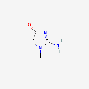
Creatine is an amino acid, chemically it is methylguanidoacetic acid. Creatine is mostly present in skeletal and heart muscles and in small quantities in the brain and testes.

Creatine


Creatinine is the metabolic end product of creatine. Creatinine is formed in the muscles from anhydrous creatine by a non-enzymatic removal of H20 and intramolecular cyclization. 

Creatinine


Blood levels of Creatinine - for men 0.75 to 1.5 mg/dL or 65 to 119 micromoles/L/L and for adult women -0.6 to 1.04 mg/dL or 52 to 92 micromoles/L.

The creation comes from two sources. A. Biosynthesis. B. Form diet.

A. Biosynthesis:

It takes place in two stages and in two places. The kidneys are the main place of biosynthesis in the first stage. However, when both kidneys are removed, about 15 % of the biosynthesis of creatine takes place in the pancreas.

The enzyme L-Arginine-Glycine-amidinotransferase catalyzes Glycine + Arginine to form Guadinoacetetic acid and Ornithine. Guadinoacetic acid, known as Glycocyamine and is transported by a carrier protein to the liver.

Control of biosynthesis.

GI source of creatine has a negative feedback effect on the renal enzyme guanidinoacetate. When GI absorption is high, renal production of guanidinoacetate is reduced proportionally but this has no effect on the production of creatine in the liver. However, creatine synthesis in the liver is dependent on kidney transamidase activity and hepatic synthesis is reduced as less glycocyamine is available in the liver.

Hormone influence.

Hyperthyroidism slows kidney transamidase activity due to high blood creatine levels.

Genetic mutation:

Mutation of the gene causing transamidase deficiency, an inborn error of creatine synthesis, is characterized by language, cognitive and behavior disorders.

B. In the liver Guadinoacetetic acid reacts with S-adenosylmethionine by enzyme methyltransferase and from Creatine and Adenosylhomocysteine. Methionine is the principal methyl donor; other minor methyl donors are choline and betaine. This reaction is not reversible. Glutathione and other reducing substances are required for the optimal activity of this enzyme.

B. Dietary source of creatine.

Food rich in guadiniacetic acid (GAA) is meat, poultry, fish, milk and milk products, and apple and loquat. Of the total turnover of 2 gm per day, a normal diet supplies only 5 % of GAA. The rest comes from biosynthesis.

Creatine in the muscles:

Creatine is transported to muscles and upon entering the muscle cells, creatine is acted upon by ATP ( adenosine triphosphate) to turn it into Creatine phosphate by ATP-creatine-phosphorylase enzyme. Creatine phosphate becomes cell bound and can not escape the cell. It is a high energy molecule, that easily transfers high energy phosphate to ADP when the muscle is contracting rapidly and exhausts all ATP. Conversion of ADP to ATP is catalyzed by adenyl kinase. Lactate and acetate block this reaction when these organic acids accumulate in muscles during prolonged activities. In the heart, the myocardium is capable of sustaining activities by utilizing fatty acids. lactate and ketone as fuels.

In the resting stage, muscle contains 6 times as much creatine-phosphate as ATP. Creatine phosphate is proportional to the body's muscle mass. In the muscles, 85 % of creatine is present as creatine phosphate. And all the creatine produced by the muscles is excreted in the urine. And the 24-hour urinary creatinine is remarkably constant in an individual.

Interest in improving muscular performance by creatine.

Sports :

Athletic performance is enhanced by a higher concentration of creatine-phosphate in the muscles and creatine is used as a nutritional supplement to that effect. Creatine supplement is available as creatine citrate, creatine monohydrate and creatine pyruvate.

Experimental use of creatine in diseases:

In ALS, Muscular dystrophy,  and Multiple sclerosis, creatine supplements are used but no significant improvement is noticeable.

Creatine ethyl ester(CEE):

A new form of creatine supplement, creatine ethyl ester, is available. It has an advantage over other forms because it resists degradation in the stomach and bioavailability is greater. It is soluble in fat and has much higher membrane permeability. It is slowly metabolized and so muscle performance can have a quicker onset and be more sustained at a high level of performance.

The half-life of creatine is 3 hrs. 3 to 6 hourly dosing is necessary in order to maintain high muscle creatine concentration. Once the supplement is stopped, the muscle creatine returns to baseline in 4 weeks.

Renal Excretion of Creatinine.

Creatinine in the blood is derived as the end product of creatine phosphate metabolism in the muscles. Creatinine is water soluble and readily filtered by the glomeruli of the kidney; about 15 % of creatinine is secreted by the cells of the proximal renal tubules in healthy adults. In renal failure, the tubular secretion may increase to 30 %, and some creatinine loss takes place through the intestine.

Effect of high protein diet.

A normal diet contains creatinine of about 1/10th of the daily requirement of creatine. When placed on a high protein diet, the fecal loss of creatinine rises sharply and creatinine only minimally.

Serum creatinine and creatinine clearance capacity of the kidney:

The Glomerular Filtration Rate (GFR) of Creatinine is considered the standard test of renal filtration capacity in health and in diseases. Any reduction in GFR is an indication of renal disease.

To detect the true glomerular filtration capacity, the test material must only be filtered by the kidney and should not be secreted either by renal tubules or the intestine. Creatinine is not ideal from that point because it is also lost in other ways as stated above.

Such an agent was Inulin. Inulin is a complex carbohydrate, obtained from the roots of the Chicory plant. In earlier times, Inulin clearance was the gold standard for renal filtration. It is now abandoned because of multiple factors including the very high cost of conducting tests requiring a hospital stay.

Then several radioactive agents were introduced. These tests were also given up due to concerns about radiation exposure to patients and clinic personnel.

Creatinine Clearance Test (CCT) is now accepted as GFR in health and in renal diseases.

Modified creatinine clearance test.

A standard CCT requires the meticulous collection of 24 hrs.' urine and several blood creatinine level determinations.

Soon a simpler test came into medical practice, which is equal to the standard CCT in every respect if not better. The modified CTT is done by deduction. The GFR is determined from one serum creatinine level.

The formula of GFR is -

For adult males:

GFR = 141 x (Scr/79.6) – 0.41 x (0.993)Age.

For adult females:

GFR = 144 x (Scr/61.9) – 1.209 x ( 0.993)Age.

Scr = serum creatinine in micromoles per liter.

[ Conversion table- mg/dL to micromoles/L of creatinine.

1mg /dL of creatinine = 0.01131222 micromoles /L.]


Correction factors are available for non-white races.

Normal GFR is above 60 ml/min and usually 80 to 160 ml/min

Other formulas:

CKG -EPI Creatinine Equation 2021.

eGFR =

142min(standardized Scr/K, 1)α * max(standardized Scr/K, 1)-1.200 * 0.9938Age * 1.012 [if female]

eGFR (estimated glomerular filtration rate) = mL/min/ 1.73 m2 ( m 2 = square meter)

Scr (serum creatinine) = mg/dL

K = 0.7 (females) or 0.9 (males)

α = -0.241 (females) or -0.302 (males)

min = indicates the minimum of Scr/K or 1

max = indicates the maximum of Scr/K or 1.

Calculate serum creatinine from GFR

eGFR= mL/min/1.73m 2 ( m 2 = square meter)

Serum Creatinine * µmol/L.

The typical range for serum creatinine is: For adult men, 0.74 to 1.35 mg/dL (65.4 to 119.3 micromoles/L) For adult women, 0.59 to 1.04 mg/dL (52.2 to 91.9 micromoles/L).

Latest method.

An iodinated compound, Iohexol, is a safe contrast agent used in radiological studies. Iohexol is now used for GFR determination. Iohexol does not combine with blood proteins and is well distributed in the body. It is excreted by glomerular filtration only. No other renal or GI process is involved in the excretion of Iohexol.

After giving a loading dose by IV at the clinic, the subject/patient is sent home to collect blood samples on supplied papers from fingertip puncture,( very much like the Glucose home test) at certain intervals. And when the test is completed, dried blood samples on paper are mailed back in a prepaid envelope. The Iohexol concentration in the dried blood samples is determined by Liquid Chromatography. And the GFR is calculated by a given formula. Recently, the  American Diabetes Association recommended the Iohexol GFR test on an annual basis in diabetics. This test is called Dried Blood Spot (DBS) for GFR.

Stress Tests for Kidneys.

Like cardiac stress tests, Kidney Stress Tests are possible. Kidney stress tests are generally not done in clinical practice but are important for drug manufacturers and researchers.

These are some of the kidney stress tests.

  1. High protein diet test. To test the GFR. 2. Creatinine load test. To test the proximal tubular cation transfer ability. 3. Water restriction test. To test the kidney's ability to concentrate urine in the collecting tubules. 4. Ammonium chloride loading test. To study H ion retention ability. 5. Oral bicarbonate load test. To study H ion handling capacity.

If one is due for a blood test, which includes serum creatinine, on making a request for a copy of test results at the time of registration, the lab will test the results. On a closer look, one will find an estimated GFR at the bottom. If GFR is a bit low, no need to be disheartened. The lab might have given the results based on a formula that takes the Body Surface Area into account. And sure enough, the lab did not take height and weight. So recalculating the result with actual height and weight might give a better GFR number.

*****************************************************************


Wednesday, April 5, 2023

Looking for Essentials Amino Acids.

                                  Searching for Essential Amino Acids.

                                     PKGhatak, MD


Essential amino acids for humans are 9 in number, namely: Histidine, Isoleucine, Leucine, Lysine, Methionine. Phenylalanine, Threonine, Tryptophan and Valine. Essential amino acids must be supplied in the food because our body can not synthesize them from other sources. Our body, however, can synthesize these non–essential amino acids, and common among them are Glycine, Alanine, Serine, Cysteine, Arginine, Aspartic acid, Glutamic acid, Arginine, Lysine, Tyrosine, Proline and Homocysteine. Amino acids are the main building blocks of protein molecules.

The human body is composed of 62 % water, 16% fat, 16 % proteins, 6 % minerals, 1 % carbohydrates and traces of vitamins and trace elements.

The function of Amino acids.

Both essential and nonessential amino acids are constituents of every cell and tissue of our body- from hair to the bone marrow cells and in between everything else. In addition, amino acids are required for growth, tissue repairs, immunity, hormone production and metabolic functions.

A few specific functions of individual amino acids are highlighted in the following paragraph.

Histidine - Blood cell formation.

Isoleucine - Increases growth hormone production. Skin and bone repairs.

Lysine - Calcium absorption, Collagen formation of tendons, cartilages, and skin.

Methionine - Antioxidant. Detoxify heavy metals.

Phenylalanine – Formation of the neurotransmitter, calcitonin and melanin.  and also the source of Tyrosine.

Threonine - Formation of elastin, the enamel of the tooth and mucin production from glands. Glucose metabolism.

Tryptophan - Formation of serotonin and melatonin, DNA repairs, Niacin    ( vitamin B3).

Valine – Muscle growth and health.

Are humans omnivorous.

No other living organism exists today that comes close to humans in choosing what to eat—from insects to the most poisonous puffer fish and snakes.

According to the WHO in the year 2021, a family of four in the USA consumed. 800 lbs of meat per month. 70 lbs of chicken per person per month was eaten during that time.

350 million tons of meat were consumed globally every year, of which pork was number one. China produced and consumed the most pork, and the USA came in third place. The USA is the largest consumer of beef and India came in 5th place; that must surprise some people. Fish consumption is highest per capita in Iceland, at about 200 lbs per year, the USA came 11th place by using 50 lbs per capita and still managed the second place by eating 290 eggs per person, only Japan consumed more, about 320 eggs per person.

The current trend in the USA.

In recent years, a vegetarian diet has gained popularity among the health and environment-conscious sections of the wealthier nations. Various degrees of vegetarianism exist. True vegetarians or vegans do not eat any meat or fish and avoid eggs or milk altogether. In India, where a major section of the population has been vegetarian for centuries, however, they consume milk from the cow, buffalo, goat and a few isolated tribes drink milk from camels. Some other Indians consider themselves vegetarian, but eat eggs and some also eat fish,  but not any animal meat.

Whatever form of vegetarian one may be, the main source of protein in their diet comes from plants. And most green parts of plants are not rich in proteins, the seeds of plants are. To meet the demands of plant seeds that are rich in proteins, more selective cultivation is required. It is becoming an important agricultural consideration for meeting this growing demand but must be met in sustainable and environmentally protective ways.

Protein requirement.

A growing child in utero needs 925 grams of protein and the mother supplies that amount during pregnancy. During lactation, an additional 1.3 grams of proteins per 100 ml of milk is needed.

Adults require 0.8 grams/Kg body weight ( stand height/weight). The elderly actually need less but defects in digestion and absorption are considered and the recommendation is like an adult - 0.8 g/Kg.

Obligatory Nitrogen Loss.

Protein turnover in the body is a continuous process of synthesis, breakdown and elimination of toxic nitrogenous waste products. When placed in a protein free but calorie sufficient diet, the body extracts the essential amino acids by breaking down proteins. This system is very efficient but the nitrogenous portion that is eliminated in urine and stool must be supplied in the food.

Effects of protein starvation.

Pictures of emaciated people ravaged by wars or severe famine need no further explanation. A short stature of a growing child, thin limbs, a pot belly, edema, fragile skin and orange hairs are the results of protein deficiency. Blood levels of albumin and hormones are low in these children. In nephrotic syndrome and severe liver cirrhosis, frequent infections and various complications are the results of derailed protein metabolic machinery of the body. Specific amino acid deficiency can occur due to inherited metabolic defects and the consequences of those can be found elsewhere.

Plant Proteins.

Many plant proteins are not available to humans due to the complex nature of the molecules, which are not easily digested or absorbed in the gut. Some plant proteins are poisonous, like in cassava.

Good source of vegetable proteins.

The high plant protein content is given here in descending order. It is the total amount of available protein but not in terms of essential amino content. The list of plant sources of proteins: Durham wheat, cashew nuts, quinoa seeds and ancient grains, dried seaweeds, rice, pumpkin seeds, beans, peas and raw soya beans.

Some individual plants with some good sources of essential amino acids are listed as -

Durham wheat supplies a fair amount of histidine, phenylalanine, tryptophan, valine and threonine.

Cashew nuts are a good source of phenylalanine, valine, leucine, lysine, Isoleucine and methionine.

Pumpkin seeds supply phenylalanine, tryptophan, Isoleucine, leucine and threonine.

Quinoa and ancient grains. Quinoa contains all 9 essential amino acids. Several other ancient grains are also good sources of proteins, chief among them are barley, farrow, buckwheat, millet, sorghum, kamut and teff. Each one has many other health benefits. Quinoa is an annual herb that belongs to the Chenopodium family. The grain contains 8 grams of protein per cup of cooked grain and is highly sought after because it supplies all nine essential amino acids. The protein of quinoa is 11s-globulin and contains no gluten. In addition, quinoa supplies unsaturated fatty acids, flavonoids, vitamins and minerals.

The rest of this group of grains contain more or less the same amount of proteins and are good sources of essential amino acids, but the amount varies from one to the other. All are good sources of micronutrients and antioxidants.

Soya beans have a good amount of leucine, lysine and phenylalanine.

Seaweeds are rich in histidine, leucine and lysine.

Peas and beans also supply fair amounts of phenylalanine, valine and threonine.

The world is always changing, and the rate of change has accelerated greatly in the last two or three centuries. The younger people of the present generation are much more concerned with animal sources of protein food along with other climate concerns. Eating proteins from nonanimal sources has come to attention lately from the processed food industries. This may be good or bad, only the future will tell.

Observing the effects of mass tourism causing accelerated degradation of the environment, like what has been done to Vienna and the Galapagos islands, should ring an alarm bell about this mass movement. No one can predict what that would do to the forested land in order to meet the demand for plant protein for human consumption.

*********************************************


Tuesday, March 28, 2023

Cholesterol and Lipoprotein

                                               Cholesterol and Lipoprotein.

                                              PKGhatak, MD

Cholesterol is a lipid. Lipids are a group of heterogeneous compounds of fatty acids and their derivatives which are not soluble in water and soluble in chloroform, ether and benzene. Oil, fat, wax and steroids belong to this group.

Protein when conjugated with lipids is called a lipoprotein.

Apo-lipoprotein:

Individual protein molecule contains a prosthetic group or a metal that gives it its special characteristic. When the prosthetic group or metal part is absent, then the protein is designated as Apo-protein. When a lipid molecule conjugates with an Apo-protein and then the conjugate combines with lipoprotein, the resulting molecule is called an Apolipoprotein.

Lipid molecules are wrapped around by proteins in order to make them soluble in the blood, making the transport of cholesterol between tissues possible. 

A diagram illustrates the makeup of lipoprotein molecules.


Chemistry of Cholesterol.

Cholesterol is designated as 3-hydroxy-5,6 cholestene.


The cholesterol molecule has a Sterol ring of 19 carbon structure and at position C-17 a side chain is attached, as illustrated in the above diagram.

 The rings A, B and C constitute a phenanthrene ring to which a cyclopentene ring D is attached. (this is cycleopenteno- perhydrophenerthrine ring or sterol ring) The double bond between position C5 and C6 gives the molecule its stability.

Source of cholesterol in humans.

Every living cell of the body can synthesize cholesterol, however, the liver is the primary organ where cholesterol is synthesized, in addition, the adrenal cortex, testis, ovaries, skin, aorta and intestine also produce cholesterol. Dietary sources supply only 15 % of cholesterol. Organ meat, egg yolk, brain and liver are good sources of cholesterol in food. The body recycles cholesterol and excess cholesterol is excreted by the liver in the bile as bile acids and neutral cholesterol. Some bile acids are reabsorbed with fat in the intestine and the rest is excreted in the stool.

The liver synthesizes about 1 gm of cholesterol a day and about 300 mg of cholesterol is obtained from food. Each 100 mg of cholesterol ingested will raise blood cholesterol by 5 mg/dL.

Synthesis of cholesterol.

The cholesterol is synthesized in the Endoplasmic Reticulum of the cytosol of the cell. Mitochondria of the cells generate energy via the Tricarboxylic acid cycle, and Acetyl CoA molecules generated in this process are available for cholesterol synthesis. Fatty acids, ketogenic amino acids and glucose are broken down to  2-carbon, Acetyl CoA molecules, and enter the tricarboxylic acid cycle.

All carbon atoms in a cholesterol molecule are obtained from Acetyl-CoA. Two molecules of Acetyl-CoA condense and form Acetoacetyl CoA by the action of the enzyme Thiolase. In the next stage, one molecule of acetoacetyl CoA condenses with another molecule of acetyl CoA forming one molecule of Beta-hydroxy-beta-methylglutaryl CoA. This synthesis is catalyzed by a rate-limiting enzyme HMG-CoA synthase (hydroxy-methylglutaryl Co-A synthase). Beta-hydroxy-beta-methylglutaryl is reduced by HGM-CoA reductase and NADPH acts as an H ion donor. In the next step, mevalonate is phosphorylated by ATP, and several steps later, condensation of several molecules of isopentyl takes place and Farnesyl pyrophosphate is generated.

Further downstream, one molecule of CO2 is lost and 5-C Isoprenoid units are formed. Six molecules of isoprenoid units condense to form Squalene. In the next step, the 19-C squalene assumes a ring form and is called Lanosterol or Steroid ring. There are more steps and at the end, a new cholesterol molecule emerges.

Regulation of cholesterol synthesis:

The rate-limiting HMG-CoA synthetase and HMG-CoA reductase enzymes are the main regulators of cholesterol synthesis. The rate of synthesis of HMG-CoA reductase- messenger- RNA is controlled by the steroid synthesis gene. Various hormones have roles in Cholesterol synthesis.

Cholesterol Esters.

Esters are formed by the conjugation of alcohol with cholesterol by the enzyme esterase reacting with the long chain fatty acid of the cholesterol linked to the hydroxyl group. Cholesterol esters are stored in the cytoplasm of the cells as oil drops. When energy is needed, esters are hydrolyzed and fatty acids are oxidized for energy production. The freed cholesterol is transported back to the liver by the Beta-lipoprotein in the form of HDL.

Cholesterol in the liver:

Cholesterol molecules go to form the cell membrane of liver cells. The rest of the cholesterol is free cholesterol, bile acids and cholesterol esters. Bile acids are Cholic acid, Chinodeoxycholic acid, deoxycholic acid and lithocholic acid. All are derived from cholesterol and each of them can combine with glycine and taurine and produce complex acids and salts.

2 gm of cholesterol is secreted by the liver into the bile. Daily fecal loss of cholesterol is 1.2 gm. Cholesterol does not supply any energy to the body and any excess cholesterol is eliminated from the body in the stool.

In clinical medicine, enzymatic analysis is used in measuring cholesterol. Gas chromatography is the gold standard of cholesterol measurement. Liquid chromatography and mass spectrometry are also used. Lipoproteins term is used for Apolipoproteins for convenience. There are four classes of Apolipoproteins based on their functions.

For the plasma membrane structural integrity: apo-B, E, A-I, A-II.

For Secretion: apo A-1, B-100, B-48.

For cofactors of enzymes: apo A-1, A-V, C-1, C-II, C-III.

For binding with receptors: apo A. apo B.

Apolipoprotein Apo-A: This is a large glycated protein of variable size.    Apo-A is a homolog of plasminogen.

Apolipoprotein B: It is perhaps more important that Apo A, is a greater atherogenic risk factor than LDL. The normal blood level of Apo B is less than 100 mg/dL. Apo B can bind with variable amounts of cholesterol. High levels of Apo B are seen in diabetes, pregnancy, and thyroid and kidney diseases. and kidney diseases. Low levels of Apo B can be associated with cirrhosis of the liver, acute hepatic necrosis, and congenital conditions.

Apolipoprotein E: Apo E is a major lipoprotein for cholesterol carriers. Genes controlling the synthesis of Apo E are responsible for the brain lipoprotein content. Alzheimer's disease and Alzheimer's disease carriers are due to carriers of the mutated gene. Apo E is also linked to cerebral angioid-angiopathy and age-related decline of cerebral functions.

Cholesterol- Esters- Transfer- Protein (CETP) facilitates the transfer of cholesterol esters and triglycerides and between LDL and HDL.

Each lipoprotein molecule is a spherical particle with a hydrophobic nucleus made with triglyceride and cholesterol ester and a peripheral envelope made by polar phospholipids, unesterified cholesterol, and one or more molecules of proteins. Proteins are bonded together with lipids in Non-Covalent bonds that make the molecule exchange its components easily.

It is customary in clinical medicine to classify lipoproteins according to their density and relative sizes. They are classified as chylomicron, very low-density lipoprotein (VLDL). Intermediate-density lipoprotein (IDLP). Low-density lipoprotein (LPL) and high-density lipoprotein (HDL)

LDL: LDL is derived from VLDL and IDL particles and carries 2/3 rd. of cholesterol in the blood. The liver turns out LDL to be transported to the muscles for energy generation and to the adipose tissue for storage of excess calories for future use. Because the LDL particles are small, they enter between endothelial cells and are deposited in the subendothelial space. Macrophages phagocytize LDL and turn themselves into ghost cells and become the site of atheroma formation. Atheroma can break and cause coagulation of blood and obstruct circulation and when this happens in the coronary arteries, a heart attack usually happens.

HDL: HDL is primarily produced in the liver and also produced in the intestine during fat digestion and absorption. In the peripheral tissue, cholesterol is released from cholesterol esters which are transported back to the liver combined with HDL. HDL plays an important role in reverse cholesterol transport from the peripheral tissue back to the liver. This property is considered Anti-atherogenesis. HDL also has anti-inflammatory, anti-oxidant, anti-thrombotic properties and inhibits macrophages from phagocytizing LDL molecules. In the liver, all extra cholesterol is secreted in bile as either free cholesterol and bile salts. 

Triglyceride:  Triglyceride is an old biochemical term. Structurally, triglyceride is Triacylglyceride. Glycerol is derived from glucose metabolism. It is esterified by fatty acids containing 16 carbon atoms or more. Triglycerides are the medical term for fat. The primary source of triglycerides is food, any extra calorie eaten is turned into triglycerides by the liver.

If the fatty acids have unsaturated bonds, the body can metabolize them rapidly. Most natural fats are mixtures of different triglycerides. The breakdown of glucose takes place in the cytosol and then the residue enters the mitochondria.

Looking at the table below, it is evident that triglyceride is a major component of Chylomicron and VLDL and IDL. It is now established that Triglyceride is an independent factor of atherosclerosis besides LDL. People with very high blood triglycerides suffer from Acute Pancreatitis. The normal blood level of triglyceride is 100 mg/dL or below in healthy adults.

Chylomicron: Triglycerides are the major component of chylomicron. After intestinal absorption, the chylomicron travels via lymphatics and then enters the portal circulation, and reaches the liver. After a high-fat meal, the plasma becomes milky opaque due to the presence of a high concentration of triglyceride. Lipoprotein lipase enzyme, present in endothelial cells of muscles, splits triglyceride and uses free fatty acids for energy. In 12 hours of fasting,  triglyceride returns to the normal level in the blood.

The table below summarizes important differences in various Lipoproteins.


Chylomicron

VLDL

IDL

LDL

HDL

Place of synthesis

Enterocytes of Intestine.

Liver cells

Liver

Endothelial cells of capillaries

Liver

Particle size

>75

25 -75

22-24

19 -23

10

Electrophoresis

None

Pro -beta

Slow- beta

Beta

Alpha

% of Triglyceride

90

54

20

4

3

% of Lipid

>75

70

24

23

10

% of Free cholesterol

1

7

9

11

5

% of cholesterol ester

2

12

35

45

30

Protein

2

10

12

20

50

Apolipoprotein

A I, II, IV,

B 48

C II, III

E


B 100

CII, III

E

B 100

C II, III

E

B 100

A I

C I, II, III

E

                             Taken from NIH publication

Cholesterol is an essential structural element of the cell wall of all living cells. Cholesterol provides stability to the cell membrane, which is primarily formed by phospholipids. All steroid hormones are derived from cholesterol, which is usually synthesized by the endocrine glands themselves. Many co-enzymes have cholesterol molecules in them. Bile salts are primary emulsifying agents for fat digestion in the intestine. The neurons of the brain have the highest concentration of lipids and cholesterol.

No discussion of cholesterol is complete without pointing out “Good and Bad cholesterol”. Cholesterol is neither bad nor good. Did anyone say glucose is bad because diabetes has high blood sugar? The word is HIGH or excess. All excesses in a biological system have a price to pay and cholesterol is no exception.
*************************************************************
Sunday, March 12, 2023

Anatomy of the Human Soul

                                                          Anatomy of the Human Soul

                                                                Pineal gland

                                                         PKGhatak, MD


                        Pineal gland.


A tiny endocrine organ in the brain that controls the circadian rhythm in humans is very much in the domain of Philosophers ever since the 17th century French nobleman Rene Descartes called the Pineal gland the site of the human soul. Today his concept of the soul residing in the pineal gland is dismissed, but the interest he generated still persists.

In the lower animal, the pineal gland acts as a light perceptive organ and is referred to as the third eye. But in the higher animals, light perception is the function of the retina. The pineal gland is also known as Conarium, Epiphysis cerebri, and Pineal body.


Descartes and the Pineal gland.

In the first book, Treatise of Man, Descartes describes a kind of conceptual model of man which consists of two ingredients, a body, and a soul. In the end, he, however, says nothing about the soul. The pineal gland plays an important role in Descartes' account. He believes body sensations, imagination and memory originate in the pineal gland and the body moves because the pineal gland directs them to do it. He sees animal spirits transformed into human bodily sensations and higher mental faculties when these animal senses reach the pineal gland via tubes, and threads, and are pressurized in ventricular cavities and directed to the pineal gland by these mechanical means.

The pineal gland, he believes, moves in three ways:

 1. By the force of the soul.

2. By the spirits randomly swirling about in the ventricles

3. As a result of the stimulation of these sense organs.

In his second book, The Passion of the Soul, published in 1649, he describes things other than the body's own parts, which are perceptually present within us, belong to the soul. The soul joins all body parts and so the soul belongs to the whole body. And the pineal gland is the only organ that joins the soul with the body with threads and spirits in the nerves. Descartes does not regard the soul as the principle of life but as the principle of thought. The ultimate and the most proximate cause of passion of the soul is simply the agitation by which the spirits move the little gland in the middle of the brain. [Please see the footnote]

Pine cone


Anatomy.

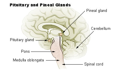
The pineal gland is located in the middle of the midbrain on the roof of the 3rd ventricle and situated below the tail end of the corpus callosum (the body of a major bundle of nerve fibers), in between the two Thalami. The pineal gland looks like a pinecone and so it was named the Pineal gland. The pineal gland is 0.8 mm in size and weighs 0.1 gm and is about the size of a rice grain. This endocrine gland is very vascular, second only to the kidneys (per each unit of mass). The blood-brain-barrier (BBB) is absent here and the hormone is secreted directly into the blood and also in the CSF. The cerebrospinal fluid bathes this gland through a small recess of the 3rd ventricle which continues within the stalk of the pineal gland.


Chemistry of Melatonin.

The pineal gland produces and releases melatonin. Melatonin is N-acetyl 5-methoxytryptamine. The amino acid Tryptophan is the source of melatonin.

Melatonin synthesis.

Tryptophan is converted to 5-hydroxytryptophan by hydroxylation. 5-hydroxytryptophan is decarboxylated to 5-hydroxytryptamine and this product is known as Serotonin.

Serotonin is converted to melatonin in two steps -

Step 1. A rate-limiting enzyme N-acetyltransferase transfers the Acetyl group from Acetyl CoA to 5-hydroxytryptamine and converts it to N-amino-5-hydroxytryptamine.

Step 2. N-amino-5-hydroxytryptamine undergoes methylation. The methyl donor is S-adenosyl methionine and the enzyme catalyzing this reaction is O-methyltransferase. And N-methyl-5-hydroxytryptamine is produced. This molecule is melatonin.

The reactions are shown as follows-

Serotonin + Acetyl CoA → N-Acetyl serotonin. This reaction is catalyzed by an enzyme N-acetyltransferase.

N-Acetyl serotonin + S- adenosylmethionine → N-acetyl 5-ydroxyserotionin.  This reaction is catalyzed by an enzyme O-methyltransferase.

Darkness induces Melatonin synthesis and release.

Darkness causes the release of Norepinephrine from the sympathetic nerve terminals of the pineal gland. The enzyme system is primed by norepinephrine and Cyclic AMP is generated. (cAMP). cAMP activates N-acetyltransferase and melatonin synthesis starts. As melatonin is forming, melatonin is secreted in the CSF and the blood. The pineal gland does not store melatonin in the gland.

If the artificial white light is of a certain strength, the effect of dark on the pineal gland ceases and no melatonin is produced or secreted. During international travel by airlines, the normal dark-light cycle is disrupted and resulting in sleep disturbances.

Breakdown of melatonin.

Melatonin is broken down in the liver by hydroxylation, then conjugated with sulfate and glucuronic acid and excreted in the urine.

Nerve supply of Pineal gland.

Somatic innervation. The 5th cranial nerve sensory nucleus, the Trigeminal ganglion, supplies nerve fibers to the stock and the gland. These fibers contain neuropeptide PACP which are vasoactive compounds. (PACP is pituitary adenylate cyclase acting polypeptide)

Autonomic innervation.

Sympathetic division nerve fibers come from the superior cervical ganglion. The parasympathetic fibers originate from the Otic and Pterygopalatine ganglia

Embryology of the Pineal gland.

In the 17th week of embryonic life, an invagination of the roof of the 3rd ventricle occurs. Initially, the pineal primordium contains Pax6 cells, arranged in a radial manner. After the neural tube fuses, the Pax6 cells rearrange into a rosette formation and then disperse in all directions. All pineal cells are derived from these progenitor Pax6 cells.

An adult pineal gland contains hormone secreting pinacocytes and microglia, astrocytes, and supporting cells. In the adult pineal gland, some progenitor cells remain. Calcium deposit in the pineal gland is common in the elderly, occasionally the entire gland may be calcified.

Role of the Photoendocrine system on the Pineal gland.

The retina of the eyes, supra-chiasmatic cells of the hypothalamus and noradrenergic sympathetic nerve fibers terminate in the pineal gland. Information about light exposure and circadian rhythmic variation is integrated into the pineal gland and regulated melatonin secretion.

Melatonin concentration in the CSF of the 3rd and 4th ventricles is higher than plasma and blood. What effect melatonin has on the neurons of the brain is not known.

Melatonin Receptors.

MT 1 and MT 2 are two types of melatonin receptors in humans. MT1 receptors are present in the suprachiasmatic cells of the hypothalamus, pituitary gland, retina and hypothalamus. When Melatonin binds with MT1, it produces inhibitory effects on the pituitary, and the release of hormones is inhibited and the blood level of Prolactin falls. Through the MT1 receptors, melatonin maintains the circadian rhythmic release of hormones of other endocrine glands.

MT2 receptors are present in the retina. When retinal receptors are stimulated, Dopamine release ceases. It also allows phase shifting of the internal circadian clock to the natural earth clock of the light and dark cycle. Other effects of MT2 receptor activation are increased phagocytosis and enhanced osteoclast activities and vasodilatation.

Hallucinogenic action.

DTM (dimethyltryptamine) is a hallucinogenic compound. Only a small amount of DMT is found in the Pineal gland. This fact might have started the notion that the pineal gland is a psychic center and controller of human behavior. 

Melatonin use.

Melatonin in the USA is an OTC drug (over the counter). It is available in 3 mg tablets, made solely in the laboratory. One melatonin compound. Ramelton is approved by the FDA for the treatment of insomnia, but the results are not consistent.

Indication of use.

Jet lag, Circadian rhythm disorder in the blind, Delayed sleep-wake phase sleep disorder in people who have delayed sleep and delayed wake time than required of them. In insomnia, melatonin reestablishes NON-RAM sleep. It is useful in shift workers and sleep disorders in children.

Adverse effects.

Melatonin is a safe supplement. However, it is a biological amine like Histamine and Dopamine. So, care should be taken when used with epileptic drugs, anti-platelet agents, BP medications, antidepressant drugs, immune modifying drugs, and anti-anxiety drugs.

____________________________________

Rene Descartes (1596 - 1650), a French mathematician, scientist and philosopher. He stated " Je pense, donc Je suis" (I think, therefore I am.)

Footnote: https://plato.stanford.edu/entries/pineal-gland/

********************************************************

Archive

  Archive. P.K.Ghatak, MD ghatak3@gmail.com 2025  March. Laser Endoscpoe 2025 February. Use of Ultrasound in Medicine MRI 2025 January. Bact...