Abnormal
Red Blood Cell Morphology and its Significance
PKGhatak, MD
Blood
is known as the vital fluid and that characteristic belongs to the
Red Blood Cells (RBCs). The hemoglobin, contained in the RBC, avidly
combines with Carbon dioxide, a waste and toxic product of cell metabolic activities; and the RBC exchanges it for Oxygen in the lungs and carries
oxygen to every living cell of the body (except the cornea).
RBC is
routinely tested for - size, shape, color, volume, uniformity of
distribution and for any structural anomalies; and all these are
reported when a CBC (complete blood count) is ordered.
No
other cell in the body has been studied
that thoroughly.
A
normal red cell (RBC) is a disc shaped and biconcave cell without a
nucleus and inclusion bodies. The red color of the cell is due to the
presence of oxyhemoglobin. RBCs appear brilliant ruby color when
allowed to settle in a test tube filled with normal saline.
RBCs
are 7 to 8 cu mm. in diameter, the central 1/3 is paler than the
periphery. Normal RBC volume (MCV) is 80 -95 fL, and the normal
number of RBCs is about 5 million per cu mm. The RBC shape is
fairly inform - the RWD (red cell width distribution) is 11 to 15 %.
RBCs
are formed in the bone marrow from the stem cells of the Myeloid line; Erythropoietin, a hormone produced in kidneys, stimulates RBC
production. In circulation, RBCs survive for 100 – 120 days. Every
part of the RBC is recycled by the spleen and other scavenger cells.
Size:
Abnormal size is called Anisocytosis.
Normal
size RBC is called Normocyte, Microcyte is < 7 cu mm (MCV<80 fL)
and Macrocyte is over 8 cu mm (MCV >95).
When
Anemia is detected, the finding is reported as either normocytic,
microcytic or macrocytic anemia. Normocytic anemia is seen after a
recent blood loss, like an accident, fracture of long bones, or surgery.
Microcytic anemia is due to iron deficiency, thalassemia, chronic
inflammation, slow GI blood loss due to cancers or bleeding
duodenal/gastric ulcers and sideroblastic anemia. Macrocytic anemia
is seen in Pernicious anemia, Folate and Vitamin B12 deficiencies,
Steatorrhea, Myelodysplastic syndrome, Liver cirrhosis, Alcoholism and Drug- use of Hydroxyurea.
Shape:
Abnormal shape is called Poikilocytosis. The abnormal shape could be
Spiculated or Non-Spiculated.
Spiculated
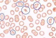
RBCs are called Burr cells, broken RBCs are known as Schistocytes resemble Napoleon hat),
Helmet cells, Acanthotic cells and Dacrocytes (tear drop shaped)
cells.
Non-spiculated
RBC are Target cells, Ovalocytes and Stomatocytes, Pencil cells, Bite
cells, Tear Drop deformities, Spherocytes and Elliptocytes.
Burr
cells are seen in renal failure.
Ovalocytes
and Elliptocytes are due to Hereditary causes; in addition,
spherocytes are also seen in severe burns, ABO incomparable blood
transfusion and acquired idiopathic hemolytic anemia.
Target
cells are common in sickle cell hemoglobinopathies, thalassemia and
iron deficiency anemia.
Bite
cells are seen in Chronic Obstructive Pulmonary Diseases (COPD).
Stomatocytes
(fish mouth like) are common in Liver disease, alcoholism, Rh-null
disease and COPD.
Teardrop deformity cells are present in Myelofibrosis and pernicious
anemia.
Helmet
cells, Broken cells or Schistocytes in the peripheral blood are ominous signs. These cells in the blood indicate active hemolysis. Usually one of the following conditions is responsible – Decimated
intravascular coagulation, TTP (thrombotic thrombocytopenic Purpura)and Thrombotic macroangiopathic anemia.
Color:
The
red color of RBCs is proportional to oxyhemoglobin concentration in
the cells. Based on the hemoglobin concentration per red cell, the
cells are called Monochromic, Hypochromic or Hyperchromic. When
color is not uniform, it is called Anisochromatic and when more than
one color is present, it is called Polychromatic RBCs. Iron deficiency is
the primary cause of hypochromia, and hyperchromatic is seen in sickle cell disease. Macrocytes in severe liver disease and Thalassemia, the RBCs can also be Hyperchromatic, this combination is called Leptocytes.
Increased
turnover of RBC in the bone marrow results in the presence of
immature RBC in the blood, these young cells retain some
remnants of RNA and appear bluish when stained and laid out as a fine network called Reticulocytes. In normal conditions, the reticulocyte count is
between 0.5 and 2 %. Reticulocytes over 2 % are seen in acute
hemolytic anemia, massive hemorrhage, and in high altitude visitors.
When bone marrow is infiltrated, as in cancer metastasis and leukemia,
Nucleated RBCs are visible in the blood and are called
Erythroblasts. In times of stress, the bone marrow also releases
nucleated RBCs, e.g., hypoxemia, and removal of the spleen.
Inclusion
Bodies:
Inclusion
bodies are iron pigment, hemoglobin, remnants of DNA and RNA
fragments in the red cell cytoplasm. Inclusion bodies are called
Basophilic Striplings and Howell jolly bodies.
Basophilic
stippling. These inclusion bodies represent defects in hemoglobin
synthesis, appear as bluish stripes, widely distributed throughout
the cell cytoplasm. Examples – Megaloblastic anemia, Post
splenectomy and sideroblastic anemia.
Howell
Jolly bodies. These bodies appear in the periphery of the cell as
purple round
dots when stained with Romanowsky stain due to the presence of fragments of
DNA.
Siderotic
granules. These granules are iron, which remains in
hemoglobin and stains blue. Example: Sideroblastic anemia.
Parasites.
The malaria parasite belongs to the Plasmodium group. Several species of Plasmodium
infect humans - mostly in people living in Sub-Saharan Africa and
Southeastern Asia. As there are different species, several parasitic forms, like gametocytes, schizonts and trophozoites, are also seen in the blood of patients infected with malaria. Considerable experience is required
to identify these intracellular malaria parasites.
Babesiosis.
Babesia infection is seen in the New England States in summer months
when tics are active. The parasites appear as ring forms, often in
groups, and in heavy infestation rings in groups appear outside the
red cells also.
Blood film in Babesiosis.
Important
Points:
To a casual reader, this article may appear confusing and overlapping.
In
clinical practice, however, the information is neatly and concisely
presented. A few
samples are presented.
Iron
deficiency anemia.
Iron
deficiency produces Hypochromic, Microcytic anemia. Besides a low
hemoglobin, HCT (hematocrit), MCV (means corpuscular volume), MCH
(mean corpuscular hemoglobin), Color index and Saturation Index are
all low. RWD
(red cell width distribution) is more than 15 %. This RWD finding is
helpful to separate iron deficiency anemia from Thalassemia where RWD
remains normal.
Macrocytic
anemia.
A
combination of low hemoglobin and low HCT in the presence of High MVH and
MCHC (mean corpuscular hemoglobin concentration) is the rule.
Hemolytic
anemia.
Intravascular
hemolysis is a vast subject as such. Features of hemolytic anemia is the presence of normocytic and normochromic anemia with a
normal RWD. If the hemolysis is less severe but recurrent then RWD
rises above normal.
Sickle
cell disease (SCD). In Benin, Senegal, and CAR (in sub-Saharan countries)
death due to childhood malaria caused havoc. A mutation of the Beta Chain
of the hemoglobin molecule at position 6 - substitution of amino acid
glutamine to valine proved to be a lifesaver. That advantage led to a wide distribution of the mutated gene in the population. However, as
people moved to the USA and Caribbean States from sub-Saharan Africa, that mutation has become
a fatal mistake. In a low oxygen environment, the RBC of SCD patients
changes from a disc to sickle form and obstructs blood flow in the
capillaries and results in severe pain and loss of function. This results in death of cells, seen from the
bone marrow to the brain.
Blood film in Sickle disease.
Thalassemia.
A growing
embryo in the mother's womb draws oxygen, due to the special characteristic
of Fetal Hemoglobin. If fetal hemoglobin persists in postnatal life, due to a gene
mutation, it is called Thalassemia. Thalassemic
patients have anemia which has the following characteristics -
Microcytic, hypochromic anemia with a high RBC count, normal MCV,
normal RWD, high reticulocyte count, basophilic striplings and
Target cells.
Peripheral blood film in Thalassemia.
TTP (thrombotic thrombocytopenic purpura).
TTP is
due to widespread platelet thrombosis in all organs producing an
alarming medical situation. Hemolytic anemia due to mechanical break
of RBC in their attempt to move forward in capillaries partially blocked by platelet thrombi, and a very low platelet count (10,000), the presence of schistocytes are
characteristic features. Direct examination of blood film by
hematologists can make a quick diagnosis.
A blood slide in TTP, the helmet cells are marked by blue circle.
Napoleon hat
*******************************