Thursday, October 13, 2022

Abnormal Red Cell Morphology and its Significance


                     Abnormal Red Blood Cell Morphology and its Significance

                                           PKGhatak, MD


Blood is known as the vital fluid and that characteristic belongs to the Red Blood Cells (RBCs). The hemoglobin, contained in the RBC, avidly combines with Carbon dioxide, a waste and toxic product of cell metabolic activities; and the RBC exchanges it for Oxygen in the lungs and carries oxygen to every living cell of the body (except the cornea).

RBC is routinely tested for - size, shape, color, volume, uniformity of distribution and for any structural anomalies; and all these are reported when a CBC (complete blood count) is ordered.

No other cell in the body has been studied that thoroughly.

A normal red cell (RBC) is a disc shaped and biconcave cell without a nucleus and inclusion bodies. The red color of the cell is due to the presence of oxyhemoglobin. RBCs appear brilliant ruby color when allowed to settle in a test tube filled with normal saline.

RBCs are 7 to 8 cu mm. in diameter, the central 1/3 is paler than the periphery. Normal RBC volume (MCV) is 80 -95 fL, and the normal number of RBCs is about 5 million per cu mm. The RBC shape is fairly inform - the RWD (red cell width distribution) is 11 to 15 %.

RBCs are formed in the bone marrow from the stem cells of the Myeloid line; Erythropoietin, a hormone produced in kidneys, stimulates RBC production. In circulation, RBCs survive for 100 – 120 days. Every part of the RBC is recycled by the spleen and other scavenger cells.

Size: Abnormal size is called Anisocytosis.

Normal size RBC is called Normocyte, Microcyte is < 7 cu mm (MCV<80 fL) and Macrocyte is over 8 cu mm (MCV >95).

When Anemia is detected, the finding is reported as either normocytic, microcytic or macrocytic anemia. Normocytic anemia is seen after a recent blood loss, like an accident, fracture of long bones, or surgery. Microcytic anemia is due to iron deficiency, thalassemia, chronic inflammation, slow GI blood loss due to cancers or bleeding duodenal/gastric ulcers and sideroblastic anemia. Macrocytic anemia is seen in Pernicious anemia, Folate and Vitamin B12 deficiencies, Steatorrhea, Myelodysplastic syndrome, Liver cirrhosis, Alcoholism and Drug- use of Hydroxyurea.

Shape: Abnormal shape is called Poikilocytosis. The abnormal shape could be Spiculated or Non-Spiculated.

Spiculated RBCs are called Burr cells, broken RBCs are known as Schistocytes resemble Napoleon hat), Helmet cells, Acanthotic cells and Dacrocytes (tear drop shaped) cells.

Non-spiculated RBC are Target cells, Ovalocytes and Stomatocytes, Pencil cells, Bite cells, Tear Drop deformities, Spherocytes and Elliptocytes.

Burr cells are seen in renal failure.

Ovalocytes and Elliptocytes are due to Hereditary causes; in addition, spherocytes are also seen in severe burns, ABO incomparable blood transfusion and acquired idiopathic hemolytic anemia.

Target cells are common in sickle cell hemoglobinopathies, thalassemia and iron deficiency anemia.

Bite cells are seen in Chronic Obstructive Pulmonary Diseases (COPD).

Stomatocytes (fish mouth like) are common in Liver disease, alcoholism, Rh-null disease and COPD.

Teardrop deformity cells are present in Myelofibrosis and pernicious anemia.

Helmet cells, Broken cells or Schistocytes in the peripheral blood are ominous signs. These cells in the blood indicate active hemolysis. Usually one of the following conditions is responsible – Decimated intravascular coagulation, TTP (thrombotic thrombocytopenic Purpura)and Thrombotic macroangiopathic anemia.

Color:

The red color of RBCs is proportional to oxyhemoglobin concentration in the cells. Based on the hemoglobin concentration per red cell, the cells are called Monochromic, Hypochromic or Hyperchromic. When color is not uniform, it is called Anisochromatic and when more than one color is present, it is called Polychromatic RBCs. Iron deficiency is the primary cause of hypochromia, and hyperchromatic is seen in sickle cell disease. Macrocytes in severe liver disease and Thalassemia, the RBCs can also be Hyperchromatic, this combination is called Leptocytes.

Increased turnover of RBC in the bone marrow results in the presence of immature RBC in the blood, these young cells retain some remnants of RNA and appear bluish when stained and laid out as a fine network called Reticulocytes. In normal conditions, the reticulocyte count is between 0.5 and 2 %. Reticulocytes over 2 % are seen in acute hemolytic anemia, massive hemorrhage, and in high altitude visitors.

When bone marrow is infiltrated, as in cancer metastasis and leukemia, Nucleated RBCs are visible in the blood and are called Erythroblasts. In times of stress,  the bone marrow also releases nucleated RBCs, e.g., hypoxemia, and removal of the spleen.

Inclusion Bodies:

Inclusion bodies are iron pigment, hemoglobin, remnants of DNA and RNA fragments in the red cell cytoplasm. Inclusion bodies are called Basophilic Striplings and Howell jolly bodies.

Basophilic stippling. These inclusion bodies represent defects in hemoglobin synthesis, appear as bluish stripes, widely distributed throughout the cell cytoplasm. Examples – Megaloblastic anemia, Post splenectomy and sideroblastic anemia.

Howell Jolly bodies. These bodies appear in the periphery of the cell as purple round dots when stained with Romanowsky stain due to the presence of fragments of DNA.

Siderotic granules. These granules are iron, which remains in hemoglobin and stains blue. Example: Sideroblastic anemia.

Parasites.

The malaria parasite belongs to the Plasmodium group. Several species of Plasmodium infect humans - mostly in people living in Sub-Saharan Africa and Southeastern Asia. As there are different species, several parasitic forms, like gametocytes, schizonts and trophozoites, are also seen in the blood of patients infected with malaria. Considerable experience is required to identify these intracellular malaria parasites.

Babesiosis. Babesia infection is seen in the New England States in summer months when tics are active. The parasites appear as ring forms, often in groups, and in heavy infestation rings in groups appear outside the red cells also.


Blood film in Babesiosis.

Important Points:

To a casual reader, this article may appear confusing and overlapping.

In clinical practice, however, the information is neatly and concisely presented. A few samples are presented.

Iron deficiency anemia.

Iron deficiency produces Hypochromic, Microcytic anemia. Besides a low hemoglobin, HCT (hematocrit), MCV (means corpuscular volume), MCH (mean corpuscular hemoglobin), Color index and Saturation Index are all low. RWD (red cell width distribution) is more than 15 %. This RWD finding is helpful to separate iron deficiency anemia from Thalassemia where RWD remains normal.

Macrocytic anemia.

A combination of low hemoglobin and low HCT in the presence of High MVH and MCHC (mean corpuscular hemoglobin concentration) is the rule.

Hemolytic anemia.

Intravascular hemolysis is a vast subject as such. Features of hemolytic anemia is the presence of normocytic and normochromic anemia with a normal RWD. If the hemolysis is less severe but recurrent then RWD rises above normal.

Sickle cell disease (SCD). In Benin, Senegal, and CAR (in sub-Saharan countries) death due to childhood malaria caused havoc. A mutation of the Beta Chain of the hemoglobin molecule at position 6 - substitution of amino acid glutamine to valine proved to be a lifesaver. That advantage led to a wide distribution of the mutated gene in the population. However, as people moved to the USA and Caribbean States from sub-Saharan Africa, that mutation has become a fatal mistake. In a low oxygen environment, the RBC of SCD patients changes from a disc to sickle form and obstructs blood flow in the capillaries and results in severe pain and loss of function. This results in death of cells, seen from the bone marrow to the brain.


                                Blood film in Sickle disease.

Thalassemia.

 A growing embryo in the mother's womb draws oxygen, due to the special characteristic of Fetal Hemoglobin. If fetal hemoglobin persists in postnatal life, due to a gene mutation, it is called Thalassemia. Thalassemic patients have anemia which has the following characteristics - Microcytic, hypochromic anemia with a high RBC count, normal MCV, normal RWD, high reticulocyte count, basophilic striplings and Target cells.

                                       Peripheral blood film in Thalassemia.

TTP (thrombotic thrombocytopenic purpura).

TTP is due to widespread platelet thrombosis in all organs producing an alarming medical situation. Hemolytic anemia due to mechanical break of RBC in their attempt to move forward in capillaries partially blocked by platelet thrombi, and a very low platelet count (10,000), the presence of schistocytes are characteristic features. Direct examination of blood film by hematologists can make a quick diagnosis. 


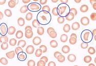
                             A blood slide in TTP, the helmet cells are marked by blue 
                                    circle.

Napoleon hat


*******************************

Monday, October 10, 2022

Eosinophil and Diseases associated with Eosinophils


                           Eosinophils and Diseases Associated with Eosinophils.

                                          PKGhatak, MD


Eosinophils are white blood cells that belong to granulocytes, other members of this group are Basophils and Neutrophils (also called polymorphs). The name, granulocyte, is derived from the presence of granules in the cell cytoplasm. The granules of eosinophils are coarse and turn red when stained with Eosin due to the presence of amino acids arginine and lysine in the protein, which reacts with Bromine in Eosin. The eosinophils are important Innate Immunocytes, involved in protecting the body from multicellular parasites, fungi, and some viruses, and are important determinants in certain diseases.

The easily identifiable feature of the eosinophils is their special characteristic, a Bilobed nucleus. 

Formation and distribution of Eosinophils.

The Hematopoietic stem cells of the bone marrow are the source of eosinophils; and in about 7 days the mature eosinophils leave the bone marrow. Most of them head for the thymus gland, spleen, lymph nodes, GI tract and the rest of the organs with the exception of the esophagus, ovaries, and uterus. Under normal health conditions, the eosinophils are not present in the Esophagus and Lungs and only a few eosinophils are present in the skin.

The Thymic Th2 cells initiate eosinophil production by secreting IL-5. (Interleukin-5).

Normal blood eosinophil count is 3 to 8 % and the total maximum count is 500 per microliter. In the peripheral blood, the eosinophils live about 1/2 days and in the tissues, they survive 8 days.

A total eosinophil count of 800 / microliter is abnormal and called hypereosinophilia, a count over 1,500 per microliter is called Hyper-eosinophilic syndrome.

There are very few conditions where the eosinophil blood counts are low. One is usually encountered when corticosteroid is administered. In Alcohol intoxication and Cushing's disease low eosinophil count are seen.

Normal functions of Eosinophil.

Eosinophils are immunocytes that act by releasing preformed substances, contained in the cytoplasmic granules. The pathophysiology of diseases is different from disease to disease. The substances carried in the granules can be grouped into – Oxygen radicals, Enzymes, Leukotrienes, Cytokines, Prostaglandins and Growth factors. In previous blogs, oxygen radicals, leukotrienes, prostaglandins and cytokines are discussed in detail and will not be repeated here.

Growth factors are TGF beta (tissue growth factor), VEGF beta (vascular endothelial growth factor), PDGE (platelet derived growth factor). In addition, the Eosinophils promote the development of post pubertal breasts, influence menstrual cycles and protect tissues from cancerous developments, prevent allograft rejection and help antigen presentation to T-Cells.

                               Hypereosinophilia

Herpesinophilia is seen in these conditions – Allergy, Asthma, Roundworm infestation of the gut, and Cutaneous larval migration like Filaria, Trichinosis, Cysticercosis. Eczema and Atrophic dermatitis, Chung-Strauss syndrome, Autoimmune diseases, Crohn's disease, Addison disease, Cancers of breasts, ovaries, and prostate. Leukemia and among the group of leukemias - Acute myelogenous leukemia (AML) and Eosinophilic leukemia are particularly striking. It is also seen in certain Fungal infections. Many prescribed medications produce allergic dermatitis and Penicillin leads the group; Penicillin may also produce Interstitial nephritis, which is characterized by the presence of eosinophils in the urine.

                                 Hypereosinophilic syndrome

When peripheral blood eosinophil count is persistently over 1,500 mcL for 6 months or longer and evidence of organ damage and allergy, parasitic infection and chromosomal abnormality are eliminated, the condition is called Hyper-eosinophilic syndrome (HES).

When the cause of high eosinophilia is known, the term Secondary Hypereosinophilic Syndrome is applied and many of the old HES are now classified under Secondary HES.

Chromosomal abnormality in hypereosinophilia.

The translocation of the gene between F1P1L1 and PDGFRA of the long arm of chromosome 4, at location 12 (4q12) produces a hybrid gene (F/P) which produces excess IL-5 causing rapid and excessive eosinophil production. Many other less prevalent Myeloid dysfunction due to chromosomal abnormalities are known, some examples are Chronic Eosinophilic Leukemia, Lymphatic HES, and Myeloproliferative HES.

Organs damaged by HES.

Esophagus, Stomach and intestine, Skin, Lungs and Heart. Multiple organs involved are the usual but only one organ or one system may be involved.

Clinical manifestation of HES.

Presentation and clinical features vary due to different organ /system involvement. Incidence in the USA is 0.3 to 6.0 per 100,000 population, including secondary EHS. General- Males are more affected by HES and ages between 20 and 50 are mostly affected. Systemic symptoms are fever, loss of weight, weakness. lethargy, pain in joints and muscles, and night sweats. Skin manifestations are itchy macular and nodular eruptions, angioedema and eczema. GI symptoms are heartburn, abdominal pain and diarrhea. Heart problems may mimic MI, endomyocardial necrosis, peripheral emboli and cardiomyopathy. Lung lesions resemble an attack of asthma, pulmonary infiltration and interstitial pneumonia and fibrosis. Unproductive cough. Hematological manifestations are many, chief among them are anemia, enlarged spleen and liver, and fibrosis of bone marrow. High serum B12 level. CNS manifestations are peripheral neuropathy, Transient ischemic attacks, strokes, and encephalopathy.

Diagnosis requires a complete blood count, serum B12 level, bone marrow biopsy and chromosome study.

Treatment of HES.

Intravenous corticosteroid is used to bring down the Eosinophil count rapidly, and Hydroxyurea is also used. Imatinib, a Tyrosine kinase inhibitor, is effective. Various other small molecules are available to suppress IL-5 production.

The prognosis is unfavorable without treatment. At present, with prompt treatment, the longevity on average is 80% in 5 years.

*************************************


Monday, October 3, 2022

Oral Ulcers


           Oral ulcers are also called Aphthous ulcers and also Canker sores.

                                     PKGhatak, MD


The word Aphthous is an adjective for the noun Aphtha. Aphtha is an old Greek word, the plural is Aphthae. Aphthae means Oral Ulcers due to any cause, that includes oral thrush and viral infections like foot and mouth disease (Picornavirus).


Oral ulcers develop in the mucous membrane (surface layer) of the gum, cheeks, roof of mouth and tongue. The ulcers have a white looking base surrounded by red inflamed borders and are painful.

Cause:

Any injury to the soft tissue of the mouth or tongue may start an ulcer. It is usually from a bite, ill fitting dentures, drinking hot drink or food, rough toothbrush, careless dental cleaning, vitamin B12, folic acid and iron deficiency, immune deficiency diseases or following chemotherapy and fungal (Candida) infection, commonly called thrush, and food allergy.

Diseases usually associated with oral ulcers:

Diabetes, Crohn's disease of the intestine, Celiac disease, Bechet's disease and Ulcerative colitis.

Other factors that favor oral ulcers are stress, hormonal changes, lack of sleep and infections.

Infection: The common virus infections are HIV and Herpes simplex; common bacteria are Staphylococcus, Actinomyces, and Helicobacter pylori. And common fungi are Candida albicans and Histoplasma capsulatum.

Course: 

Oral ulcers can be recurrent if the associated disease or conditions are not properly treated. Oral ulcers usually heal in 10 to 14 days.

When ulcers persist for more than 3 weeks, one of these conditions are responsible: - Fever, diarrhea, new ulcers appear before old ulcers heal, large size ulcers, ulcers on the outer lip surface are associated with oral ulcers.

Clinical types of oral ulcers:

There are two types - usual oral ulcers and herpetiform oral ulcers.

The usual ulcers are small to moderate sizes, oval to round shaped and have defined red edges. These ulcers heal without scarring.

Herpetiform oral ulcers are clusters of pinpoint ulcers, 10 to 100 ulcers and appear in groups. Later in the course, it may merge to become a large ulcer. These ulcers have a close resemblance to Dermatitis Herpetiformis of the skin. These lesions are not due to herpes virus infection.

Treatment:

Home recedes. Plenty of free advice is given over social media. Most of them include - frequent warm water salt gurgle several times a day; applying Back Tea bags directly over the ulcers - the tannin of the tea improves pain and accelerates the healing of ulcers. Similarly, powdered Alum (potassium ammonium sulfate), Baking soda, Orejel, Ambesol, honey, Milk of Magnesia and many other remedies are advocated.

Any food, having salt sprinkled over should be strictly avoided – salt crystal lodged in the ulcer crater is very painful and causes more tissue damage. Also, to be avoided are very hot food or drinks, spicy, difficult to chew food items and sour fruits and drinks.

Medication:

The most useful medicine is the Colchicine tablet. Use of Colchicine 0.5 to 2 mg a day for 2 days is enough to stop oral ulcers appearing and speeds up ulcer healing. At times Colchicine needs to be taken for longer periods of time, in reduced doses, to prevent a recurrence. Thalidomide 200 mg twice a day for 3 to 8 weeks is advised in immune related Aphthous ulcers. For bacterial infection, Minocycline or Tetracycline 250 mg in a cup of water(180ml) swishes around in the mouth and then spits out, three times a day for 3 to 5 days is also used.

At the same time, any underlying condition and illness should be adequately addressed.

*********************************

Saturday, October 1, 2022

Gut Bacteria and Human Health


                                              Gut Bacteria and Human Health

                                                PKGhatak, MD


Every surface of the human body harbors microorganisms of various kinds, some are just parasites, others live in symbiosis. When the symbiotic relationship breaks down for one reason or another, the health of the body is adversely affected. Recent studies on Gut bacteria show these actions are both local and systemic, including immune mediated diseases. Dominant gut bacteria are Firmicutes, Bacteroides, Actinobacteria, Proteobacteria, Fusobacteria and Verrucomicrobia. However, Firmicutes and Bacteroides account for 90 % of the gut bacteria. About 10 to the power 10 number of bacteria are present per gram of feces in a normal state of health.

Firmicutes:

Firmicutes phylum is composed of more than 200 different genera such as Lactobacillus, Enterococcus, Clostridium, and Ruminicoccus.

Firmicutes bacteria produce fermentation of undigested carbohydrates and cellulose and produce intestinal gases and Lactic acid. It releases antioxidants and micronutrients. The growth of colonies of Firmicutes in the colon plays an important symbiotic relationship. Humans provide food for the bacteria and Firmicutes in return supply micronutrients, suppress the growth of disease causing bacteria, namely enteric staph, anaerobic bacteria and Clostridium difficile.

Bacteroidetes:

Bacteroidetes are gram negative pleomorphic rods, non-spore forming, anaerobes and are both helpful bacteria of the gut but also may cause anaerobic infection under certain conditions. In a normal symbiotic existence, the Bacteroidetes break down undigested simple sugar, complex sugar, fat and protein in the gut and support other gut bacteria and also supply the human hosts with vitamins and micronutrients. A diet rich in oats, onion, ginger, flax seeds, and apples is a good food source for the Bacteroidetes.

The ratio of 20: 620 between Firmicutes and Bacteroidetes indicates a normal ratio and indicates good health of the individual. When the ratio tilts toward Firmicutes, it is generally associated with increased calorie absorption from the gut, insulin resistance and obesity, indicating a chronic inflammatory environment leading to cardiovascular disease propensity.

Immunoglobulin A.

Peyer's patches of the small intestine and B lymphocytes in the lamina propria of the colon produce IgA antibodies. Gut bacteria are in a seesaw relationship with gut IgA.  Bacterial antigen stimulates IgA antibody production and antibodies keep bacterial growth in check.

How symbiotic relationships develop.

The foreign antigen detection cells are Dendritic cells. In the gut, the dendritic cells are present in a loose connective tissue layer just beneath the gut epithelium and are in close proximity with the gut bacterial colonies. This constant contact has produced a state of "Inflammation Anergy" and the dendritic cells do not collect bacterial antigens and as a result, the macrophages do not produce pro- inflammatory cytokines.                 

IgA associated diseases:

Though IgA antibody production is highest among all Immunoglobulins, very little IgA antibody enters the blood; IgA is almost exclusively used up in the gut. In diseases due to IgA deposits in tissues, the IgA can be detected by Immunohistochemical staining of tissues.

COVID-19. The covid virus infects the gut mucosa, and the IgA antibodies try to eliminate the virus by its neutralizing action, however, in a small number of cases the elimination is only partial and COVID-19 virus is detected in feces for a long time.

IgA Nephropathy (IgA N).  It used to be known as Berger's disease.

IgA Nephropathy is the most common type of Nephropathy in Asia and also in the world.

It is postulated that the respiratory tract bacteria produce Degalactosylation of IgA molecule. This degalactosyzed IgA (dIgA) becomes an antigen and antibodies are produced. Then antibodies and dIgA combine and this Antigen + Antibody complex is deposited in the extracellular matrix of the Glomeruli of the kidney leading to IgA Nephropathy. The previously held theory was that aberrant glycosylation of IgA leads to polymerization of IgA and because of the complex's large size, these complexes are deposited in the mesangium of glomeruli. Inflammation starts as a result and glomerulonephritis progresses to membranous glomerulonephritis with crescent formation.


Henoch Schönlein purpura.

Purpuric eruptions develop following a URI. The purpura is mostly seen in the legs, buttocks and sometimes in the hands, face and trunk. The purpura is more abundant along with the pressure points like the sock line and the waistband. Purpura is palpable and does not fade away on pressure. Children between 2 and 6 are mostly affected. Male children and white and Asian children are more vulnerable to HS purpura. The underlying pathology is a vasculitis of small vessels of the skin, mucous membrane and joints. Lesions in the mouth and arthritis may develop. GI symptoms are common and some also develop IgA nephropathy.

Skin lesions.

Dermatitis herpetiformis.

Clusters of raised red, intensely itchy lesions develop on elbows, knees, buttocks, occasionally on arms and on the scalp. The lesions are intensely itchy. Often blisters develop from scratching and scars form when they are opened up from scratching.

People of the age group 30 to 40 of European heritage develop recurrent lesions. Oral and GI lesions also occur. Aphthous ulcers in the mouth and pits and fissures on the enamel of teeth may appear, GI manifestations are bloating, cramps and diarrhea.

Cause.

IgA antibodies develop against the skin epidermal transglutaminase. It is the skin manifestation of Gluten enteropathy from gluten sensitivity.

Diagnosis.

Demonstration of IgA deposition in the upper layers of the dermis by immunofluorescence.

Dysmorphic Epidermolysis Bullosa.

IgA deposition at the junction of the basal layer of the epidermis and papillary cells of dermal held by collagen fibrils are damaged by the process. The epidermis is easily lifted off from the dermis with minor trauma. Blister formation and secondary infections are common. Epidermolysis Bullosa is an inherited disease. It is inherited by autosomal recessive and autosomal dominant modes and multiple gene mutations are present.

There are several clinical types based on clinical presentation and the associated involvement of other systems. GI, GU, Respiratory, ENT may variously be involved.  It may be present at birth or at anytime later in life. It is a serious disease. There are serious complications. even when patients initially respond to treatment. Esophageal stricture and cancer are usual.  Diagnosis is made by immunofluorescence staining of the biopsy tissues. The prognosis is variable but not good in general.

Summary.

IgA is the principal immunoglobulin in keeping the GI tract and other mucous membranes of hollow organs free of pathogens. Because of constant contact with bacteria, some of the bacterial degradation products/secretory enzymes become antigenic and antibodies are produced. Antibodies can react with normal tissues like nerve cells, GI cells, etc. This hypothesis has generated a new look into the pathogenesis of Multiple sclerosis, Parkinson's disease, etc. In the future, more and more laboratory confirmations will be available.

Lately, many attempts have been made worldwide to limit the use of antibiotics in supposed bacterial infections or recurrence, which are in fact infections from viruses. Also cut down the duration of antibiotics use from 15 to 5 days for an infection, like walking pneumonia and many other minor ailments. This practice will result in the return of a healthy symbiotic relationship between the gut bacteria and humans.

***************************


Saturday, September 3, 2022

Osteoarthritis


                                                          Osteoarthritis

                                                PKGhatak, MD


Osteoarthritis is a degenerative disease followed by inflammatory disease of the joints.

In the initial stage of Osteoarthritis (OA), the wear and tear produce damage to the cartilage covering the ends of the bones forming a joint. When cartilage is destroyed, the bones are exposed and rub against each other, causing pain. Subsequently, the bones and joint capsule also undergo inflammatory changes. Osteoarthritis (OA) is distinct from Rheumatoid Arthritis (RA) where inflammation primarily is in the synovial membrane of the joints and then spreads to bones and the rest of the joint structures. RA is an autoimmune disease and affects the lungs and other collagen vascular structures of organs.

OA is a disease of the elderly. Young people also develop OA from sports injuries, in addition, OA develops secondary to other diseases and a few hereditary diseases which damage the cartilage.

Cartilage.

The ends of bones forming a joint are covered by a layer of cartilage called Articular cartilage. Articular cartilage has a unique ability to withstand high loads with little damage. Cartilage is connective tissue and provides structural support like bone. In fact, in the animal kingdom, the development of the backbone, the cartilaginous vertebral column appeared first, then came the bony vertebrae, even then, currently the oceans are full of cartilaginous fishes, sharks for example.

The cartilage is composed of a dense extracellular matrix. In the matrix a few sparsely distributed cells called Chondrocytes are present. The extracellular matrix is composed of water, collagen(protein), proteoglycans (protein+carbohydrate) and other glycoproteins. These structural components make cartilage retain water and provide a cushion and absorb the body weight and pressure during joint movements.

Blood supply to cartilage is absent, this results in poor healing properties of cartilage. It is devoid of nerves and so insensitive to pain.

The part of the bone next to the cartilage layer is known as the subchondral plate and is very vascular. Nutrition to articular cartilage is provided by synovial fluid.


Commonly affected Joints in OA.

OA can start in any joint, but the joints commonly affected joints are Knee, Lumbosacral and Cervical Spine, Hips and thumb.

The thumb and distal finger joints are commonly affected in elderly females. Knee, Lumbosacral, Cervical Vertebrae and Hip joints are equally affected in both sexes. OA of shoulders and fingers are generally spared, unless overused, for example, manual laborers and coal miners.

The middle joints of the fingers are typically affected in RA. This is an important distinction between the RA and OA. RA patients show associated systemic symptoms and x-ray evidence of erosion of bone ends of the finger joints and elevated acute phase reactants in the blood.

Risk factors to OA.

Injury, obesity, manual workers, sports injuries, diabetes, in addition, OA develops secondary to several diseases. Closely resembles but distinct from OA are Gout, Pseudo gout, and Peripheral sensory neuropathic arthritis.

Secondary causes of OA.

Metabolic defects of cartilages:  Ochronotic arthropathy, Alkaptonuria, chondrocalcinosis, or pseudo gout.

Connective tissue metabolism:    Hurler syndrome, Morquio disease, Marfan syndrome, Poly-epiphyseal dyslexia.

Congenital bone defects:   Scoliosis, Spina bifida, Club foot, Leg-Parthese disease.

Hereditary gene mutation:    Hemochromatosis. Sickle Cell Anemia.

Blood clotting abnormality:   Hemophilia.

Autoimmune disease:   RA, Psoriatic arthritis. Crohn's disease, Ulcerative colitis,

Leukocyte Antigen gene mutation:    Ankylosing spondylitis due to inherited HLA-B27 gene.

Endocrine disease:    Acromegaly, Cretinism, Dwarfism, Hypothyroidism, Menopause.

Neurogenic:   Muscular dystrophies. Syringomyelia. Charcot arthropathy. Reflex sympathetic dystrophy.

Vascular:    Aseptic necrosis of hips, Caisson disease

Symptoms of OA.

All different types of arthritis produce similar symptoms like pain, swelling, muscle stiffness, and limitation of range of motion. OA is a local disease and produces very few systemic effects. OA of the vertebral column has some unique features. In cervical spine OA - the wry neck may start even before the radiological evidence of OA. Thorns like outgrowths of bone spicules produce sharp jabbing pain. OA of facet joints limits turning of head and torso side to side and specially backward. Stenosis of the cervical spine can produce spastic paralysis of the legs. In the lumbar area, OA produces muscle spasms. Lumbar stenosis can interfere with bladder and bowel functions, in addition to muscle weakness of lower legs, often seen as foot drop and marked limitation of climbing stairs.

Diagnosis.

History and clinical examination are sufficient to make a diagnosis of OA. There is no blood test to confirm OA, however, blood tests are done to exclude other forms of arthritis. X-rays are an essential part of diagnosis showing loss of joint space, subchondral bone sclerosis and small outgrowths from the ends of bones in OA.

In the weight bearing joints, like knees, an x-ray is used to stratify OA into early, intermediate and advanced stages. In the advanced stage of OA knee, surgical replacement of the knee is advocated.

Medical treatment of OA.

There is no cure for OA. The non-steroidal anti-inflammatory drugs are the mainstay of the treatment of OA. These drugs produce relief of pain and limit bone inflammation.

In addition to medication, physical therapy, daily exercise and weight reduction, where applicable, are advised. When pain is acute, rest of that joint is essential till the pain is tolerable then physiotherapy is helpful. Use of other anti-inflammatory drugs, narcotics and muscle relaxants are discouraged.

In some European countries, intra-articular lubricant, a visco-supplementation containing Hyaluronic acid, is injected into the knee to lessen friction and pain. Use of it in the USA is generally not recommended because the beneficial effects last only a few months, if at all. and a chance of infections in repeated injections is a real possibility.

Surgical Treatment.

In advanced knee OA, joint emplacement is commonly advised. Corrective surgery of various kinds is done for cervical and lumber vertebral stenosis.

********************************


Friday, August 26, 2022

Long Covid

                                                          Long Covid

                                              PKGhatak, MD


Long covid is a new disease. The precise definition of Long covid is lacking. A cornucopia of symptoms lasting for weeks to 18 months or more experienced by people who had a covid illness and recovered are thought to be the sequel to covid infection and named Long Covid.

There is no reliable way to determine the percentage of the world population that developed Covid-19. In January 2022, one estimated the rate was 57 %, another source reported it was over 75% in January 2022 (6+ billion out of 8 billion).

The vast majority of covid-19 illness produces symptoms that are very similar to the Common cold and symptoms last only 3 to 5 days. The number of seriously ill patients worldwide will remain unknown because the variable of public health standards prevails in the vast number of developing countries and local politics prevented notification.

Who developed Long Covid.

The wide spectrum of people with mild symptoms or no symptoms but who had a positive PCR test to more seriously ill patients with a variety of symptoms are considered to have Long Covid. Even those who were fully vaccinated and boosted, contact with the virus also reports long lasting symptoms. It appears severity of symptoms is not a factor in acquiring Long Covid. It is estimated that 35 to 20 % of patients who recovered from covid-19 infection developed Long Covid.

What are the symptoms of Long Covid.

General. Loss of weight. Lethargy. Palpitations, Pain in joints, skin rashes,

Respiratory. Unproductive cough, shortness of breath, worsening existing asthma and COPD

Neurological. Loss of smell or taste, both, weakness, numbness, lightheartedness, brain fog (difficulty in thinking and concentration), disturbed sleep,

Psychological. Depressive mood, lack of energy, pain and tenderness in many areas of the body, anxiety.

GI. Lack of appetite, intermittent diarrhea, vague abdominal pain, bloating sensation.

Cardiac. Variable BP and sudden surge of BP, palpitation, chest pain.

Urinary. Dysuria and frequency.

Metabolic/ Hormonal. Out of control blood sugar, thyroid function, menopausal symptoms, lack of libido. 

Worsening of symptoms that remained after recovering from severe COVID-19, like heart failure, asthma, paralysis of limbs, etc. Weakness and brain fog are mostly responsible for absenteeism at the workplace in the long covid.

Many researchers noted that these symptoms are not unlike symptoms of chronic fatigue syndrome, also known as Myalgic encephalomyelitis and PTSD (post-traumatic stress syndrome)

What are the reason/ reasons for Long Covid.

No one reason stands out among several put forward so far. In general, people who are immunodeficient are more prone to Long Covid. It is theorized that immunocytes in these groups fail to clear the virus but the virus is weakened and continues to infect new tissues. This model fits with the observation that patients treated with a cocktail of antibodies during the early phase of infection have higher instances of Long Covid.

Another interesting theory is the virus takes sanctuary within the immunocytes and waits for an opportunity to re-emerge like the HIV/AIDS virus and Paxlovid breakthrough infection. Recent discovery points to Langerhans cells of the skin, which engulf the virus but fail to digest because the virus directs anti-enzyme to neutralize the digestive enzymes of the Langerhans cells.

Long covid has emerged as a major disabling illness putting a drag on the economy and untold suffering for people eager to return to work but unable to.

Initially, it appeared booster covid shots were beneficial in terms of lessening the symptoms and the length of the period of disability. But subsequent studies failed to find any benefit of the booster shots.

Covid -19 pandemic caused havoc to humanity. It continues to evolve as a major health risk factor worldwide. One reliable source report 6 million deaths from covid-19, others think that number is way short of the actual number, citing India as an example. The WHO reports one million covid deaths since January 2022 alone.

Sooner or later a plateau will be reached in the rate of new covid-19 infections which can be managed like Influenza outbreaks with vaccines given every year. Antiviral drugs against the covid-19 virus may prove to be as elusive as HIV/AIDS virus. And hopefully, the Long Covid will be just another cause of Chronic Fatigue Syndrome or Myalgic Encephalomyelitis for those who prefer Latin names.

***************************************************

Wednesday, August 24, 2022

Bell's Palsy

                                                                        Bell's Palsy.

                                                              PKGhatak, MD


Sudden onset of paralysis of one half of the entire face is called Bell's palsy. In medical terms, it is a lower motor neuron paralysis of the facial muscles of one side. Bell is Sir Charles Bell of Scotland (1774-1842). He was a Neuroanatomist cum surgeon. He gained first-hand knowledge of facial injuries while working as a surgeon in the Battle of Waterloo (1815).

The term “Palsy” is derived from the Anglo-French word “Palseie”. The Greek word “Paralysis” means loosening, it is derived from the verb “Paralysia” meaning to disable/ enfeeble.

Although Sir Charles Bell was the first to provide the anatomic basis for the condition that bears his name, in recent years researchers have shown that other European physicians provided earlier clinical descriptions of peripheral cranial nerve 7 palsy. History of facial distortion by Greek, Roman, and Persian physicians, culminating in Razi's detailed description in al-Hawi (9th century CE). Razi distinguished facial muscle spasm from paralysis, distinguished central from peripheral lesions, gave the earliest description of loss of forehead wrinkling, and gave the earliest known description of bilateral facial palsy. In doing so, he accurately described the clinical hallmarks of a condition that we recognize as Bell palsy.

Dr. Bell, of Bell's palsy fame, had suffered a right sided facial paralysis of his own before he became famous.

A question:

Mona Lisa. Whoever has seen the world famous painting of Mona Lisa by Leonardo da Vinci must have pondered what that smile is all about. At a conference in Vienna, a group of researchers said Mona Lisa had right facial paralysis during her pregnancy and recovered after childbirth.

Anatomy of motor nerve of the face.

The above diagram is an outline of the 7th nerve motor nerve supply to facial muscles. The pyramidal cortex sends signals to the upper division nucleus of both 7th cranial nerve motor nuclei. (Facial nerve). The lower division of 7th nerve motor nucleus, on the other hand, receives signals from one side only (contralateral side). In a stroke due to pyramidal cortex disease, only contralateral side of the face is paralyzed and muscles of forehead remain functional.

In Bell's palsy, the entire facial muscles, including the forehead muscles, are paralyzed.

Common causes of Bell's Palsy.

Chill wind blowing against the face in a long drive.

Herpes zoster. Herpes simplex. Epstein-Barr and COVID-19 virus infection.

Lyme disease from spirochete infection.

Acoustic neuroma (tumor of 7th nerve fibers in the ear), Schwannoma (cells that make myelin) and cholesteatoma.

Injury to the Facial nerve.

During surgery of the parotid gland, a Knife wound behind the ear. Fracture of the base of skull. Nerve block during dental procedures.

Pregnancy and diabetes mellitus.

Critical areas of the path, the Facial nerve, where disease is likely to produce Bell's palsy.

1. Narrow canal inside the Temporal bone. 

2. The neck of the mandible.

 3.Parotid salivary gland.

Causes of paralysis of the Lower part of the face only.

A.  Strokes from blocked blood supply to the mid brain.

B. Blood clot emboli originate from heart valves, heart chambers, or atrium in atrial fibrillation.

C. Infectious vegetation from heart valve or artificial valve disease.

D. Intracerebral bleeding from use of anticoagulants.


************************************************************


.


Sunday, August 21, 2022

Dizziness and Vertigo.

                                                          Dizziness and Vertigo

                                                    PKGhatak, MD


Dizziness is not an uncommon symptom, specially in the elderly. Patients describe the symptoms in various ways - lightheartedness, feeling unsteady while walking, a sense of not being able to keep an upright posture while trying to get up from a bed or a chair, head spinning, about to faint and like.

Vertigo symptoms are related to a false sensation of motion of the surroundings.  Generally, head movements precipitate an attack of vertigo. Fear of falling on the ground from a bed or an upright position is often associated with nausea and jerky movements of the eyes.

The distinction between dizziness and vertigo is important from diagnostic and treatment points of view. But often one term is used for the other. The task falls on the physician to get a clear history from the patient and only then proceed with the investigation.


What causes dizziness:

Many medical conditions may cause dizziness, chief among them are postural hypotension, irregular cardiac rhythm, disturbed blood flow to the hindbrain (lower posterior part of the brain) and low blood sugar.

Other conditions less commonly associated with dizziness are alcohol intoxication, antidepressant drugs, migraine, and carbon monoxide poisoning.


What causes vertigo:

Vertigo is due to abnormal signals fed to the brain from the semicircular canals of the inner ear called the vestibule.

Each vestibule contains 3 semicircular canals, positioned at 90 degrees to each other. The canals are filled with a viscous fluid called endolymph, floating on the endolymph there are tiny pebble like structures called otoconia, connected with the vestibular division of the 8th cranial nerve. Any movement of the head displaces the otoconia and initiate signals from both ears and the signals are reciprocal to each other. The brain integrates these signals and accurately calculates the coordinates of the head position in relation to the horizon.

Diseases affecting the inner ear are the cause of Vertigo.

Common conditions causing vertigo:

Viral infection of the balance organ is commonly known as Labyrinthitis, motion sickness due to overstimulation of the brain from the constant change of head position – a common experience of passengers on a boat in a choppy sea, benign paroxysmal positional vertigo, and head injury. Other conditions are:-

   Neuronitis, Meniere's disease, Tumors like meningioma pressing on the hindbrain, Vestibular migraine, and Brain cancers. Cerebral strokes and Multiple sclerosis.

The medication can also cause Vertigo, a few examples are certain antibiotics, water pills, cancer drugs, and aspirin in heavy doses.

Meniere's disease:

A combination of tinnitus (low volume high pitched noise like the chirping of crickets), some loss of hearing, and vertigo is known as Meniere's disease. The reason for this disease is not known. Symptoms are generally episodic but tend to be recurrent. After each attack, the hearing loss increases.

Benign Paroxysmal Positional Vertigo (BPPV):

Sudden head movement or turning the head to a certain position causes BPPV. It is very disturbing to the patients but it causes no permanent harm to the body. The sensation of the horizon tilting up and down causes the patient to grab whatever they can to avoid falling to the ground. The condition is self-limited but may be prolonged. A certain maneuver of the head position can stop vertigo and the patient can learn the technique and can self-treat. 

******************************************

Sunday, July 24, 2022

Sudden Death


                                                                  Sudden Death

                                                        PKGhatak, MD


In human experience, an unexpected sudden death must be the most devastating and sad event for the family members.

When death finally comes to a person with an incurable chronic illness, some may say “Good is merciful and he/she is in peace. But what one can say to a mother when a child is killed in a school by gunshot or a newborn dies from sudden infant death syndrome. Is the same God merciful here also?

In the USA, about 200,000 deaths are happening every year from unintentional causes, chief among them are drug overdose, motor vehicle accidents, falls, and drowning. Deaths from gun violence and suicide are 45,000 per year for each. The remaining deaths are from accidents. heart attacks and undetermined causes. The deaths from undetermined causes pose another dimension to the suffering for the family – did they miss something to spot in time and take proper action to prevent death.

Sudden Infant Death Syndrome.

Healthy infants below 1 year. old people were dying suddenly for no apparent reason. This was a major concern of health professionals and in 1969, 'Sudden Infant Deaths Syndrome (SIDS)' was coined to study these cases and find the cause and prevent such deaths. In the USA in the year 2020, 3,400 babies below one year of age, died suddenly. Of these, about 40 % were SIDS, 30 % were accidental suffocation and 25% were unknown causes. The present incidence of SIDS is about 96 deaths per 100,000 live births.

Various theories were put forward to explain the cause but most of them were not substantiated by scientific studies. Placing babies on their stomachs in the crib was the main common factor. Since this practice was abandoned, the incidence fell and held steady.

Cardiac Arrest:

'The heart stops pumping blood' is the simplest way to define the term cardiac arrest. As the heart stops so also does the circulation of blood and within a few minutes living cells begin to die. If resuscitation can be started early and properly the heart can be revived within several minutes.

Mechanism of cardiac arrest:

Lack of oxygen

Oxygen supply to the heart muscles depends on patent coronary arteries. If a coronary artery is blocked by a rupture of an atheromatous plaque, blood cannot go past the blocked area, and lack of oxygen makes heart muscle hyperexcitable, and abnormal ventricular contraction begins. If the situation does not reverse, then ventricular tachycardia (VT) and ventricular fibrillation (VF) precipitate. In ventricular fibrillation, blood virtually remains in one place, and no pulse is generated. This is the usual cause of death.

Ventricular Tachycardia (VT):

In ventricular tachycardia, the ventricles contract 120 to 180 times a minute. At this rate, ventricular chambers have only 0.25 seconds to 0.125 seconds to fill (normal ventricular filling time is 0.5 sec). This results in a precipitous drop in cardiac output and blood pressure, and the patient goes into shock.


In right coronary obstruction, the blood supply to the Sinus Node, the normal heart pacemaker of the heart, is cut off. After the right coronary artery is blocked, the heart rate slows (sinus bradycardia) and A-V(atrioventricular) nodal rhythm and various forms of heart blocks and complete heart block may develop. Complete heart block generally produces convulsions and loss of consciousness. It can also produce ventricular fibrillation, shock and death.

Uncommon but significant other causes of VF.

Hypokalemia, hypocalcemia and hypomagnesemia are known to produce VF.  Special importance in clinical practice is the condition called prolongation of Q-T interval which can precipitate a fatal multifocal VT. Prolonged Q-T interval results in enhanced automaticity, which most commonly originates in the right ventriclar conductive tissue. At the cellular level, the VT is caused by electrical re-entry. Myocardial scarring from the previous heart attack increases the likelihood of electrical reentrant circuits.

VT from a prolonged Q-T interval is called "Torsade de Pointes(meaning twisting around a point). It is an uncommon and distinctive form of polymorphic VT, characterized by a gradual change in the amplitude and twisting of QRS complexes around the isoelectric line.


Determination of Q-T interval.

[The linear regression model yielded a correction formula (for a reference RR interval of 1 second): QTLC = QT + 0.154 (1-RR) that applies to men and women. This equation corrects QT more reliably than Bazett's formula, which overcorrects the QT interval at fast heart rates and undercorrects it at low heart rates. Lower and upper limits of normal QT values concerning RR were generated.]

The normal Q-T interval in men is 350 to 450 milliseconds and in females is 360 to 460 milliseconds. 

Many medications can prolong Q-T interval. A short list of drugs is:

A cardiac drug - Quinidine. 

Antihistamine – Astemizole, Diphenhydramine.

Antibiotics – Macrolide antibiotics, Quinoline group of antibiotics.

Antihypertensive – Nicardipine.

Antidepressants – Serotonin receptor blockers, Amitriptyline, Lithium

Anti-malarial – Chloroquine.

Anti-Fungal – Ranolazine

Anti-cancer – Tamoxifen

Anti-convulsant – Felbamate, Fosphenytoin.

Bronchodilators – Albuterol. Salmeterol.

CNS stimulants – Amphetamine.

Muscle relaxants – Tizanidine.

Many people die in their sleep who took a cold or allergy medication before going to bed. Torsade de Pointes is likely the main reason for such deaths. 

Incidence of heart attacks and fatality in the USA:

In 2018, about 30 million incidences of heart related conditions were recorded. The common heart conditions are angina, heart attacks, cardiac arrest and heart failure.  And 650,000 deaths are due to heart conditions per year. The cost of treating heart related diseases, including loss of wages, totaled about $ 260 billion each year in the US.

 Incidence of Out of Hospital Cardiac Arrest:

On average 360,000 incidents per year are recorded in the USA alone, which is about 1000/day. 80% of cardiac arrests happen at home, 15 % in public places, and 10% in nursing homes. 2 incidences of cardiac arrest happen out of 10 million athletic events per year. The recurrent cardiac arrest rate is 20%. Males suffer much more heart attacks and the number in females increases sharply after menopause. Males in 50 -59 yr. have the highest rate - 27 % of heart attack, followed by 25 % in 60 -69 yr. age group, below 50 years. of age is 18 %. Looking at this in another way - the incidence of first heart attack per 100, 000 population in the 40 - 49 years age group is 98. After the first heart attack, life expectancy is cut short by 18 years.

Opioid Overdose:

Opium and its semisynthetic derivatives act as powerful inhibitors in the respiratory centers (located in the hindbrain). Opium causes a diminution of the respiratory drive and depth of breathing. Accumulated Carbon Dioxide (CO2) and Acid blood pH (pH less than 7.4) are powerful respiratory stimulants but fail to overcome opioids' depressive effect. The person who overdosed slips into a coma and dies. These deaths occur at night during sleep and are not noticed by family members or friends.

Strangulation, Aspiration of solids in the major airway:

Accidental swallowing of a solid object or a large piece of meat completely blocks the airway above the larynx. The person struggles when the airway is blocked, but he/she is unable to clear the throat, often because of associated alcohol intoxication, which is commonly seen in adults. Lack of oxygen produces ventricular arrhythmia and death due to ventricular fibrillation.

Road Accident and Gunshot Wound:

Loss of blood is the primary cause of shock and death. In some cases, direct injury to the heart results in cardiac arrest.

Pulmonary Embolism and Ruptured Arterial Aneurysm:

Massive pulmonary embolism very rarely happens in an otherwise healthy individual. Medical conditions change certain coagulation proteins favoring intravascular clotting. To name a few conditions - myeloma, polycythemia, malignancy and leg vein thrombosis. Massive pulmonary embolism prevents blood from reaching the gas exchange portion of the lung (alveoli). Lack of oxygen produces bluish discoloration of the face, lips and tongue (central cyanosis), the breathing rate increases to 30 -35/ minute, BP falls and the patient goes into shock. High flow oxygen fails to correct low oxygen in the blood. Death from massive pulmonary emboli usually happens within hours.

Aortic and abdominal aneurism, in the early stages, may remain silent. During a routine physical examination and also chest x-ray can detect silent aneurysms. When an undetected aneurysm ruptures no time should be lost in order to stop internal bleeding. Otherwise, it will be fatal.

The unexpected sudden death of a middle aged family man with young children at home brings massive grief to the family members and many problems arise from unfinished work left behind by the departed. Even with expert help, no one can adequately prepare and leave things in the proper places. Humans have yet to discover what is waiting for him/her tomorrow.


 Written in Memoriam of Shomik Roy, my nephew. 1971 - 2022.

*************************************************


Sunday, June 5, 2022

Epilepsy

                                                                 Epilepsy

                                                  PKGhatak, MD


Epilepsy is abnormal electrical activity of the brain producing alteration of muscle tone or convulsions, and changes in behavior and consciousness of an individual. Any one of these symptoms or various combinations of symptoms are the features of epilepsy. Convulsions are commonly known as seizures. Seizures and epilepsy are often used interchangeably, but strictly speaking, when two or more seizures occur at least 24 hours apart, then it is called epilepsy.

Previously seizures were classified by various names and at one time 30 to 40 different names were used. At present, there are two main groups of seizures

1. Primary generalized seizures. 2. Partial or limited seizures.

1. Primary generalized seizures.  Previously this was called Grand Mal seizures. In primary generalized seizures, abnormal electrical activities originate from both sides of the brain.

 2. Partial limited seizures originate from one limited area of the brain and remain localized to one half of the brain.

Commonly used terms for seizures are:

  1. Simple focal. 2. Complex focal. 3. Absence of seizures. 4. Atonic seizures. 5. Tonic-Clonic seizures. 6. Myoclonic seizures.

This classification is not hard and fast because one type of seizure may change to another, for example, a simple focal seizure may turn into a complex focal, or a tonic seizure turns into a tonic-clonic seizure.

A list of medical conditions that may cause seizures.

  1. Alcohol withdrawal.

  2. Hypoglycemia (very low blood sugar)

  3. Cerebral anoxia

  4. Strokes

  5. Certain medications

  6. Encephalitis and meningitis

  7. Sleep deprivation

  8. Sustained exposure to flashing bright light

  9. High fever and teething in children

  10. Drug abuse, usually cocaine and amphetamine.

  11. A-V malformation of the brain

  12. Eclampsia of pregnancy

  13. Hypothyroidism

  14. Electrolyte imbalance.

  15. Brain tumors.

Common causes of seizures according to the age of patients.

In young children. Brain injury during birth, Congenital abnormalities of the brain, Fever.

Young adults. Traumatic brain injury. Surgical scar of the brain, Drug abuse.

Adults. Brain injury and tumors.

Elderly. Strokes, Alzheimer's disease, Hypoglycemia, electrolyte imbalance.

CNS infection is an additional cause for all groups.

Clinical Presentation.

  1. Absence or Petit Mal seizure.

It is commonly seen in children between 4 and 14 years of age. Sudden onset of a blank look in the middle of a conversation, smacking of lips and chewing motion. The seizures generally last only 10 to 15 seconds. The cortico-thalamic tract propagates abnormal impulses. Inheritance of GABRG2, GABRG3 and CACN gene mutations produces malfunction of energy dependent T calcium channels, which produce hyperexcitable brain cells and initiate seizures. This condition generally resolves at puberty.

  1. Atonic seizure or Drop Attack. Sudden loss of muscle tone or strength causes the patient to fall on the ground, attacks lasting 15 to 30 seconds. The patients retain consciousness during the attack. The cause of the origin of this seizure is unknown.

  2. Simple Local and Complex Seizures.

In a simple local seizure, it is also called an Aura. The seizures are either a movement abnormality, sensory or autonomic or psychological abnormal experiences. Muscle tightening, rolling eyeballs head movements are some of the common movement abnormalities. Numbness or crawling of insects under the skin is often mentioned by the patients. Hallucination involving hearing or vision is common. The patients may experience unfounded fear or anxiety. The patients are fully cognizant of abnormal muscle movements or sensory experiences and can fully recall incidents.

Complex seizures are like Simple Local seizures but the patients are not aware of the seizures during the attack or do not remember anything after the seizures stop. This seizure is also known as Temporal lobe epilepsy.

    4. Tonic-Clonic seizures.

Violent muscle contractions and fall on the ground and complete loss of consciousness, often a shrill cry is heard due to laryngeal muscle contractions, air entry to trachea may be prevented producing cyanosis, foaming of the mouth, chattering of teeth or forceful mouth closure, involuntary urination, arching backward due to back muscle contractions are some of the features of grand mal seizures. This seizure, the general public understands to be epilepsy. The seizures last several minutes and when exceeding 5 minutes, this becomes an emergency known as Status Epilepticus.

   5. Myoclonic seizure.

The muscle contractions are tonic-clonic but limited to a small number of muscles. Convulsions last only 2 to 3 seconds and the patient remains conscious.

Diagnosis of seizures.

An eyewitness account of a tonic-clonic attack is as good as a diagnosis that can be made on clinical grounds only to be confirmed by an Electroencephalogram (EEG; see footnote) and MRI. In many cases, photic stimulation is used for additional EEG recording. Other provocation tests may be required for localizing.

EEG in a normal person shows wave patterns classified on the frequency of waves per second. Normal waves are Alpha - 8 to 13 waves per second, Beta -13 per second, Theta - 4 to 7 per second, Delta -up to 4 per second, Gamma - 25 to 100 per second. The waves are not synchronized.

EEG in epilepsy shows synchronized spikes waves, each wave lasting 1/12 second, followed by delta waves.


MRI in epilepsy.  MRI shows structural abnormalities of the brain. In epilepsy, the MRI should be normal unless the epilepsy is due to other medical conditions deforming the brain. Other forms of MRI, like functional MRI, are useful for research purposes. 

In suspected CNS infection, spinal fluid examination and proper cultures and immunological studies are needed.

Treatment.

Status Epilepticus is an emergency. After securing the patient on a stable surface, the airway is protected and supplemental oxygen is administered.

IV Lorazepam 2 mg is given slowly, repeated after 1 minute in steps till the seizure stops or the maximum dose of 0.1 mg/Kg is given. If after 30 minutes the seizure continues, then Levetiracetam 20 mg IV is given. Alternatively, Valproic acid 40 mg IV may be used. If seizures continue then the Anesthesiology team is called and general anesthesia is induced to control convulsions.

Oral medication.

There are several effective oral medications available. The choice depends on the type of seizure, age and sex of the patients. In pregnancy and breastfeeding, special attention is required.

Commonly used oral drugs are Levetiracetam, Phenytoin, Valproic acid, Lamotrigine, Topiramate, Phenobarbital, Zonisamide,  Carbamazepine and Gabapentin.

In most cases one drug is not enough to control seizures, two or more drugs are required. Levetiracetam is well tolerated and has few side effects. It works differently from other anticonvulsant drugs. Levetiracetam combines with SV2 protein and prevents the release of neurotransmitter that is responsible for the epileptic electric discharge.

Duration of therapy.

Oral medications are continuously used for months and unless patients have no seizures for two consecutive years, the drugs are not stopped. In most patients, drugs are required for life. Therapeutic blood levels of many drugs are available and should be utilized to adjust the dosage. Phenytoin therapeutic level is 10 to 20 mcg/ml. Care should be taken in adding an additional dose of phenytoin at the higher range of therapeutic level because a small addition may push blood levels well past the normal range.

Surgery.

Removal of a single scar of the brain provides the best surgical outcome in uncontrollable seizures. The use of Laser ablation has increased safety. In recent years increasing numbers of drug resistant patients are opting for surgery. Many additional images, provocation tests and on the operating table brain mapping provide a more detailed location of the origin of the seizure and spare the functional areas of the brain.

Footnote:

Brain cells generate tiny electrical charges from various metabolic processes. These electrical activities can be recorded by placing electrodes on the head attached to a recording machine, unlike an EKG machine. Up to 64 electrodes and several hours of recording may be necessary to detect epileptic spikes. Each electrode records the activities of the cells directly underneath it and graphs are inscribed on a moving paper and all the leads record at the same time and each lead is identified by its position on the head.

In normal conditions, the brain cells are at different levels of activity and their activities are not synchronized. EEG of an epileptic discharge stands out as large waves and many adjoining leads record the same waveform synchronously.

*******************************

Archive

  Archive. P.K.Ghatak, MD 2025  March. Laser Endoscpoe 2025 February. Use of Ultrasound in Medicine MRI 2025 January. Bacteriology and Rober...