Monday, June 28, 2021

Cough.


                                                                   Cough

                                                     PKGhatak, MD


Cough is a protective action, either voluntary or reflex, to clear the airway of secretion.

The pharynx is a Greek word meaning the throat. The throat is a hollow muscular structure, stretches from the base of the skull to the beginning of the larynx in front and the esophagus behind. Epiglottis is a cartilaginous structure guarding the openings of the larynx and esophagus. The upper posterior part of the pharynx is the nasopharynx where nasal passages open. The middle part is the oropharynx, an extension of the oral cavity and the lower part is the laryngeal pharynx.
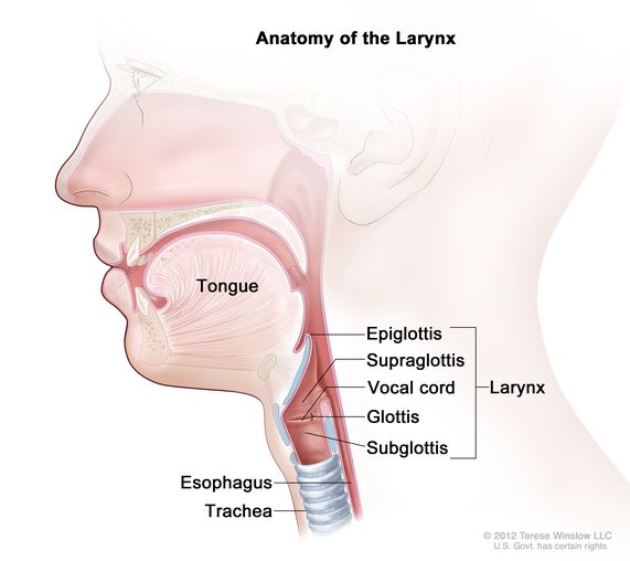


The sensation from the nasopharynx is carried by the maxillary branch of the trigeminal nerve (cranial nerve 5). The oropharynx is innervated by the sensory division of the glossopharyngeal nerve. The larynx, vocal cords and nasopharynx are innervated by the sensory fibers of the Vagus nerve –the internal division of the superior laryngeal nerve. A sensory branch of the Vagus also supplies a portion of the external auditory canal and Eustachian tube. Branches from these nerves form a tangle of nerves called the Pharyngeal plexus. The trachea and bronchus down to the respiratory bronchioles are innervated by the recurrent laryngeal nerve of the vagus. These nerves carry sensation to the brain. But the cough center in the brain is not a localized center.

The motor impulse to the muscles involved in the cough reflex is supplied by several nerves. The main muscle is the diaphragm: The phrenic nerve innervates the diaphragm. The intrinsic muscles of the Pharynx are supplied by the recurrent laryngeal nerve, and only the stylopharyngeus muscle is innervated by the motor fibers of the glossopharyngeal nerve. The external intercostal muscles, anterior abdominal muscles, are innervated by the intercostal nerves of the thoracic segments of the spinal motor nerves of the abdomen.

Mechanism of cough.

The initial action is the closure of the glottis (the vocal cords and the space between them), the epiglottis closes the opening to the larynx. The next act is a forceful contraction of all muscles, resulting in positive pressure in the thoracic cavity. The trachea and bronchus become shorter and narrower. The last act is the sudden relaxation of the vocal cord and throat. That results in a sudden gush of air expelled from the airways carrying all irritants captured in the bronchial secretion.

Though coughing and sneezing are triggered simultaneously with some common noxious stimuli, these two reflexes are different. A person can cough in sleep or under light anesthesia. On the other hand, a person who has to be awake to sneeze at the same time cannot sneeze voluntarily.

The force generated by a reflex cough can reach 100 miles/hr. The voluntary cough can vary greatly from just a short repeated cough commonly called a nervous cough to a forceful cough with the use of all muscles including accessory muscles of inspiration as in cases of bringing up thick tenacious sputum. When a cough brings up blood - is called Hemoptysis.

Factors triggering coughs.

Irritants: Irritation of the pharynx, trachea-bronchus from irritant gas, or fine sprays of chemicals like perfume, incense, household cleaners, and detergents.

Infectious agents: respiratory viruses, Covid-19, influenza, bacterial, fungal, mycobacteria.

Allergy and asthma.

Sinus and nasal discharge: Postnasal drip and vasomotor rhinitis, allergic rhinitis are some examples.

Medication: ACE inhibitors.

Pulmonary fibrosis: Idiopathic pulmonary fibrosis, post viral pneumonia fibrosis. Rheumatoid arthritis, SLE and scleroderma.

Malignant lesions: larynx, vocal cord, and Lung.

Aspiration of gastric acid: gastric reflux and aspiration.

Foreign body in airways.

Anxiety and psychosocial: also called nervous cough.

Putting a Q-tip in the ear.

Voluntarily or on request by the examining physician.

Types of Coughs:

Wet cough. In infection and allergy, where a lot of nasal secretion is generated.

Dry cough. Commonly seen in the use of ACE inhibitors, Pulmonary fibrosis.

The patients cough repeatedly, a short unproductive cough, involuntarily, and at times disturbs sleep.

Paroxysmal cough. The patient coughs uncontrollably and violently followed by a whooping sound in an attempt to resume breathing. Patients soon feel exhausted from coughing as seen in whooping cough(pertussis)

Tickle cough. In vasomotor rhinitis and allergic rhinitis, sudden onset of cough occurs and continues till secretions are completely cleared from the airway.

Nervous cough: repeated attempts to clear the throat when there is no secretion.

Croup. This is seen in children under 5 yrs. old in viral infection because of narrow airways. The swelling in and around the larynx produces raspy and squawking sounds.

Persistent cough. When cough persists over 6-8 weeks, as often seen in allergy and gastric acid aspiration. In delayed diagnosis like eosinophilic bronchitis, carcinoma of the lung, TB, fungal infection, etc.

Night cough. It is usually seen in gastric reflux and asthma.

Features of vasomotor rhinitis:

The precise cause is unknown, various agents are suspected including weather, perfumes, alcohol and spicy food. The cough follows a sudden gush of nasal watery discharge. The disincentive signs are the absence of an itchy nose, watery eyes, or scratchy throat.

Complications of persistent cough:

Most coughs are episodic, related to the upper respiratory tract infection, and are controlled easily. In prolonged vigorous cough, some of the following complications may be seen.

Fainting spells, Subconjunctival hemorrhage, small urine leaks, Inguinal and femoral hernias, Fractured ribs, Sore chest wall and costochondritis. Exhaustion, Sleep deprivation.


*****************************


Saturday, June 26, 2021

I Can't Breathe

                                                            Dyspnea or Shortness of Breath

                                                           PKGhatak, MD


“ I can't breathe” has become a worldwide cry for social justice after the killing of George Floyd in Minnesota.

I can't breathe, difficulty in breathing, sense of suffocation, air hunger, shortness of breath, short of wind, tightness in the chest, uncomfortable in breathing, not getting enough air, are some of the ways patients describe their difficulty - in medicine known as dyspnea.

The word dyspnea is derived from the Greek word dyspnoia – meaning breathing disorder.

The American Thoracic Society calls dyspnea” a subjective experience of breathing discomfort that consists of qualitatively distinct sensations that vary in intensity.”

Can't breathe is one of the most frightening symptoms of human experiences. A sense of impending doom overshadows all other concerns. The patient fights with all his might to right the situation.

A healthy person may experience shortness of breath during strenuous exercise like climbing several flights of stairs or hiking mountains and visiting high altitude places.

Shortness of breath in all other cases is due to an abnormal condition/condition in the lungs or heart or neuropsychiatric causes.

Points to remember that dyspnea is subjective and varies in intensity.

When patients present with dyspnea the doctor categorizes them into groups according to 1. its severity. 2. episodic or chronic.

Some of the conditions of life threatening acute shortness of breath (SOB) are.

  1. Allergic and anaphylactic. 2. Asthma. 3. Acute left ventricular failure (commonly called Pulmonary edema). 4. Cardiac tamponade (pericardial effusion or bleeding) 5. Rupture of the lung (pneumothorax). 5. Broken ribs and bloody pleural effusion 6. Pulmonary emboli. 7. Massive hemorrhage. 8. Choking. 9. Bilateral pneumonia like COVID-19. 10. Carbon monoxide poisoning.

Chronic cases of SOB are 1. COPD. 2. Congestive heart failure. 3. Chronic anemia is secondary to systemic diseases. 4. Neuromuscular diseases like ALS, paralysis of the diaphragm, myasthenia gravis. 5. Morbid obesity. 6. Ascites. 7. Large intra-abdominal mass like Wilms tumor. 8. Chronic bilateral pleural effusion. 9. Interstitial pulmonary fibrosis. 9. Deformity of chest wall e.g. Kyphoscoliosis.10. Subphrenic abscess.11. Chronic illness or disability leading to deconditioning.12. Anxiety and psychosis.

Treatment:

The goal is to restore the normal functioning of the lung, heart, blood and brain as soon as possible.

Pulse oximetry is in wide use since the covid pandemic, it is the initial test and any lack of oxygen should be immediately corrected by giving oxygen.

The other conditions are just too many to discuss here. But the best place is the emergency room for severe acute situations and the doctor's office for non-life-threatening conditions.

*************************************************

Thursday, June 24, 2021

Chest Pain.


                                                               Chest Pain.

                                                (Pain Pathways of the Heart).

                                                 PKGhatak, MD


The American Heart Association and its affiliates have been very successful in educating people on the significance of chest pain and its relation to an acute heart attack.

Heart muscles are not richly supplied with pain nerve fibers (called Afferent or sensory fibers) compared with the pericardium (fibrous covering of the heart). A wider area of the heart muscles has to be injured to elicit pain sensation. Even then, the pain is not felt in the heart but projected on the Central chest wall, neck, Lower jaw, and arms down to the fingers, upper abdomen, or on the back in between the shoulder blades.

This peculiarity of projection of visceral pain onto the skin is limited to organs innervated by the Vagus nerve.

In the early stage of fetal development, the human body was a segmented tube; each segment had its own pair of blood vessels and pair of nerves. In the later phase of development, as limbs and internal organs developed, that representation remained intact.

In the case of the heart, the location of pain felt on the skin is due to the fact that the heart developed from the pair of blood vessels from Cervical 7 to Thoracic 3 segments. The sensory nerve fibers of the heart join the vagus and sympathetic nerves to carry pain sensation to the brain.

In 1991 Dr. Armor described a new concept – Heart-Brain, consisting of 40,000 neurons situated in the heart and functioning as a mini brain. Nerve fibers from these neurons make direct connections with the amygdala, hypothalamus, thalamus and relay information to the cerebral cortex. The sensation from these neurons modifies effects of both the sympathetic and parasympathetic nervous system, and according to Dr. Armor, generates memory, emotion and modulates pain sensation.

Sensory nerves of the heart.

The Vagus nerve and nerves of the sympathetic nervous system carry Pain sensation to the brain.

The sensory nervous system of the heart is distinct. The nerve cells, nerve fibers, and ganglions are separate from the outflow tracts of both the parasympathetic (vagus) and sympathetic systems.

Vagus nerve.

The sensory fibers of the heart originated in the Nodose Ganglion of the Vagus, situated in the Jugular foramen. These fibers travel with the vagus nerve into the chest. Fibers destined to innervate the heart pass through the cardiac plexus without making any connection. The pain sensation from the heart is carried to the sensory nucleus of the vagus – Nucleus Tactus Soliterious, situated in the dorsomedial medulla. From there, the 2nd order neuron carries the sensation to the thalamus. From the thalamus, the pain sensation reaches the cerebral cortex.

80 % of pain sensation is carried by a pair of the vagus nerve. The right vagus nerve innervates the sinoatrial node, atrioventricular node, and atrial walls. The left vagus nerve innervates these structures and also from the rest of the heart. There is a considerable overlap of innervation of the heart from both vagus nerves.

Sympathetic innervation of the heart.

The neurons, that carry the pain sensation from the heat in the sympathetic nervous system, are situated in the Dorsal Ganglion from the 8th cervical to the 3rd thoracic segment. The axon of the nerve enters the spinal cord via the Dorsal root and makes a connection with the neuron situated in the dorsal horn of the spinal cord. The second order neuron carries the pain sensation upward to the Thalamus. And from the Thalamus the sensation carries to the cerebral cortex by the 3rd order neuron.

Nerve supply to the pericardium.

The pericardium has two sets of innervations.

The surface or parietal pericardium is supplied with neurons situated in cervical 3 to cervical 5 segments. The pain fibers travel via the Phrenic nerve.

The visceral pericardium is supplied by nerves that travel to the brain via both sympathetic and parasympathetic nerves.

******************************************


Tuesday, June 22, 2021

Single Pulmonary Nodule


                                       Single Pulmonary Nodule.

                                          PKGhatak, MD


In certain circumstances, a Chest CT scan is obtained as a part of workup - like a seat belt injury in an automobile accident or an undetermined cause of chest pain or shortness of breath, and to the surprise of the patients and doctors, a single pulmonary nodule is detected on the CT scan.

The incidence of an unsuspected Single Pulmonary Nodule (SPN) is 1 million / year in the USA. The attending doctor must give the patient the most recent information about the possible cause of the SPN. In the USA, there is a 40 % chance that the SPN is a growth at the initial time of detection. The vast majority of these are benign lesions, if the nodule is less than 3 cm, then only about 1 % of all the nodules so detected turn out to be malignant. Whether the SPN is malignant or benign, that depends on many factors including the radiological features of the SPL.

The first question the doctor asks the patient is whether he/she had a CT scan of the chest or chest x-ray previously, if so, that x-ray/CT scan must be reviewed and compared with the recent CT of the chest. Say, 2 years earlier CT, the SPN was of the same size - it was most likely a benign lesion.

This is important because no one wants to miss a diagnosis, Carcinoma of the lung in a very early stage. After all, the cure rate is 90 % at that stage compared with 30 % in stage II CA lung.

How to define a Single Pulmonary Nodule.

This definition is radiological and strictly adhered to.

These are the features: a discrete, well-marginated, rounded opacity, not exceeding 3 cm in diameter, surrounded by normal lung tissue; away from the hilum and mediastinum of the lung and no sign of pleural effusion, atelectasis and lymph node enlargement.

Nodules over 3 cm are called pulmonary masses. It takes 10 years for a nodule to reach the first 1 cm size.

What are common causes of SPN.

The three most common categories of SPN are benign adenoma, granuloma, and malignant tumor. The percentage of these conditions varies according to counties and even different regions of the same country. For example, in the desert region of the USA granulomas from fungal infections are more frequent than tumors, in developing counties granuloma of Tuberculosis is more prevalent.

What are granulomas.

A granuloma is a localized collection of inflammatory cells surrounded by blood vessels.

Granulomas are of two kinds: Inflammatory and Non-Inflammatory.

Infectious causes are Tuberculosis, Histoplasmosis, Coccidioidomycosis, Cryptococcus, Blastomycosis, Nocardiosis, and Hydatid cysts.

Non-infectious granulomas.

Rheumatoid nodules, Granulomatous angiomatosis, Sarcoidosis.

What are benign lung tumors.

Bronchial adenomas, hamartomas, benign tumors originate from cartilage, connective tissues, muscles, etc. And a special group of bronchial adenomas called Carcinoids.

What are the radiological signs of SPN being cancer,

SPN larger than 3 cm in diameter, irregular margin, ground-glass appearance (structure of lung is visible through the lesion), stipple calcification, eccentric calcification; doubling time is short (less than 120 to 600 days). Care must be taken not to call an opacity a nodule if it is located outside the lungs, artifacts, foreign bodies, and nipple shadow.

What are the risk factors for malignancy.

Chance of malignancy increases with 30 + years of smoking history, older age; exposure to radon, asbestos, nickel, chromium, vinyl chloride, polycyclic hydrocarbon; history of previous malignancy and chest radiation.

Nodule size and a chance of malignancy.

 5 cm nodules have a 60 % chance of malignancy, nodules 8 cm are 80-90% malignant.

What lab tests are required to diagnose benign lesions.

Serology test for collagen vascular disease, Rh factor. Serology for fungal infections, and fungal antigen tests where possible. Angiotensin converting enzyme for sarcoidosis., p ANCA and cANCA for angiitis. For Pulmonary Tuberculosis - interferon gamma release assay.

Follow up CT scan when initial CT and other tests are inconclusive.

For less than 3 cm lesions, a yearly CT scan. If stable for 3 years, then no further scans.

For 3 to less than 5 cm lesions, 3 to 6 months interval CT scans.

For 5 to 8 cm lesions an initial biopsy is called for.

How to proceed with SPL.

If history, radiological scans including PET scans, and various blood tests are inconclusive, then a Biopsy of the SPL is called for.

To improve the chance of a better yield by biopsy and with minimum damage of normal lungs, these important additions have taken place.

Skinny needle biopsy (SNB) with fluoroscopy, ultrasound, or CT guidance. Video-assisted thoracoscopic biopsy, GPS guided biopsy.

Bronchoscopic biopsy has also evolved into endobronchial ultrasound (ERUS) biopsy, electromagnetic navigation bronchoscopic biopsy.

At the present time, these modalities of SLN biopsy produced 80 % improvements over skinny needle biopsy.

What to do if SPN is a biopsy proven malignant tumor.

Surgical resection is the treatment of choice.

If the patient is not a candidate for surgical resection but has biopsy proven malignancy, then the following options are available.

External radiation therapy, Stereotactic radiosurgery, Percutaneous radiofrequency ablation.

Single Pulmonary Nodules are mostly asymptomatic when initially detected for unrelated health reasons or during lung cancer surveillance programs for chronic smokers, and the patients are more anxious than sick. A timely investigation and proper counseling must be provided. Most SLNs are benign, but benign lung tumors can cause pneumonia, lung abscess or hemoptysis and in rare instances turn malignant over years if not properly followed.

***********************************************************


Monday, June 21, 2021

Psoriasis.

                                        Psoriasis

                                   PKGhatak, MD


Psoriasis is a skin disease that manifests as red, itchy scaly patches. Psoriatic lesions commonly appear over the knees, elbows, trunk and scalp. Psoriasis patches can range from a few spots of dandruff-like lesions to scaling lesions covering a large area of the body.

Types of psoriasis:

For descriptive purposes, the psoriatic lesions are called:

Plaque psoriasis, Nail Psoriasis, Guttate psoriasis, Inverse psoriasis, Pustular psoriasis, Erythrodermic psoriasis, and Psoriatic arthritis.

The most common type of psoriasis is plaque psoriasis.

What causes psoriasis.

It is a multifactorial disorder caused by an interaction between Inherited Genetic Mutations and environmental risk factors. The abnormal Immune reaction manifested by very rapid turnover of skin cells, dead skin shedding lags behind the new cells generated, resulting in itching and redness of the skin.

What are the risk factors.

Some of the known triggers are – Streptococcal infection of the skin and throat, dry and cold weather, cuts and bruises of the skin, insect bites, stress, certain medications like - antimalaria drugs, High BP drugs, lithium, etc., sunburns, and rapid withdrawal of prednisone.

Is vaccination safe in Psoriasis.

Childhood vaccination should proceed as usual. All vaccinations, skin injury, and surgery may produce a flare up of psoriasis, this is known as Kobener Phenomenon.

What genetic mutations are associated with Psoriasis.

Genome-wide association studies have identified more than 60 susceptibility regions, those genes are related to Thymic 17(Th-17) cell action. Interleukin-36 -36(IL-36) is an important amplifier of Th-17 signaling.

In addition, Linkage analysis identified 9 different regions - known as PSORS 1 to PSORS 9 - associated with psoriasis.

In North American PSORS 2 is identified as a common variant and psoriasis is inherited as an Autosomal Dominant condition. Mutation of CARD 14 gene identified as the molecular defect underlying cause of psoriasis. Card 14 encodes an adaptor protein that is highly expressed in skin cells known as keratinocytes.

In worldwide studies, PSORS1 locus maps detected an association of PSORS1 to the Major Histocompatibility Complex (MHC) on chromosome 6p21.

Signs and symptoms of psoriasis.

Psoriasis is a chronic remitting skin disease, generally worse in the winter months and improves in summer. About 8 million people in the USA have psoriasis; 40 % of them may also develop arthritis.

Psoriatic skin lesions appear as red patches of skin with thick silvery scales. Psoriasis may appear as raised red or pink plaques with silvery scales called guttate psoriasis. Guttate psoriasis is common in children. In addition, the following symptoms and signs may be present.

Dry, cracked skin that may bleed or itch.

Itching, burning, or soreness.

Thickened, pitted, or ridged nails.

Swollen and stiff joints in Psoriatic arthritis. The clinical picture of psoriatic arthritis resembles Rheumatoid arthritis; arthritis may precede skin lesions and over time becomes resistant to therapy.

Treatment of Psoriasis.

It can be discussed in three categories.

1. Topical 2. Phototherapy, 3. Systemic.

Topical.

Emollients.

Local skin emollients are moisturizers and are effective in early cases involving limited areas of the body. It retains moisture in the skin, thereby reducing itching and scaling.

Hydrocortisone cream/ointment.

Steroid creams of various strengths are available by prescription and the over-the-counter 1% hydrocortisone creams are useful for reducing irritated skin lesions and reducing flaking and itching.

Vitamin D analogs.

It slows down skin cell production and has an anti-inflammatory effect. The ointment can be used on the scalp, the trunk, limbs, and other areas.

Calcineurin inhibitors.

Several compounds are available. They reduce immune reactions. And produce remission of skin lesions, and is useful in lesions over sensitive areas of the body and also the scalp.

Coal tar.

Heavy oil obtained from coal processing is effective in psoriasis. The mechanism of action is unknown. The oil has an odor and can stain clothing and bedding.

Dithranol.

It is used only under medical supervision. Dithranol ointment can be applied over a wide area and allowed to act for 10 to 60 minutes, then washed off. It may burn the skin if used inappropriately.

Phototherapy.

Special light therapy centers use various forms of light therapy. A wide area of the body can be treated. Each session lasts only a few minutes but is repeated 3 -4 times a week and stretched over 6 to 8 weeks. UVB light is invisible to the eyes and is commonly used in phototherapy. Phototherapy can be combined with Dithranol and Coal tar skin ointment.

Phototherapy is effective when other treatments fail.

PUVA.

Psoralen is combined with Ultra Violet A light.

This combination makes the UV A light penetrate deep into the skin. It is useful in cases where other forms of therapy have failed. But repeated use may cause skin cancer.

Systemic therapy.

Drugs are classified as Non-Biological and Biological agents.

Non-Biological.

All these drugs are useful in reducing skin cell proliferation, controlling inflammation, itching, and scaling. Many of them also have significant side effects that require regular follow ups.

Methotrexate.

Initially, the methotrexate is administered IM injections, then switched to oral therapy. Usually given once a week, periodic blood count and renal function are checked for side effects.

Cyclosporine.

This medication is an anti-rejection drug; newer drugs of this category are Sirolimus and Tacrolimus. Drugs are given orally and also locally on the skin as an ointment. Blood levels of these drugs are checked to adjust the dose to keep in the therapeutic range. All of these drugs are useful in suppressing immune reactions.

Acitretin.

It is a retinoid compound and is taken orally. It is widely used but also has side effects including liver damage.

Apremilast.

Apremilast is a Phosphodiesterase inhibitor and the pill is taken by mouth daily. It is effective in both skin lesions and psoriatic arthritis.

Dimethyl fumarate.

This drug is primarily used in Multiple sclerosis. It is also used in Psoriasis.

Biological agents.

Bioactive immunosuppressants are given by injections. Each drug is administered by a given protocol. If significant improvement of psoriasis is not seen in 8 to 12 weeks, then these drugs are discontinued. Since these are used in advanced and resistant cases, the outcomes of use in psoriasis are also variable.

Commonly used biological drugs are -

Etanercept. Administered weekly by IM injection.

Adalimumab. Given every 2 weeks by injection.

Infliximab. Given by intravenous injection.

Ustekinumab. This drug can be given subcutaneously or by IV infusions. Usually given every 4 weeks. It is effective in severe plaque psoriasis.

*************************************************


Psoriasis  usually appears as red or pink plaques of raised, thick, scaly skin. However, it can also appear as small, flat bumps or large, thick plaques

Sunday, March 7, 2021

Lung Abscess


                                                             Lung Abscess

                                                  PKGhatak, MD


In any infection, the body tries to limit the spread of infection by pouring in inflammatory cells at the site of entry of the infective organisms. A dense layer of inflammatory cells, white cells and their products are formed around the infection, which is known as a pyogenic membrane. In the center of it, the dead and liquefied cells accumulate. In the lungs, a few smaller branches of bronchioles remain open and air enters the liquefied center giving a distinct picture of air-fluid level on the x-rays. These bronchioles, however, are too narrow to drain the thick accumulated secretion. Unless the cellular derbies are cleared from the site, the repair and healing cannot begin. This may lead to one or more complications - enlargement of abscess, breaking open into a larger bronchiole forming a pulmonary fistula, opening in the pleural space forming empyema, eroding into a blood vessel producing hemoptysis, the distant heterogeneous spread of infection and abscess formation in other organs.

The lung abscess is called primary when the lung is directly infected. An abscess is called a secondary abscess when the lung infection comes from an infected blood clot from below the diaphragm via the inferior vena cava and infected cardiac vegetation. From direct contact with the lung as it happens in amoebic liver abscess, and subphrenic abscess.

The usual source of lung abscess is the aspiration of anaerobic bacteria of the mouth from choking, general anesthesia, dental procedures, alcoholism, seizures, reflux esophagitis, hiatal hernia, esophageal-pulmonary fistula, head and neck cancers, strokes with dysphagia, bulbar palsy, Parkinson's disease, ALS, etc.

Abscess formation can also develop from preexisting cavities in the lungs as cystic fibrosis, emphysema bullae, bronchiectasis, tuberculosis, carcinoma, foreign body- specially partial dentures, loose tooth, small toys in children, and intra- pulmonary sequestration and bronchial cysts - are congenital conditions. Narcotizing vasculitis and infarction of lung from Pulmonary emboli.

Common Bacteria.

Anaerobic gram negative and anaerobic gram positive bacteria predominate in adults. Staphylococcus aureus, including MRSA, is a common bacterium in children. Other rare infections are fungus, nocardia, mycobacteria, actinomyces, lung parasites, and amoeba.

Bacteroides fragilis, Fusobacterium capsulatum, and F. necrophorum are the most frequent causes of anaerobic lung abscess. Occasionally gram positive Pepto streptococcus and microaerophilic streptococcus infections are seen. Mixed bacterial infection is also frequent. In rare instances, Gram negative rods like E. coli, Hemophilus influenza, Pseudomonas aeruginosa are seen specially in hospitalized patients with ventilator associated lung abscesses.

Pathology.

In the first 48 hrs. following aspiration, inflammatory exudate takes place. Toxins released by the bacteria initiate vasculitis and venous thrombosis and necrosis of lung tissues.

Common areas of the lung are prone to abscess formation.

Lobes involved in abscesses are the result of aspiration to the most dependent part of the lung. In a bedridden patient, the posterior segment of the right middle lobe generally develops an abscess. In alcoholic and unconscious patients' apical segments of the lower lobes are involved.

Signs and symptoms.

Patients are sick and a distinct foul odor due to anaerobic bacteria is often detected. Chills, fever, night sweats, loss of significant weight are common. Chest pain and shortness of breath are usual. Cough may start as non-productive but soon becomes productive thick pus, at times blood mixed or frank hemoptysis is seen.

Clubbing of fingers is often seen. Fixed rales and other auscultation features may be present based on how close the abscess is to the chest wall.

Diagnosis.

Chest x-ray shows air-fluid level in a thick-walled cavity, rarely, more than one abscess is present. CT scans often are not required for diagnosis but are needed in case surgical treatment is deemed necessary.

Bronchoscopy is mandatory to detect bronchial obstruction by a tumor or foreign body. And to obtain bronchial secretion/pus for both anaerobic and aerobic cultures, gram stain, fungal and mycobacteria cultures, Bronchoscopic biopsy, brushing and cytology are routinely performed.

Cultures.

Aerobic and anaerobic cultures and sensitivity tests are most valuable in selecting proper antibiotics.

Treatment.

As soon as a lung abscess is suspected or detected on x-rays, intravenous antibiotics are initiated. The initial choice of antibiotic is Clindamycin 600 mg given every 8 hrs.; some physicians also add another agent like metronidazole and beta lactam cephalosporin, Fluoroquinolone. Macrolide antibiotic is used in mixed bacterial infections. Antibiotic coverage depends very much on the prevalence of common lung abscess bacteria of the local community and reports of culture and drug sensitivity tests.

It is to be noted that if proper antibiotics are chosen, a clinical improvement is expected in 3 to 5 days – lowering of temperature, less sputum production and a general sense of wellbeing.

The antibiotic coverage should be continued till the patient is afebrile and nearly symptoms free. That may take 3 to 6 weeks. At that stage, antibiotics are usually switched to orally.

In addition to IV antibiotics, chest physiotherapy, postural drainage, chest percussion, bronchodilator therapy, etc. are prescribed, including the attention paid to improve nutrition.

Switching mode of therapy.

If the expected improvement is not forthcoming at the end of 2nd week of therapy, then further assessment is necessary with repeat CT scans and cultures.

If the size of the abscess is seen to have enlarged to 6 cm or more, then some forms of surgical interventions are required. The less invasive procedure was tried first. CT guided or Ultrasound guided transcutaneous drainage of the abscess is performed under local anesthesia.

If more than 12 weeks have passed and the abscess remains, then lobectomy is generally required.

Those patients respond well to the initial therapy, and a complete resolution of abscess takes place in 3 to 6 weeks. Complete resolution of abscess by chest x-ray usually takes 12 weeks, even though the patient is recovered clinically and symptoms free from having an abscess.

****************************************

Saturday, March 6, 2021

Empyema


                                                         Empyema

                                             PKGhatak, MD


Empyema is defined as an accumulation of pus in the pleural space.

The surface of the lung and the inner surface of the chest wall is lined with a thin layer of serous tissue, leaving a minimal space in between known as the pleural space. The pleural membrane is covered by just one layer of mesodermal epithelium upon a connective tissue layer consisting of collagen and elastic fibers. A thin film of pleural fluid bathes both the parietal (chest wall) and visceral(lung) pleura and acts as a lubricant; that makes the lungs expand and contract without any friction during breathing. The pleural fluid is formed by the parietal pleura and the fluid is removed by the lymphatics of this layer, so pleural fluid formation and removal go on continuously. The visceral pleura gets its blood supply from the bronchial arteries and has no sensory nerve supply, so the visceral pleura is pain insensitive, whereas the parietal pleura is richly supplied with pain fibers from the intercostal nerves and also receives blood supply from the intercostal blood vessels.

In pneumonia, usually, more fluid is formed in the pleural space, but the presence of fluid is not easily detected by examination or by simple chest x-rays. CT scan can detect in 15 to 30 % of cases minimal pleural effusion called Parapneumonic pleural effusion. As pneumonia resolves, so also the parapneumonic pleural effusion.

About 6 % of children and 5% of adults with community acquired pneumonia develop parapneumonic empyema. When pleural effusion is infected by the extension of infection, the fluid becomes cloudy and thick. If the infected fluid has the following characteristics, then the effusion is called Empyema: fluid pH is less than 7.2 (normal blood pH is 7.4), glucose less than 60 mg/dl. LDH is three times the upper limit of serum LDH or LDH over 1000 IU/dl, WBC over 50,000/ml, protein over 3,000 mg/dl. Gram stains identify the presence of bacteria.

Risk factors.

In children, empyema often results when pneumonia is treated with inappropriate antibiotics. For those who are not fully immunized, chickenpox is followed by pneumonia, and the use of Ibuprofen in pneumonia.

In adults, the risk factors are homelessness, pulmonary emphysema, Pulmonary tuberculosis, IV drug use, not receiving the pneumonia vaccine, alcoholics, HIV infection, and immunosuppression by disease or therapy, and lung abscess. 35 % of anaerobic lung access is associated with empyema.

Both adults and children are at risk of empyema following prolonged chest tube drainage of bloody pleural effusion from a gunshot wound of the chest/ lung, knife wound, or automobile accident resulting in lung contusion, rib fracture, or pneumothorax.

Post-Surgical Empyema.

Following lobectomy and pneumonectomy due to malignancy or other diseases, if the lung fails to fill up the pleural cavity, then prone to empyema. This may be a special problem in the removal of bullous lesions in emphysema patients. Other causes of Empyema are Bronchopleural fistula, esophageal pleural fistula, perforated duodenal ulcer, amoebic liver abscess, perforated colon diverticulitis, and peritonitis, the infection may spread to the pleural cavity and foreign bodies in the pleural space like infected fragments of a projectile.

Common bacterial empyema.

The common bacteria is Streptococcus pneumonae type1. In community acquired pneumonia, the Streptococcus pneumonae type1 accounts for nearly 50% of postpneumonic empyema, Staphylococcus aureus followed by anaerobic bacteria infections. Viral parapneumonic and tubercular pleural effusion may also lead to empyema.

Symptoms and Diagnosis.

Persistent temperature elevation- often to 101F or higher, shortness of breath, chest pain, weight loss, anorexia, and general debility are common symptoms. On examination decreased or absent breath sound on one side of the chest, dullness on percussion. Leukocytosis with a shift to the left, toxin granules and Doehle bodies in the neutrophils are present. Chest x-ray shows pleural fluid. Ultrasound detects and delineates empyema easily and is usually repeated for monitoring purposes. CT chest is helpful in trauma cases and also in difficult cases.

Diagnostic thoracentesis is almost universal in all cases of more than minimal pleural effusion. In empyema, the pleural fluid is thick, difficult to aspirate, and cloudy to milky white. The other features will match the characteristics listed above.

Empyema fluid is sent to the lab for aerobic and anaerobic cultures and also for fungi and mycobacteria cultures. Gram stain of empyema fluid shows the presence of bacteria.

Treatment.

Closed tube drainage with a vacuum assisted closure device is inserted by Thoracoscopy. A wide bore chest tube Malicote 10-14F, Pigtail 8 -12F, or 28-32F French catheter is generally suited. Thoracoscopy can also detect septa, loculation, compartmentalization of fluid and that can be removed at the same time. That makes empyema drainage easier. Thoracoscopic lysis of adhesion is generally repeated.

Fibrinolytic enzymes like deoxyribonuclease DNase or tissue plasminogen activator tPA are injected into the pleural cavity and usually repeated to liquefy thick fluid.

Antibiotics are administered by IV and guided by culture and sensitivity tests.

In closed tube drainage of empyema, the lung should expand fully in 14 days.

If the lung fails to expand, then re-culture of fluid, review of the entire case, use of multiple antibiotics, or other surgical measures are called for.

Open drainage.

If the empyema fluid is so thick that even the French 32 size catheters are not draining fluid, then a wide area of the wound is created over the empyema. A large bore chest tube is inserted in the dependent part of the empyema and the other end of the tube is drained into a colostomy bag. The cavity is irrigated daily with mild antiseptic fluid, or the cavity is packed with saline moistened surgical gauge and changed daily. The cavity size should decrease gradually and the wound should seal in 4 to 5 months.

Open flap or Eloesser flap.

In this procedure, a wide incision is made just over the diaphragm, the most dependent part of the pleural cavity. A large skin and muscle flap is created. 1 to 3 ribs are resected to make a large opening into the chest cavity, and the skin-muscle flap is stitched in a way that creates a one-way opening for drainage, eg, making drainage of empyema easier. One way flap prevents air entry at the same time empyema is drained.

Decortication.

In the previous generation, tubercular empyema was the primary reason for this surgical operation. But occasionally some empyema fails to heal by the above methods and decortication is required.

If the lung does not expand to fill the chest cavity in 140 days. This procedure is considered. It is a major open thoracic surgery procedure, and many sick patients may not be able to withstand this procedure.

In this procedure, the entire pleura is stripped away and all cellular/fibrinous materials are removed. The lung with stripped away thick covering expands readily and the operative wound heals in 3 to 4 weeks.

The incidence of empyema has been declining over the past decades but in recent years,  the empyema incidence began creeping upwards. Early diagnosis and proper closed drainage and antibiotic therapy should resolve empyema in 2 to 3 weeks. The decision to open drainage of Empyema should be made after a thorough review of therapy and should be delayed until it is proven to be the only option available.

*************************************************


Thursday, March 4, 2021

Science of High Altitude Pulmonary Edema.


                                             Science of High Altitude Pulmonary Edema

                                                      PKGhatak, MD


Airlines are carrying a large number of people to high altitude sky resorts and mountain climbing facilities. These popular recreational activities also put people at risk of mountain sickness and acute pulmonary edema.

At sea level, the partial pressure of oxygen (Pio2) in the air is 160 mm Hg. At 8,000 ft the Pio2 is 120 mmHg, at 11,000 ft. above sea level the Pio2 is 87 mmHg, and at extreme high altitude, 18000 ft., the Pio2 is only 50 mm Hg.

At any altitude above 4,000 ft. the blood level of oxygen is low and becomes lower at higher and higher altitudes.

Air enters the alveoli of the lungs with inhalation. On the way to the alveoli, the air becomes fully saturated with water vapor. At a body temperature of 98.8 degrees F, the partial pressure of H2O is 47 mmHg, PiO2 is diluted as air enters the alveoli and Pi02 becomes (160-47) 113 mmHg. The blood in the capillary is separated from the alveolar air by only one layer of cells. The oxygen(O2) of air first gets dissolved in the alveolar fluid and then rapidly binds with the hemoglobin of blood because of the high affinity of oxygen to hemoglobin and forms a loose compound oxyhemoglobin. The mixed venous blood Pvo2 is 40mmHg, favoring oxygen flow toward the blood due to a pressure gradient of 83 mmHg (113- 40).

When this is happening, carbon dioxide (CO2) of blood is moving in the opposite direction – from blood into the alveoli, again due to the pressure gradient of 40 mmHg of blood to the + 0 mmHg of alveolar air. The CO2 binding is accelerated by the presence of enzymes, carbonic acid anhydrase. As little as 2 to 10 mmHg pressure gradient CO2 can move rapidly and completely.

As the blood leaves the alveoli and returns to the left side of the heart, the O2 carrying capacity of blood is at maximum – the PaO2(partial pressure of arterial blood) is over 90 mmHg and the O2 saturation (O2 sat) is 98 to 100 %.

Let's look at what happens at 8,000 ft. The Pio2 in the alveoli is 73 mmHg (120 – 47). The O2 gradient is (73-40) 43 mmHg. At 43 mmHg, the O2 sat is 75 %. This is significant. Hypoxemia and patients immediately develop symptoms.

Effects of Hypoxia on Local Population.

The people living in the high plateau of Tibet for over 7,000 years. The average altitude of the Tibetan plateau is 11,000 ft. They have developed an advanced adaptive mechanism, and it stands in contrast to the rest of high-altitude residents of Ethiopia and Andes Mountain people. In Peru and Bolivia, the present population moved in about 1,100 to 800 years ago and has yet to adopt the full high-altitude mechanisms of the Tibetan population.

In Peruvians and Bolivians, the hemoglobin level is higher due to the increased Erythropoietin activity, the minute ventilation is decreased, pulmonary arterial pressure is higher, right ventricular hypertrophy is present, Nitric Oxide (NO) levels are lower compared with Tibetans. They have more complications during pregnancy and childbirth, and newborns have lower weight at birth.

In Tibetan, the mutation of EGLN1 and EPAS1 genes carries the adaptive oxygen sensing and high altitude hypoxic adaptive mechanisms. The hypoxic stimuli of carotid and aortic bodies are muted. There is no hyperventilation. At 85 mmHg, the O2 saturation of the air is over 87%, the nitrogen oxide levels in the lungs and blood are high. The hemoglobin levels are not elevated. The resting ventilation is high, and lung volumes are larger. The smooth muscles of the pulmonary artery are not hypertrophic, pulmonary hypertension does not happen. The weight of newborns is normal.

Effect of Hypoxia on Recent Arrivals.

Acute Mountain Sickness.

On the very day of arrival, symptoms of cerebral anoxia develop. The main symptoms are headaches, dizziness, nausea, anorexia, fatigue and shortness of breath on minimal activities, difficulty sleeping and a mild dry cough. Swelling of the face, hands, and feet develops.

High Altitude Pulmonary Edema (HAPE).

From day 2 to day 4 of arrival, the symptoms are more severe. The shortness of breath at rest develops. The temperature rises to 101F degrees. Palpitation and rapid breathing, mental confusion, incoordination and prostration soon appear. The cough becomes productive of pink frothy sputum and occasionally becomes bloody, labored breathing with gurgling sounds on breathing is heard, cyanosis, a slow heart rate and BP began to fall. Acute respiratory distress and Acute Pulmonary edema set in.

High Altitude Cerebral Edema (HACE).

Anoxia of the brain cells is responsible for increased fluid accumulation in between brain cells and produces persistent headache, dizziness, nausea, the sensation of pins and needles, lethargy, incoordination, loss of sleep, tinnitus, violent behavior and loss of consciousness and seizures.

Pathophysiology of HAPE.

1. Hypoxia producing capillary leaks.

The tight junctions between the type I alveolar cells prevent excess fluid accumulation in the alveolar space. A thin layer of fluid is essential for the normal function of ventilation and gas exchanges. The integrity of the alveolar membrane depends on oxygen; in the hypoxic condition, the alveolar type I cells develop gaps between cell junctions and fluid escapes in the alveolar space. Nitric oxide production is reduced, lack of nitric oxide in the lung results in smooth muscle contraction of the pulmonary arteries and the Pulmonary arterial BP (PBP) is elevated. That produces Right heart strain and hypertrophy. Dr. J.B. Web described the genesis of the capillary leak “The hydrostatic stress that exceeds the load bearing limits of the collagen network [of alveoli] results in rupture of the basement membrane.”

2. Derailed removal of fluid.

Two separate but interrelated paths are present in the lung to ensure fluid removal, these are

A. Starling force driven sodium channels.

B. Na and K iron ATPase dependent trans-epithelial sodium channels.

The functioning of both these channels is defective in hypoxic conditions.

If the foramen ovale was not completely closed the opening of the foramen ovale enlarges and the Right to Left shunt takes place which makes hypoxia more profound and early right heart failure develops. Endothelin-1 activities are reduced and contribute to Pulmonary BP elevation.

 C. Increase in pro-inflammatory markers in the lung.

Analysis of bronchial fluid obtained during the Intubation of HAPE patients shows an increase in pro-inflammatory markers, namely interleukin IL-1 beta, IL-6, IL-8, TNF alpha. In addition, the total cell counts are elevated, and increased lung macrophages are present. Blood levels of LDH, CRP, and total proteins are increased. The monocytes attractive protein1alpha (MIPA1alpha) and monocytes chemo-attractant protein1 and IL8 are significantly higher in Chronic Mountain sickness.

D. The anti-inflammatory markers, IL alpha, IL-10, show no change or levels are decreased.

Chronic Mountain Sickness.

Prolonged stay at high altitude results in the continuation of acute symptoms, in addition, the patients develop sleep disorders. Mental confusion, abnormal behavior, cognitive impairment and various neurological symptoms.

Returnee of a Native of High Altitude.

High altitude people living on a lower plane for an extended period and then on returning to high altitude experience a more severe form of acute Pulmonary edema. The basic reason for this disease is not known.

Diagnosis of HAPE.

Respiratory symptoms in recent arrivals at high altitudes should always alert the possibility of acute mountain sickness or HAPE. This is the primary diagnosis and other possibilities like cardiac failure, PE, and pneumonia should come later.

Shortness of breath at minimal effort, or at rest, temperate elevation, frothy pink sputum, air hunger, cyanosis, tachycardia, and increased rate of respiration are universally present. Moist sounds, rales, on auscultation on both sides, chest x-rays- bilateral mid-zone, bat wing shaped infiltrates, and a low O2 saturation by pulse oximetry should be diagnostic of HAPE.


Immediate oxygen is administered by any available source to keep the O2 sat over 90%. Portable Hyperbaric oxygen chambers are generally available or should be made available as soon as possible. An arrangement must be made to take the patients to a lower altitude to a better medical facility.

Medications.

Nifedipine 20 mg tablets are given at 4 hourly to lower Pulmonary hypertension (PAP). Acetazolamide, a diuretic, also lowers PAP by blocking calcium channels, Tadalafil, a phosphodiesterase inhibitor, is also used. A bronchodilator Salmeterol is used to ease breathing.

Dexamethasone is used when cerebral edema is suspected in HAPE with predominant cerebral symptoms.

Prevention of acute mountain sickness and HEPA.

People should consult mountaineering schools/ training centers at the time of preparation for such events. The day before arrival, Nifedipine 80 mg long acting tablets one tablet and on arrival day and onwards one tablet should be taken. No physical activities on the 1st day. Sleeping at a lower altitude should be considered. Daily hiking/climbing should be limited to 1500 feet.

Use portable oxygen when the pulse oximeter shows saturation below 90 %. And seek advice from the team leader or the guide.

*************************************************


Sunday, February 28, 2021

Non Alcoholic Fatty Liver Disease


                                          Non-Alcoholic Fatty Liver

                                        PKGhatak, MD


An ultrasound scan of the abdomen is an unusually simple procedure for investigating abdominal and pelvic conditions. Many patients were found to have fatty liver unexpectedly and unrelated to their symptoms for which ultrasound scans were performed.

Incidence is quite high and is rising not only in the USA but also worldwide. In the USA, about 25 % of people have a fatty liver on ultrasound scan, in India, it is about 35 %.

Chronic alcoholism is a common cause of fatty liver. But it is also detected in high numbers in non-alcoholics. This is now designated as Non-Alcoholic Fatty Liver Disease (NAFLD).

The cause of NAFLD is unknown. People with NAFLD have a high association with Diabetes II, hepatitis C and HIV infections. More causal associations are seen in hepatitis B and A, obesity, physical inactivity, metabolic syndrome - the combination of high BP, obesity around the waist, high triglyceride & LDL cholesterol. Psoriasis, Hypothyroidism, hypopituitarism, Hemochromatosis, sleep apnea, polycystic disease of the ovary, and high fructose soft drinks and prepacked food and frozen TV dinners.

The adverse effect of the medication is known to produce NAFLD -   Acetaminophen. A heavy dose of tetracycline, oral hormone therapy, amiodarone, valproic acid, certain cancer drugs, some HIV drugs.

NAFLD is the initial stage of fatty liver, a more advanced stage of inflammation of the liver leads to a state called Non-Alcoholic Steatohepatitis (NASH). The incidence of NASH in the USA is about 6% of the population and in India, it is about 12 %.

Symptoms.

The majority of patients are symptoms free. Some complaints of vague upper abdominal discomfort, right upper abdominal pain, bloating, and anorexia. Some other patients with more advanced diseases have a loss of weight, enlargement of the liver, anemia, frequent infections, and gastrointestinal bleeding.

Investigation and diagnosis.

Liver enzymes - ALT and AST are elevated, due to liver damage. Platelet count falls due to decreased production of Thrombopoietin, a hormone produced in the liver and needed for platelet generation in the bone marrow.

Later, vitamin K-dependent clotting factor deficiency leads to the prolongation of Prothrombin time. Serum albumin level falls.

MRI of the liver clearly delineates pathological changes in liver cells. A new generation of Ultrasound scans – Fibroscan of the liver is also very sensitive like MRI and detects the degree of fibrosis. Fibrosis is the result of deaths of hepatocytes and inflammatory cell infiltration of the liver. Hepatic cirrhosis is the end stage.

Fibrosis Calculation.

Several institutions have developed a Fibrosis score, corroborated by liver biopsy, based on Age, ASL and ALT, and Platelet count. It is very useful for patients and the treating physician for monitoring progress. This practice has decreased the need for liver biopsy.

Formula : 

 Age ([yr] x AST [U/L])   /   ((PLT [10(9)/L]) x (ALT [U/L])(1/2)). 

Using this formula, patients with a score > 3.25 are likely to have advanced fibrosis whereas those with a score < 1.45 are unlikely to have advanced fibrosis.

A biopsy of the liver is the final confirmatory test, but biopsy is not necessary in the majority of cases.

Complications.

As the NAFLD progresses, the inflammation leads to the development of Steatohepatitis, the next step and ultimately to hepatic cirrhosis. Complications of cirrhosis are bleeding esophageal varices, hypoalbuminemia, ascites, hypersplenism, thrombocytopenia, and hepatic failure.

About 5 % of NAFLD patients with hepatic cirrhosis develop Hepatocellular carcinoma.

Treatment.

No effective drug treatment is available.

A weight reduction of 5 to 7 % of body weight is associated with improvement.

Daily physical exercise or 45 45-minute walk is advocated.

Elimination of Fructose from food and drinks should be done.

Vitamin E as high as 700 IU was advised, but worsening Diabetes and increased prostate cancer risk should be taken into consideration before undertaking such therapy.

Liraglutide with vitamin E is also tried.

Aramchol a combined fatty acid and bile acid conjugate is approved in Israel and in India for use in NASH.

Several anti-fibrosis agents are undergoing trial in the final stages.

******************************************************

Archive

  Archive. P.K.Ghatak, MD ghatak3@gmail.com 2025  March. Laser Endoscpoe 2025 February. Use of Ultrasound in Medicine MRI 2025 January. Bact...