Use of Ultrasound in Medicine
P.K. Ghatak, M.D.
Ultrasound
is a portion of sound that is beyond human ears can hear. The human hearing range is 20 to 20,000 Hz, and the intensity 2 to 120
decibels (dB). A decibel named after Alexander Graham Bell, the telephone, telegram, and audiometer inverter.
Ultrasound
is used in medicine in two separate fields.
Imaging.
Therapeutic.
The
images are called Sonograms, and the process of generating the images is
called Sonography or Ultrasound study.
There
are many ways Ultrasound is utilized in the medical field. It will be
discussed later in this article.
What
is Sonogram:
The
sound waves are generated by vibrating crystals. The desired
diagnostic sound waves are directed to the organ or organs under
examination. The sound waves after encountering a barrier reflect
back some or nearly all sound waves back. These reflected waves are
collected and a computer generates pictures in great detail.
The
reflected sound waves are often called Echo and the images generated are called Echogram, for example, Echocardiogram. The Echo has a special
quality, among the reflected sound waves, which must reach the
human ears after some delays for it to be registered as a separate
sound. This delay is called Impedance and the impedance is variable
depending on the tissue, air and fluid; this characteristic
allows sonography to construct an image.
Who
introduced Sonography in medicine:
Dr.
Karl Dusskil, a Neurologist used sound waves in 1942, in his attempts
to locate brain tumors. He took this idea from studying a publication
of an Italian scientist, Spallanzani who discovered bats use
ultrasound to locate insects and this process is called Echolocation.
How it
is possible to image brain matter which is encased in a cranium, made
of bones.
It
uses the same principle as is used in the Subtraction Angiography.
If the parameters of ultrasound of the cranial bones are known then,
they are subtracted from the final scan and the true image of the
brain emerges.
The science behind the conversion of electricity to sound.
When
asymmetrical crystals, eg, Quartz or Polycrystalline Ceramics are
subjected to physical stress, they are polarized and generate
electricity which is called Piezoelectric.
In a
reversed situation, when these crystals are placed in an electric
field, they begin to vibrate and generate sound waves. By varying the
crystals, frequency and amplitude of the electrical field, the sound
waves of various frequencies and variable strength can be generated.
Components
of Ultrasound (US) instrument:
The
basic components are ultrasound the Generator, also called the Transducer
or Probe and the Receiver. The receiver is also incorporated into the
transducer. The other parts are the monitor and controls. The
transducer contains an assortment of crystals, and it comes in various
shapes and sizes to fit the part of the body to be examined, eg, the neck
is a small saddle-shaped gadget and a large gently curved transducer is used in the abdomen and pelvis examination.
In
addition, there are specially designed probes for special locations
to name a few - transthoracic esophageal ultrasound for examination
of the left atrial chamber of the heart, and transrectal for
examination of the prostate gland.
Ultrasound
images:
US
imaging is used for every organ system of the entire body, beginning
from the time of conception to the last illness. US study is also
useful for the evaluation of the functional status of an organ,
detection of motion and direction of blood flow.
The
special uses are called by different nomenclatures, eg,
Echocardiogram, Doppler study of carotids or aortic valve, etc.
Advantages
of Sonography:
1. The
instrument is not expensive and the machine is portable.
2. There
is no special reparation required for most studies and repeated
examinations can be done to watch the progress of a disease or the state
of recovery with treatment.
3.
Patients are not exposed to radiation.
4. The
technology is easy to teach and teaching facilities are locally
available and not expensive.
5. The patient does not require to lie down for a US scan, in fact it is an advatade to scan when the patient is sitting or standing to detect small plural effusion or ascites, also, the venous obstructions in legs are better imaged is upright position.
Disadvantage:
1. Air
does not produce echo and any structures next to a pocket of air in
the body like the Pancreas and Lungs are difficult to visualize.
2. The
image quality is very much dependent on the technical skill of the
operator. It is like a good professional photographer setting up his
camera for a portrait – the aperture, duration of exposure, power of
the flash or lighting, etc. The image quality of US varies greatly on
the frequency and strength of US generated by different probes, the
depth and direction and the focal point; the angle the transducer
makes with the body at the contact point. The direct end-on-view
makes a clear image and increasing the angle makes the image fainter.
The echo and artifacts can be manipulated to enhance the image
brighter.
To
minimize the adverse effect of the presence of air, in between the
transducer and the skin of the patients, a water-based jelly is used.
This jelly contains glycerin and propylene glycol in addition to
water. It also provides a smooth surface for the probe to glide
against the body and minimize patients's discomfort.
Types
of Sonography.
A-
Mode. A stands for Amplitude. A single pulse is transmitted through
the body and the echo is collected and a linear image is produced. It
is a one dimensional image.
B-
Mode. B stands for Brightness. B-mode images are two dimensional,
also called 2-D echo. The amplitude is in the X-axis, Y-axis is time.
Using a proper probe and selecting the correct frequency of sound,
depending on the depth of the organ located in the body, the technician
should able to select echo of adequate amplitude to generate a good
picture.
M-Mode.
M is for Motion. Over a time period, successive B-mode echos are
recorded and placed next to the previous one. Any moving part of an
organ, eg, a beating heart, can be imaged like a moving picture taken
by a camera put on sport mode.
3-D
and 4-D Echo. The 3-D echogram is generated by taking the echo of the
same organ or tissue from many angles and then stitching all of them
together as one picture. It is like drawing a box on a flat sheet
of paper, showing length, breadth and height. A 4-D echogram is a
series of 3-D echograms over a time, depicting changes in volume or
size over a specific time frame. The time is the 4th
dimension.
Other
echograms for special purposes.
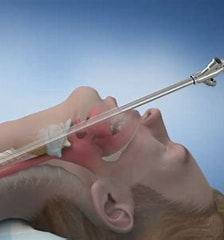
Trans
Thoracic Echocardiogram (TTE). A narrow probe is introduced through
the mouth into the esophagus and positioned just behind the Left
Atrium and various aspects of this chamber are examined. This is
useful in atrial fibrillation, pulmonary embolism, infra-atrial shunt
and heart failure.
Trans-rectal.
This is a standard for evaluation of the size, volume and consistency of
the prostate gland and is utilized in transrectal biopsy of the
prostate gland.
Transvaginal
and or, Pelvic ultrasound: This is a standard imaging technology for
the evaluation of every aspect of pregnancy, size and shape and growth
growth of the fetus, placental health, blood flow, status of amniotic
fluid and ectopic pregnancy. In other situations, any pelvic mass,
ovarian pathology and ascites can be imaged with clarity.
Doppler
Ultrasound:
Vesto
Melvin Slipher discovered when a planet was moving away from the
Milky Way, the light waves emitted from the planet, were less frequent
and appeared Red than when the planet was moving towards the
telescope of the observatory and the light waves had higher
frequencies and appeared Blue.
In an Ultrasound study, the same phenomenon is utilized to detect whether
the blood is moving forward in the blood vessels or falling
backward. The Red Blood Cells (RBC) scattered US waves when hit
by the US and the receiver records reflected waves and plots the blood
movement towards the probe or away from the probe.
The
Doppler US is useful in evaluating Aortic stenosis, Aortic and Mitral
valve incompetence, Deep vein thrombosis of thigh and pelvic veins,
Inferior Vena cava obstruction, the patency of the bile duct and many other
uses.
Echocardiography:
A
Swedish cardiac surgeon, Dr. Inge Edler, was searching for ways to
investigate the Mitral valve function before surgical repair of
mitral stenosis, which was a common malady of that time. A scientist
named Carl Hellmuth Hertz was using Refected Palatinoscope for
testing metal properties. These two scientists teamed together and
took a moving echocardiogram of the Mitral valve. They published their
findings in 1953, the echocardiogram generated was a 2-D motion
image, and with this, the Echocardiography was born. They were awarded
the Lasker Prize in 1977 but many scientists lamented that they
deserved the Nobel prize in medicine.
What
echocardiogram is used for:
This
is a versatile machine for evaluating both the structure and
functions of the heart as a whole and of its parts. The wall motion
abnormality, following a suspected coronary event, indicates possible
MI. Excess blood left in ventricular chambers indicates poor
contralie function of ventricluar muscles as happens in heart
failure. The functional status of all cardiac valves, the size and shape of
valves, and the prolapse of valves due to papillary muscle damage can be
easily detected. Individual ventricular systolic output or ejection
can be studied by moment by moment and used every day for follow-up care in coronary artery disease and following bypass surgery.
Interested readers may read many excellent articles published by the
American College of Cardiology.
Medical
Uses of Ultrasounds.
The
use of the US is very extensive in daily medical practice. It can be
summarized as
Diagnostic
Therapeutic.
Combination of the two.
Many
of the diagnostic uses are already mentioned, in short, any diagnosis
requiring an image can be accomplished by the use of a US study.
Physiotherapy:
Spain
ankles, frozen shoulders, tendinitis, and tennis elbow are treated with
low frequency US to increase the blood flow. The same principle is
utilized in poor healing of bone fractures.
Tissue
Biopsy:
Skinny
needle biopsy for any nodules or growth, whether in a deep organ like the kidney or near the shin surface like the thyroid gland and breast
tumors, the intervention radiologist accurately maps the tumor and its
location then under its guidance introduces the needle and performs
biopsy.
Stent
placement:
In
cases of deepening jaundice due to bile duct obstruction from any
cause, including hepatocellular carcinoma, to immediately relieve
unremitting symptoms of patients, the doctor locates the intrahepatic
dilated hepatic duct by using the US and then threads a probe into it
then inserts a stent to drain bile in the duodenum. This kind of
procedure is done for obstruction of Cerebrospinal fluid circulation
after an attack of meningitis /encephalitis. Renal pelvic
obstructions or ureteral obstruction either from tumors or stones.
Aspiration of pleura fluid and drainage, pericardial effusion,etc.
Central
venous access:
Instead
of a blind procedure of placing a Central Venous Line, using the US to
locate the subclavian vein or innominate vein is much safer and less
likely to produce any complications. In “hard to find veins” US helps
to find a vein in the forearm to start an IV line.
Intra-arterial injection of thrombolytic agents:
In
cerebral embolism or thrombosis, a Doppler study of the diseased vessel
is used and then under US guidance the vessel is catheterized and the thrombolytic agent is infused.
Transdermal
administration of medication:
The US is
used to improve the absorptive capacity of a patch of skin and then a
patch incorporated with medication is applied over it.
US in
Surgery:
US
energy is used in the ablation of tissues is called High Intensity Focus
Ultrasound (HIFU). The high energy sound heats up the target tissue
and destroys it, eg, aberrant ectopic focus in atrial fibrillation,
Uterine fibroid and Prostatic hypertrophy. Other examples are
Tracheal, Laryngeal papillomas, nasal polyps, vocal cord growth, etc
Skin
Incision by using the US. Just like a laser beam in making a skin incision,
US energy is also used for the same purpose.
Cauterization
of bleeding vessels. An easy way to control bleeding is by using US
energy.
In Eye
surgery:
Phocoemulsifier-
In cataract surgery, a needle is inserted into the cataract and it
vibrates at US speed and emulsifies the lens. Then the derbies are
suctioned out, then an artificial lens is inserted.
In
Kidney stone and Gall stones: Extracorporeal
shock wave Lithriopsy.
The
machine is called Lithotriptor and the process is Lithotriopsy. The
body is immersed in water and shock waves are applied to the body to
break up stones in smaller fragments and the gravels then pass out
easily. The US waves are generated in several ways.
1. Electrohydraulic.
The shock waves are created by making tiny sparks underwater between
two metal points.
Electromagnetic.
An electromagnatic coil generates shock waves.
Piezoelectric.
Quartz or ceramic crystals generate the waves.
The other is light. Laser energy is directed toward the stones by breaking them up into small piec
During 1920 to 1940, European soccer teams used Ultrasound as a
physiotherapy agent for the treatment of the injuries of their players.
In 1958, the US was used in the OBGyn and soon became a standard
technology used for various aspects of pregnancy, fetal development
and ovarian pathology. The field became wide open with the
introduction of Echocardiography and Doppler ultrasound.
Today,
the US is the most versatile and often utilized technology in the medical
field. Portable US instruments carried by the medical personnel on the
nursing floors like a NoteBook and used for diagnostic purposes and
also in the therapeutic armament, In a urologists office, the post
voided residual urine is determined in patients having voiding problems, just before the urologist sees the
patient. Many cardiologist carry similar portable machine, at the
point of service, for the evaluation of the many aspects of cardiac
functions of their patients, whereas, the stethoscope is
progressively becoming a museum piece. In the near future, all medical
offices will be using a miniature portable Ultrasound like nowadays
the weatherman talks about the weather on TV carrying a small
NoteBook in his hand.
************************************************************