Saturday, November 25, 2023

Oriental Sore / Cutaneous Leishmaniasis.

                                                Cutaneous Leishmaniasis

                                                  P.K. Ghatak, M.D.


Oriental sore is present in a wide area of the world. The majority of the cases are from the New World countries - Brazil, Bolivia, Peru, Panama, and countries around the Mediterranean Sea. CL (Cutaneous Leishmaniasis) is also prevalent in the Old World, the Middle East and Central Africa.

CL is a parasitic disease of the skin and the mucous membranes of the nose, mouth and throat. The parasite is Leishmania, which belongs to the Trypanosomatidae family and the Kinetoplastids order. CT is transmitted to humans by Sandflies.
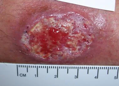


Incidence of CL.

About 600,000 to 1 million new cases of CL occur each year. The mortality is few and comes from secondary bacterial or fungal infections, but the morbidity is a serious problem.

Anatomy of Leishmania.

Leishmania is a unicellular organism that exists in two different anatomical forms. In the insect vector, Leishmania is an oblong, flagellated form called Promastigotes, which reside in the proboscis of the sandfly. A small, round, non-flagellated form called Amastigote is ingested by the sandfly from humans during feeding. In the gut of the sandfly, the parasites change to flagellated form.

The nucleus of Leishmania is present in the center of the cell; in front of the nucleus in the cytoplasm, a mass of mitochondrial DNA is present called the Kinetoplast. From the Kinetoplast a flagellum originates and extends outside. In the cytoplasm, a single strand of mitochondrion and Golgi apparatus are also present.

Leishmania have a unique DNA and glycosomes (peroxisome-like organelles). It was believed that Leishmania did not propagate by sexual union, but recent studies provided contrary evidence that amastigotes exchange nuclear material during binary fission. Leishmania has 34 to 36 chromosomes and during cell division, the chromosomes do not condense.

Life Cycle of Leishmania.

The flagellated Promastigotes infect humans. Sandfly bites introduce the Promastigotes to the human victims. The macrophages, neutrophils and phagocytes engulf the parasite but are unable to break down its surface membrane. Inside the cells, the Promastigotes change into Amastigotes. In the macrophages, the amastigotes multiply rapidly to such a number that the cell bursts open and the newly released amastigotes infect other tissues.

Inside the sandfly.

The sandfly takes a blood meal and swallows macrophages loaded with amastigotes, inside the gut, the macrophage wall breaks down, releasing the amastigotes. Amastigotes transform into Promastigotes and divides and move to proboscis of the sandfly.

 20 different species of Leishmania produce CL., and 90 different species of sandflies are the vectors. The important Leishmania species in the Old World are L. infantum, L. donovani, L. major, L. aethiopia. In the New world's species are L.mexicana, L.infuntum, L.guyanensis, L.amazonensis, L.braziliensis, and L.panamanensis.

The Phlebotomy sandfly is the vector in the old world and Lutzomyia sandfly in the new world. A species of sandfly harbors only certain Leishmania species and that limits one type of CL lesion confined to a local area and absent from the adjoining village or locality.

Development of the Skin lesions and Clinical Types.

The sandfly bites on the face, forearms, hands and legs. The bites are painful. A red papule develops at the bite site, which turns into a nodule. Several days or weeks later, the nodule ulcerates. This is the beginning of the chronic indolent Oriental sore.

The subsequent healing or progression of CL depends on (a) Leishmania species.(b) victims' immune status - both the innate and acquired immunity. (c) Intercurrent immune diseases. (d) suppressed immunity by drugs, which lowers cellular immune functions or delays and diminishes immune globulin synthesis.

Clinical types of CL. (a) Limited Cutaneous Leishmaniasis.(b) Recidivans Leishmaniasis.(c) Mucocutaneous Leishmaniasis. (d) Anergic Diffuse Dermal Leishmaniasis. (e) Post Kala-azar Dermal Leishmaniasis.

Clinical features.

(a) Limited Cutaneous Leishmania. The initial lesion does not progress to ulcers because the victims have good levels of immunity. The lesions heal spontaneously with scars, and patients develop permanent immunity.

(b) Recidivans Leishmaniasis. The initial lesion heals with scars, and months or years may pass without further disease activities. Then new skin lesions begin to appear at or near the scars. The process goes on for a while, and ultimately the skin lesions heal. Generally, no medication is required.

 (c) Mucocutaneous Leishmaniasis. It is the most devastating form of CL. The initial lesions develop around the nostrils, mouth, and eyes. Satellite lesions may develop in the throat. The mucosal lesions produce tissue necrosis and usually penetrate into the deeper layers of tissues and erode the muscles, cartilages and bones. Without medical therapy, no recovery is possible. The WHO recommends that all Mucocutaneous Leishmaniasis should be treated and the WHO provides guidance and medications in participating countries.

 (d) Anergic Diffuse Dermal Leishmaniasis. The patients are immune deficient and usually have HIV infection. The initial lesions are papules and nodules, but lesions do ulcerate. Because of the lack of immune cellular response, multiple lesions appear, usually on the face, arms and buttocks. Later, the entire body is covered by several forms of skin lesions – plaques, papules and nodules. The appearance of the patient resembles nodular leprosy, however, no nerve damage occurs in this form of leishmaniasis. Treatment with medications is not very effective and where initial success is achieved, the recurrence of new lesions is common.

 (e). Post Kala-azar Dermal Leishmaniasis. Visceral leishmaniasis is better known as Kala-azar. 2 to 20 years after the cure of Kala-azar, some patients develop hypopigmented macules, nodules, plaques and erythematous plaques on the face and other areas of the body. The lesions are recurrent and may persist for years.

Diagnosis of CL.

Biopsy. The demonstration of amastigotes within the macrophages is the standard diagnostic method. If there is no blood in the smear and stained with Geismar or Wright stains, a trained person can identify amastigotes without difficulty. An alternative to a biopsy is the aspiration of clear serum from the lesions. This method is equally sensitive. However, in certain forms of CL, the parasite numbers are few. So more and more reliance is shifting towards antibody detection by ELISA or antigen DNA detection by PCR test.

Treatment of CL.

The drug treatment is standardized by the WHO and the CDC in the USA. Few cases of CL were detected in the returning veterans from Afghanistan and Syria. In addition to standard therapy, each country has a host of local treatment protocols which are not effective but popular among the locals. Some of them are Cryotherapy, Heat therapy at 40 to 42 degrees C, Paromomycin topical preparations, infiltration of the ulcers with the solution of Sodium Stibogluconate, and urea 10 -15 % solutions. Oral dapsone, Oral Allopurinol and Oral antifungal agents

Standard therapy.

Pentavalent Antimony agents. 

Two commercial preparations are available -Sodium stibogluconate (pentostam) and pentostam eglumine Antimonate (Glucantime). Both of these are given intravenously for 10 to 20 days

Liposomal amphotericin B. It is an antifungal drug and is also effective in leishmaniasis but expensive.

Oral Pentostam, pentostam

 Oral Miltefosine. It is a wide spectrum antibiotic also effective against Leishmania. Initially, it was approved for cancer therapy.

Various combinations of drug therapy are practiced based on the leishmania species and the degree of tissue damage.

Prognosis.

Mucocutaneous and Anergic diffuse dermal leishmaniasis have poorer prognosis because of destructive facial lesions and disfigurements. Mortality is negligible as such, but secondary infection may lead to septicemia and deaths.

Vaccine: No commercial vaccine is produced for Leishmania disease. Russia made a vaccine using attenuated L. major and used in Russian endemic areas with 50 % success in controlling new skin lesions, but the vaccine does not protect against infection from other species than L. major, and also has some troublesome side effects.                           

*************************************************


Tuesday, November 21, 2023

Chagas Disease


                                                             Chagas Disease

                                                   American Trypanosomiasis.

                                                         PKGhatak, MD.


Chagas disease is named after Dr. Carlos Chagas of Brazil. Trypanosoma cruzi causes Chagas disease in South America, and Trypanosoma gambiese causes Sleeping Sickness in Africa. Several species of Leishmania are pathogenic to humans, and both Trypanosoma and Leishmania belong to one family.

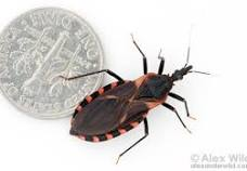
In 1909, Dr. Chagas investigated an unknown illness that was common in the rural areas of Brazil. He found a blood sucking insect feeding on humans and leaving behind feces and urine near the wound. The excreta of the kissing bug were loaded with the infective form of the parasite. The victims, while sleeping, rubbed the feces and contaminated the wound and the parasite also entered the body through the conjunctiva of the eyes.

Trypanosoma cruzi can infect in other ways - Contaminated food and drink by vectors' feces and urine often cause local endemics, contaminated blood transmission and organ transplantation are less common.  Mother to child transmission during pregnancy is a major concern, and the children in subsequent pregnancies are also infected.

The victims developed illness in two phases – 1. The initial or the first stage. 2. The chronic stage.

The initial stage.

The initial illness is a mild local inflammation and the regional lymphadenitis and in a few instances only mild systemic symptoms. Occasionally, a furuncle, called Chagoma, develops at the wound site. The illness is self limited. About 50 % of new infections may remain symptom-free.

In the 2nd or Chronic phase.

Years may pass without any signs of illness, and then patients present with symptoms of serious heart, gastrointestinal and neurological diseases.

The parasite Dr. Chagas discovered was a flagellated Protozoa in the blood of the victims. He conclusively proved the pathogenicity of the protozoa by recovering the same organism is susceptible animals. Dr Chagas named the parasite Trypanosoma cruzi to honor his mentor Dr. Cruz.

The Life Cycle of the Protozoa Parasite:

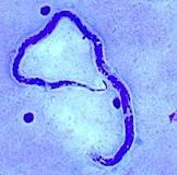
The organism is a motile, unicellular organism having one flagellum. The protozoa have 5 stages of development and two methods of propagation - one by binary fission and the other by sexual union. The different stages of life of the protozoa are named according to the point of attachment of the flagellum to its body or the absence of the flagella. The Greek word for the flagella is mastigot. The infective form that circulates in the blood of the victims is called Trypomastigotes.

Inside the bug.

In the midgut of the insect, the trypomastigotes transform into Epimastigotes, having the flagellum attached to the end of the body. Here epimastogotes multiply, and the epimastogotes enter the hind gut and transform back to Trypanostigotes, and infect humans when the bug takes a bloody meal.

In the victim.

The Trypomastigotes transform into a round, small unicellular organism inside the victims' cells. The parasite has no visible flagella and is called Amastigotes. Amastigotes multiply by binary fission. Amastigotes transform into trypomastigotes and burst open the victim's cells. The released Trypomastigotes infect more tissue, and also enter the bloodstream and lymphatics and are widely distributed in the body. Some of the Trypomastigotes switch back to Amastigotes,  divide again and keep on multiplying by binary division. And the cycle repeats again and again.

Besides humans, small animals like monkeys, armadillos and dogs are also infected.

The insect vector. 


The bug that transmits Chagas disease is a Triatomine bug, a variety of Reduviid bug, locally known as the Kissing bug, as they find it on the faces of victims.

There are several species of the Triatomine bug, one species is active in a certain locality, or another. The bug is active at night. They come out of cracks of the wall, fall from the ceiling, or just crawl on the bed from outside. The bugs bite on the faces of humans (part not covered during cold nights). A recent report says the oral route of infection, in recent days, has been the dominant path of infection of Chagas disease.

Chagas Disease:

The incubation period is short.

The initial illness is mild and self limited, and often remains asymptomatic.

The chronic phase of Chagas disease is a protracted severe symptomatic disease of the cardiovascular, gastrointestinal, and Neurovascular systems. The mortality rate is high and the disability is severe.

Pathophysiology:

The amastigotes invade muscle cells of the heart and the smooth muscles of the esophagus, and colon, and also invade the brain. The inflammatory reactions are mediated by acute phase Cytokines. The initial myocarditis is followed by necrosis of the muscles, which are replaced by fibrous tissues. The cardiac and smooth muscles of the GI tract become weak and flabby. The heart is the most common organ affected by Chagas disease. Cardiac impulse propagation along the His bundle due to fibrosis manifests as heart block, branch block, cardiac arrhythmia and systemic embolism. Massive dilated cardiomegaly and heart failure are the prominent features of this disease. Megaesophagus leads to difficulty in swallowing solid food, and megacolon leads to chronic constipation, abdominal distention and discomfort.

The CNS infection results in strokes and many other neurological manifestations.

Diagnosis:

The mainstay of diagnosis of Chagas disease is the demonstration of moving Trypomastigotes under the coverslip of a thick smear of blood. Under the trained eyes, the movement of the transparent parasite around stationary red cells is not difficult. It is a cost-effective test and practical in the rural communities.

Other confirmatory tests and PCR antigen detection and indirect ELISA antibody detection.

Medical treatment:

Two oral medications, Benznidazole and Nifurtimox, are effective in killing the parasite but the drugs must be given early in the acute phase of the disease in children 2 to 18 years of age. The older adults develop toxic side effects more frequently, and drug therapy is not recommended in adults. Women of reproductive age are tested and treated with drugs to prevent congenital Chagas disease. 

Benznidazole is an imidazole derivative, active against intracellular parasites. In Chagas disease, the drug is prescribed for 60 consecutive days. The drug inactivates parasitic enzymes and damages DNA and large protein molecules by generating cation radicals. Common side effects of this drug are various GI symptoms, skin rashes,  peripheral neuritis, thrombocytopenia, leukopenia, and anemia. It is potentially carcinogenic.

Nifurtimox is a nitrofuran drug. The mode of action of Nifurtimox against the parasite is similar to Benznidazole and it generates radicals through its action on nitrogenous enzymes. The adverse reactions are also like Benznidazole. But when prolonged treatment is necessary, Nitrofurans is not suitable because of its adverse effects, which are more intense than Benznidazole. Both drugs are contraindicated in pregnancy.

Surgery: 

In the past, various types of cardiac surgery were tried to improve the function of the failing, frail and flabby heart muscles. In the long run, none of those procedures proved to be beneficial and subsequently abandoned. Heart block is effectively treated with cardiac pacemakers. Only cardiac transplantation is a solution for severe heart failure.

Megaesophagus.

Laparoscopic myomectomy, esophageal mucosectomy are less involved procedures. Heller-Pinotti procedure is a more involved operation and is done in early swallowing difficulties. It is a modified Fundoplication operation.

In advanced cases, the Thal-Hatakufu operation is done. In this operation, Esophagogastricplasty is performed. The success of the Thal-Hatakufu operation is moderate.

Megacolon.

A Total Colectomy operation is required with anastomosis with -(a) ileorectal, or, (b) ileoanal, or, simply a colostomy.

Prognosis of Chagas disease depends on the stage of the disease. In the early active phase, the drug treatment is curative.

The survival in the chronic phase is poor.

Incidence and prevention of Chagas disease.

18 million people in 21 countries of Central and South America are infected with Trypanosoma cruzi and 100 million people are at risk of infection.

 Insecticide is used to control the insect vector. Better housing and public education have been successful in bringing down the spread of Chagas disease.


***************************************************


Tuesday, November 14, 2023

Dracunculiasis / Guinea Worm Disease


                                                            Dracunculiasis

                                                     Guinea worm Disease

                                                      PKGhatak, MD


Guinea worm disease and the name of President Jimmy Carter are intertwined, to recall one, the other name pops up immediately. President Jimmy Carter single-handedly brought the misery of African people suffering from Guinea worm disease to our consciousness.

Dracunculus medinences, the parasitic nematode (roundworm) has been a curse of humanity known since 1000 BCE. The worm lives a symbiotic life with the water fleas called Copepods, which are abundant in the small pools of water in Sub-Saharan Africa. Farmers and others drink water from these pools during hot summer months and ingest Copepods and become infected with the Guinea worm larva.

Life cycle of Dracunculus medinences:

Humans, domesticated dogs, and cattle are victims of the roundworm. Dogs and cattle are infected because of their closeness with their masters. People develop incessant burning pain during the release of larvae by the gravid female. Farmers dip their feet in the pool in order to get some relief from the burning sensation. The worm releases thousands of larvae, which are food for the Copepods.

Thirsty men drink water from the pool during summer days, working in their fields under the hot sun. The outer cuticle of the copepods dissolves by the gastric juices. The released larvae enter the small intestine and begin to move along the tissue planes to the abdominal and thoracic muscles. In about 3 months, the worms reach sexual maturity. There, the male and female worms mate, and soon the male worm dies. The female starts her migration towards inferior extremities and finally settles underneath the skin of one leg. It takes 12 to 14 months to complete this migration. Once the right moment arises, the worm breaks through the skin of the foot or lower leg, forming an ulcer, and continues to deliver larvae for 10 weeks.

The victim suffers burning pain during the entire period. There are no medications to kill the worm or any vaccine to prevent infection. The only solution, as President Carter saw, was to provide people with simple water filters and educate, educate and educate people on how to protect themselves. And he nearly achieved his goal.

Guinea worm diseases:

Nausea, vomiting, and diarrhea following drinking contaminated water are common.

Painful blisters on the legs, leg ulcers, secondary bacterial infection, draining wounds, abscesses, and gangrene of limbs are similar symptoms in all the villagers.

About 1 % fatality from septicemia.

Control of Guinea worm:

This task is a WHO project.

People at risk of Guinea worm infestation:

People living in the countries located in sub-Saharan Africa from Angola to South Sudan are at risk.

The success story:

In 1986, 3.5 million people had draculiasis and in 2022 only 13 people were found with guinea worm disease. 17 counties out of 21 were free of guinea worm. That is a 99.99 % success rate.

Treatment :

No improvement has taken place over the age-old custom of grabbing the worm with a tweezer, as it breaks the skin. Tie the worm to a small stick and periodically twist the stick with the worm. Slowly and bit by bit, the entire I meter long and 1 to 2 mm wide Drancanculia will be out of the leg. But multiple worm infestations are the norm, and so the misery of the sufferers continues.

The name Drancunculus is a misnomer, the worm does not drink blood like Dracula or another worm – Hookworm.

***********************************************************************


Monday, November 13, 2023

Elephantiasis and Tropical Pulmonary Eosinophilia


                              Elephantiasis and Tropical Pulmonary Eosinophilia.

                                                  PKGhatak, MD


Round worm infestation of people living along the coast of the Bay of Bengal causes Filariasis. The common nematodes are Wuchereria bancrofti, Brugia malayi, and Brugia timori. Elephantiasis is the result of Lymphatic channels obstruction by the adult nematodes producing gross deformation of the legs of the victims, resembling elephants' legs. Tropical Pulmonary Eosinophilia is produced by the type I hypersensitive reaction to microfilaria antigen, which is released intermittently from the trapped microfilaria in the lung parenchyma.

Life story of the filaria worm:

All the nematodes have a similar life cycle. It consists of 5 stages, part of them in humans and the rest in mosquitoes. A wider variety of mosquitoes - Culex, Anopheles, and Aedes are vectors of human filariasis. The female mosquitoes are infected at the time of feeding on the blood of the infected patients. In the gut and thoracic muscles of the mosquitoes, the microfilaria molt twice and the 3rd stage larvae are infective microfilariae which move to the salivary apparatus of the mosquitoes and wait for the opportunity to infect humans and carry on to complete two more molting and take up permanent residence in the lymphatic channels, lymph nodes, and spleen of victims as adults worms. The male and female worms unite and a female gives birth to thousands of larvae every day. These microfilaria come out at night and circulate in the systemic blood, hoping to be ingested by a mosquito and to continue the life cycle.

Elephantiasis:

120 million people in a wide area of the world, spanning from India, South Asian countries, Western Pacific islands, Tropical Africa, Brazil, Haiti, Dominican Republic and Guyana are at risk of filariasis.

The adult filaria worms preferentially reside in lymph nodes of the groin and neck. The female worms remain fertile for 5 years out of 9 years of their lives. Lymphatic obstruction produces repeated Staphylococcus and fungal infections and scarring. The lymph flow disruption causes the thickening of the skin, and the skin turns hard and lumpy, and the legs become enormous. In W. Bancrofti infection, the skin of the perineum thickens and causes disfigurement and deformities of the genitalia. The lymph edema that develops from Brugia infection spares the perineum and external genitalia.

Obstruction of the thoracic duct produces bilateral pleural effusion, the fluid is turbid due to the presence of high fat content, specially after a fatty meal. Abdominal pain and Chylous ascites result from abdominal lymphatic obstruction.

Complications: Ulceration and abscess formation, sinus formation from chronic ulcers develop in patients who are not properly cared for. Depression and loss of employment are generally common.

Tropical Pulmonary Eosinophilia (TPE):

Tropical Pulmonary Eosinophilia is much more common in India and in the adjoining countries than Elephantiasis.

TPE is a hypersensitivity eosinophilic inflammation of the respiratory organs. Nocturnal cough, wheezing, fever, loss of weight, blood stained sputum and eosinophilia, at one time thought to be Psudopulmonary eosinophilic tuberculosis. Dr. Weingarten was the first to use the term Tropical Pulmonary Eosinophilia in 1943. The eosinophil count is generally over 3,000/ml. Serum IgE over 1000 mg/dl.

Chest x-ray shows interstitial infiltrates to reticular interstitial pulmonary fibrosis.

Pathology of TPE.

An eosinophils release basic and acidic proteins, peroxide and neurotoxic chemicals in the tissues around the larvae. This weakens the microfilaria and restricts their activities. Complement activation increases opsonization and destruction of microfilaria. The Thymic Lymphocytes type II activation produces IL-4 and IL-5, filaria specific IgM, IgG and IgE and eosinophils. IL-4 potentiates inflammation and Interferon-gamma suppresses inflammation.

Diagnosis of filariasis:

Old standard diagnostic test of direct visualization of microfilaria in the nocturnal blood samples is difficult to identify and often negative, specially in Elephantiasis. Various methods of concentration of blood for easier detection of microfilaria are practically replaced by the PCR test to detect filarial antigen and indirect ELISA antibodies are more in use at present.

Aspiration of lymph nodes and detection of microfilaria in the fluid occasionally provide positive results. Also, in some cases, microfilaria are detected in ascites and pleural fluids.

Treatment of Elephantiasis:

Adult worms are difficult to remove, even by surgery. Ulcerated skin and gross deformed skin segments are removed by surgery.

Treatment of TPE:

In India, where more TPE is seen than Elephantiasis, it is customary to use steroids initially for a few days, then Diethylcarbamazine is used for 21 days. The results are excellent. Recurrence of TPE is due to reinfection rather than failure of treatment.

Albendazole and Ivermectin are also used, but on a limited scale and on a case-by-case basis.

****************************************

Sunday, November 12, 2023

Onchocerciasis


                                                                       Onchocerciasis

                                                                       PKGhatak, MD.


Onchocerciasis is known as River Blindness. 31 nations in Africa, Yemen and several countries in South and Central America where Onchocerciasis is endemic.

A black fly of the Simulium group is the vector, the worm is Onchocerca volvulus and humans are victims and harbor this Nematode worm. In 1915, Dr. Rodolfo Robles found the worm and linked it to eye diseases.

The life cycle of Onchocerca is almost identical to that of Loia loia worm. The areas of exception are the vector is black fly, the habitat of black fly is the fast running rivers and the nematode is Onchocerca volvulus.

The important difference in the pathogenicity of human illness is that the microfilaria are allergenic to humans, while the adult worms are not. The microfilaria wanders around the body underneath the skin in the subcutaneous tissue and produces several different types of skin lesions. The eye diseases produced by Onchocerca are conjunctivitis, corneal scar, uveitis, glaucoma, macular edema and optic atrophy and blindness. Chronic sclerosing keratitis is the main cause of blindness. Onchocerca is the second most common cause of  blindness. 17 million people are at risk and 800, 000 have already lost their eyesight.

Onchocerca microfilaria is in symbiotic relation with the bacteria Walachia group. The dying microfilaria releases bacterial antigen that produces sensitization and allergic reaction, and when Ivermectin produces mass killing of microfilaria, the overabundance of antigen produces anaphylactic shock and deaths.

WHO has elimination programs for this illness. WHO distributes Ivermectin tablets to the participating nations. And has already eliminated it from several countries in South America, Colombia being the first. Ivermectin kills the microfilaria but not the adult worm, as a result, Ivermectin had to be repeated every 6 to 12 months intervals.

Serological tests and PCR tests are available for diagnosis but visualization of microfilaria in the blood is the mainstay locally.

******************************************************

Loiasis


                                                               Loiasis

                                                        PKGhatak, MD.


Loiasis is a human parasitic disease produced by a nematode - Loa loa. It belongs to the Filaria group of round worms. Loiasis is endemic to 11 counties of Central and West Africa. In the rainy season, the disease activity is maximum, which coincides with the breeding season of the Chrysops fly. The fly is a deer fly, locally known as Mango fly or Mangrove fly.

20 million people are at risk of Loiasis and annual incidence is 3 to 10 million. The illness was thought to be benign, now, that is questioned by the finding that the mortality reaches 14 % in local areas where parasitemia is unusually high – over 30,000 microfilaria /ml of blood. Another worm disease, Onchocerciasis, is also endemic in several countries in the very area; and the treatment of Onchocerca by Ivermectin leads to the development of encephalitis and deaths of unsuspected patients having both these two diseases simultaneously.

The first case of Loiasis was reported from San Domingo in 1770, by a French surgeon Mongin who saw the Loa loa worm in the eye of a woman but was unsuccessful in removing it.


The Chrysops fly is unusually aggressive and determined. It lacerates the skin of its victim with its sharp saw like proboscis and then licks the blood from the wound. The bites are quite painful and attempts to drive the fly away, lead to more bites by the same determined fly, who must have a bloody meal for her egg development.

The life cycle of Loa loa:

In the gut of the fly, blood containing microfilaria undergoes development to a 3rd stage of infective microfilaria and in 10 days the microfilaria moves to the proboscis of the fly and is ready to begin its life in humans.

The skin wound and the draining lymph nodes swell and become tender. In 6 months to a year, the worm becomes an adult. The adult worm moves around in the subcutaneous tissue and the sexually mature worms unite and the female worm gives birth to about 20,000 microfilaria every day. The microfilaria move into the pulmonary circulation, and from the lungs, they enter the systemic circulation every day during 10 AM and 3 PM. There they wait for the fly bite and begin their lives inside the fly. Then the cycle repeats. An adult worm can live up to 15 years.

Symptoms produced by the parasite:

Both the adult worm and microfilaria are allergenic to humans.

Most victims, however, are asymptomatic. Generalized itching, urticaria, recurrent muscle and joint pain and tender lumps on the skin over the underlying worm develop. These lumps are common around the knees, ankles and other joints and are called Calabar swellings. The migrating adult worm in the subconjunctiva of the eye and eyelids is a characteristic feature of Loiasis and is an African Eye Worm Disease. Adult worm in the eyes occasionally enters the vitreous humor of the eye, and secondary infection may lead to blindness. The risk of encephalitis when Invective is given is not to be underestimated.

Diagnosis requires visualization of Microfilaria in the blood, collected during daytime, and blood smears are stained with Giemsa stain. Serological tests and PCR antigen recognition tests are neither locally available nor standardized.


Treatment: Three medications, namely, Diethylcarbamazine, Albendazole, Ivermectin, are used in the elimination of both the adult worms and microfilaria. The selection of a particular drug based on the microfilaria load, the patient's symptoms, and allergic history.

**************************************
Saturday, November 11, 2023

Myiasis


                                                                         Myiasis

                                                                  PKGhatak,MD.


Several species of flies seek living animals, including humans, for laying eggs, so that the newly hatched maggots will have an instant source of food to feed on and grow rapidly. Maggot infestation of humans was first described by Frederick William Hope of Jamaica in 1840 when he described a man with maggots eating away his flesh.

The term Myiasis is reversed to invasion by maggots of otherwise normal individuals. Maggots deliberately applied for cleaning and debridement of chronic wounds are not included under this term.

An outline of the life of maggots: Two species of flies - Blowfly and Housefly, seek out humans to lay eggs on any exposed part of the body, from the scalp to the sole of the feet. Each female fly lays about 100 to 300 eggs. Depending on the outside temperature and the type of fly, these eggs hatch in 8 to 12 hours and immediately burrow under the skin and begin feeding and growing. In about 50 to 60 hours, they are fully grown and all the maggots are similar in size and maturity. They stop feeding and fall off the body and pupate. They later emerge as adult flies.

These three species of fly are responsible for most Myiasis – the Botfly, the Tumbu fly and the Screwworm fly.

Myiasis is described under several categories based on the location of the maggots and the symptoms they produce. These categories are Cutaneous, Creeping, Wound, Body cavities, and Accidental Myiasis.

Types of Myiasis.

Cutaneous Myiasis: The back of the head and skin of the back are the favorite places for this fly to lay eggs. Growing maggots produce bumps on the skin. These lesions are painful and itchy. On close examination, a hole is visible on the top of a bump, through which the maggot gets its air to breathe. These holes are used to pull maggots out with a pair of forceps.

Creeping Myiasis: Humans are an accidental host for this parasitic maggot. Maggots can not develop in humans, so the maggots move around underneath the skin and give the victims a creepy sensation. Surgical removal of maggots is necessary.

Body cavity Myiasis: The fly deliberately targets the ear canals, nose, mouth, and eyes. Growing maggots produce secondary infection and usually lead to serious respiratory, gastrointestinal and neurological complications. From the roof of the nose or eye sockets, the maggots penetrate the base of the brain. Meningitis, encephalitis and brain abscesses are usual complications. Surgical removal of maggots is often required.

Wound Myiasis: Open wounds are within easy reach of flies. Maggots eat away dead and dying tissue and, in general, do not invade normal living tissues.

Accidental Myiasis: Farmers in Africa and South American countries, at times, have to drink water from the nearby streams, which are usually contaminated with fly eggs. These eggs hatch in the stomach of the victims. Growing maggots produce nausea, vomiting and diarrhea. Maggots die due to low oxygen in the G-I tract or are removed by administration of medication and purgation.

In the USA, Myiasis is not seen in the local population.

South American countries and tropical Africa are endemic to Myiasis.


**********************************


Friday, November 10, 2023

Rickettsiapox


                                                           Rickettsiapox

                                                        PKGhatak, MD


Rickettsiapox is a milder form of systemic disease, the predominant lesion is the skin infection produced by Rickettsia akari. This bacterium is a parasite of the house mouse mite. In an overcrowded squalid apartment building infested with mice harboring R. akari as a parasite.

A cluster of cases in New York City in 1946 led to the discovery of the mite and bacterium by an amateur entomologist, Charles Pomerantz. Earlier, investigators considered rickettsiapox as modified chicken pox.

The mite bite produces a red papule, which turns into a vesicle and heals, leaving a black eschar. A week later, the patient develops a sudden onset of chills, fever, headaches, diffuse body aches and pain and photophobia. After 2 to 4 days, the entire body is covered with red maculopapular eruptions, soon they turn into vesicles. The skin lesions heal in 10 days and the scabs are shed.

Skin biopsy when treated with conjugated antirickettsia globulin can detect rickettsia antigen. PCR tests are also developed. The 4-fold rise in antibody titer is a standard initial diagnostic test. Antibiotic Doxycycline is given for 7 days.

The disease is milder in comparison with other forms of rickettsiosis and is usually self-limited.

Rickettsiapox is endemic in the Balkan States, Korea, Ukraine, South Africa, and major US cities. The average incidence of rickettsial is about 30 /year in the USA.

***************************************************************

Epidemic Typhus


                                             Rickettsia prowazekii.

                                                PKGhatak, MD


Rickettsia genera have many pathogenic species and out of them, Rickettsia prowazekii is the deadliest for humans. During WWI, a score of soldiers fought and died in the trenches and another score died in the field hospitals because the soldiers were infested by a parasite, body lice loaded with R. prowazekii.

The US army developed a vaccine, using inactivated R. prowazekii, that saved many soldiers on the US side, but the vaccine was abandoned because of toxicity. No new or effective vaccine has been produced since then.


The disease is known as Epidemic Typhus. Typhus means hazy – the term aptly describes the mental conditions of the soldiers. There are two other forms of the same illness present, and fortunately, both of them produce milder symptoms and fewer fatalities. The initial infection is followed by several months or years of normal health, then R. prowazekii, which had remained dormant in the lymphatic tissue, reemerges and produces illness. The disease is called Brill Zinsser Disease. In the southern states of the USA, flying squirrels are harbors of R. prowazekii and humans are accidental victims.

Body lice are infected by sucking patients blood. R. prowazekii multiplies in the gut of the louse and then bursts open. The bacteria remain alive in the dead lice and in the feces of lice. As patients itch, the bite sites get smeared with the bacteria. Dried feces along with the bacteria can float in the air and infect people as they inhale the contaminated air. This characteristic of R. prowazekii leads to certain countries to use these bacteria as a terrorist weapon. Consequently, the US government prohibited the culture of R. prowazekii in laboratories. Only in government facilities, under strict conditions, culture is permitted.

The incubation period is 10 to 14 days. The skin bite sites and the local lymph nodes may become tender. Sudden fever, conjunctivitis, headaches and mental confusion are usual initial symptoms. Deafness due to the 8th cranial nerve lesion, macular skin rashes which spread centrifugally from the axilla but spare the palms and soles and later become confluent and hemorrhagic, and enlarged spleen are characteristic features.

The pathology of the Epidemic typhus is vasculitis. This results in multiorgan infection. Renal failure, pneumonia, myocarditis, gangrene of extremities, encephalitis, and death.

In epidemics, the diagnosis is based on clinical grounds and treatment is started immediately without waiting for any laboratory test results. The choice of antibiotic is Doxycycline, but Chloramphenicol or Riphampine can be used as a substitute in special circumstances.

In Brill Zinsser disease, the presence of serum IgG antibodies is common. In Endemic typhus, IgM antibodies appear in the blood in 5 to 12 days. The PCR test is rarely used, because as of now, 7 different genotypes are in circulation.

The mortality is 10%. Morbidity is significant, with most having an amputation or organ impairment.

Recent endemics:

Isolated Epidemic Typhus occurs in Siberia, but Russia experienced a local outbreak of Epidemic Typhus. Peru and Burundi also had Epidemic Typhus recently. The endemic areas of Epidemic Typhus are Central and Northeast Africa, Central and South America

****************************************************


Rocky Mountain Spotted Fever

                                     Rocky Mountain Spotted Fever

                              P K Ghatak, MD


Rocky Mountain spotted fever is a catchy name but unfortunately, this disease is neither that common in the Rocky Mountain area, nor the only spotty fever. This illness is caused by an unusual bacterium, Rickettsia rickettsii, and transmitted to humans by a dog tick.


Skin rashes of various types are common findings in diseases transmitted by tick bite, flea bite, and bites from lice and mites.

The Rickettsia family of organisms has many characteristics similar to viruses and other features like bacteria. Rickettsia lives as a parasite in the arthropods without harming them. Humans and other animals are susceptible to illness and if the treatment is delayed, deaths generally follow. In 1896, a US Army major Dr. Marshall Wood described Rocky Mountain Spotted Fever (RMSF). In 1899, the first description of a RMSF was published in a medical journal. The case came from the Snake River valley of Idaho. In 1906, Dr. Howard Ricketts identified the pathogen in the blood of a patient; also recovered the same organism from a guinea pig, after inoculating it with the eggs of infected ticks.


At risk people:

The majority of RMSF occur in the US States east of the Mississippi River, most frequently from the Carolinas and Virginia. Arkansas, Oklahoma, and Tennessee. In Arizona, the brown dog tick is the vector. In recent years, the incidence of RMSF is on the rise in Arizona.

The annual incidence of RMSF in the USA is 2.2 per million people.

Clinical feature:

The incubation period is 2 to 14 days. The initial symptoms are like any other viral illness. The skin rash generally appears on the 2nd day onwards and by 5 days the majority will develop red petechia which gave the disease its name. The petechiae start on the wrist, they appear successively on the forearms, ankles, legs and toes. These rashes are tiny, flat pink colored macules and nonpruritic. Rashes also appear on palms and soles. The rashes change color to brawn and towards the end of the illness turn to black eschars and finally fall off the body.  


                                        Skin rashes on the ankle.

If the patient remains untreated just for a few days, the bacteria spread rapidly through the entire body. The patient becomes deadly sick and develops multisystem failure.

Pathology:

Rickettsia rickettsii invade directly the endothelial cells of blood vessels. The organism rapidly multiplies and spreads. Just in a day or two, all the major organs of the body are inflamed.

Diagnosis:

Blood cultures are difficult to grow in the laboratory. Cultures medium must contain nucleated living cells.

Rise of antibody titer 4 times over the base value, is too late for the patients to wait for treatment. The treatment must begin with the suspicion of RMSF. Skin biopsy is very valuable. Identifying the Rickettsia with immuno-histologic staining is relied upon, but a skin biopsy must be obtained before starting antibiotics. Antibody level does not rise till the 2nd week of the illness, so it is not helpful in clinical situations, the PCR test is the other diagnostic test.

Treatment:

Doxycycline is the preferred antibiotic. The treatment must be continued till the patient is febrile. In pregnancy, chloramphenicol or Rifampin can be used as an alternative.

A report says

RMSF has become increasingly more common in certain areas of Arizona. Between 2003 and 2018, approximately 430 cases were reported, with an associated case-fatality rate of approximately 5%The mortality rate in untreated cases of RMSF is 20-25%. Mortality rates can be as low as 5% with proper antibiotic therapy and as high as 70% in untreated elderly individuals. Death in 5 days can be expected in fulminant casesThe classic clinical triad of fever, headache, and rash may be present in less than 5% of patients in the first 3 days of illness but increases to 60-70% by the second week after tick exposure. The absence or delayed appearance of a rash increases the difficulty of diagnosis”.

 edited: Dec .2025
************************************************************
Thursday, November 9, 2023

Lyme Disease

                                                         Lyme Disease 

                                   P.K.Ghatak, M.D.


Lyme disease was unknown in the USA, but in 1970 an outbreak of a tick borne disease in Lyme, Connecticut established its name and proved to be the most common vector borne disease in the USA. About 30,000 cases of Lyme disease are reported each year. The bacterium belongs to the Spirochete family, and Lyme disease is due to Borrelia burgdorferi. The other important member of this family is syphilis.

Spirochetes are gram negative, spiral in shape, measuring 3 to 500 micrometers long and 0.03 to 3 micrometers in diameter and are motile due to the presence of variable numbers of flagella arranged along the axis

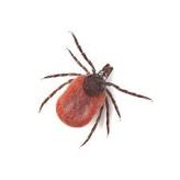
The vector is a tick – Ixodes scapularis, rarely another tick - Amblyomma americium (Lone star tick) transmit the spirochetes in the southern states.

Population from Maine to Maryland and New Jersey, Pennsylvania, Wisconsin is prone to increase tick bites due to the increasing number of deer in the woods.

The adult tick must feed 48–72 hours during that time, B. Burgdorferi enters the wound from the tick's saliva contamination.

The incubation varies from a few days to several weeks following the tick bite.

Based on the time of presentation and symptoms, the disease is conveniently separated into 3 groups.

  1. Initial. Skin rash and slight fever. The skin rash is the hallmark of Lyme disease at this stage. The rash is red to violet in color, target like lesions called Erythema marginatum.

  2. Early disseminated disease usually developed in 2 -3 weeks but may be late, as late as 10 weeks. The presenting symptoms are painful swelling of one knee or ankle, musculoskeletal pain, conjunctivitis, heart block, and Bell's palsy.

  3. Late or chronic. After months of quiet period, the asymmetrical arthritis of hands and spine, headaches, peripheral neuropathy, muscle weakness, cranial nerve palsy develop and persist for a long time despite treatment.

The skin rashes are due to an inflammatory reaction from the presence of spirochetes in the skin. The spirochete may enter fibroblasts and live in them permanently. In the early state, 50% of patients are seropositive, it becomes 100 % as weeks and months progress. Arthritis develops from cross-reaction of the spirochetal membrane protein with the host's connective tissue and neural tissues. The newly formed complex is antigenic. The antibodies react with the complex, and organ damages take place from released pro-inflammatory cytokines. People having HLA-DR$ and HLA-DR2 are likely to suffer most.

Chronic neurological manifestations are the results from the presence of the remnant DNA of the spirochetes in the nervous system of the victims, which triggers an excess production of Interferon alpha.

CDC recommends the following procedures.

Initial stage: ELISA immunoassay of IgM and IgG.

Confirmatory stage: Western blot testing.

a). Symptomatic for less than 30 days. Perform both IgM and IgG Western blot tests.

b). Symptomatic for more than 30 days, only IgG Western blot test is advised.

c). A newer test -  the C6 peptide test, which was prevalent in Europe, has been approved as an alternative to the Western blot test. C6 peptide test is less expensive and equally sensitive as the Western blot test.

Initial stage: The preferred antibiotic is Doxycycline by mouth for 10 to 21 days. Children, pregnant and lactating women should have Amoxicillin or Cefuroxime axetil PO for 10 to 21 days.

With cardiac, neurological or musculoskeletal involvement, the treatment should be continued for 28 days.

In late stage: Parenteral Ceftriaxone, or Cefuroxime or Penicillin G in high doses given for 14 to 28 days.

Post-treatment Lyme Disease:

About 20% of patients who completed recommended treatment continue to be moderately symptomatic for 6 months or longer. The common symptoms are fatigue, musculoskeletal pain, hearing loss, headaches, ambulatory and balance problems, paresthesias, depression and sleeping difficulties, and other symptoms.

************************************************************

Archive

  Archive. P.K.Ghatak, MD ghatak3@gmail.com 2025  March. Laser Endoscpoe 2025 February. Use of Ultrasound in Medicine MRI 2025 January. Bact...