Thursday, August 6, 2020

Advances in the Treatment of Parkinson's Disease

                            Advances in the Treatment of Parkinson's Disease

                                        PKGhatak, MD


Parkinson's Disease (PD) is a progressive degenerating disease of the central nervous system (CNS). The cause of PD is unknown. When other conditions of the body lead to the development of symptoms similar to PD, then the condition is called Parkinson syndrome or Parkinsonism. The main causes of Parkinson's syndrome are cerebrovascular disease, small and major strokes, toxic chemicals and medications generally used to treat schizophrenia, and repeated trauma to the brain as happens in boxing and football games.
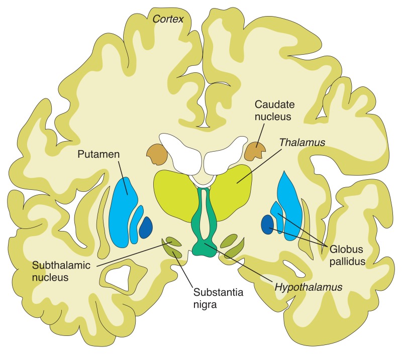
Figure 8.1. Structures of the basal ganglia.


The cells in the Substantial Nigra secrete a neurotransmitter, called Dopamine, which is responsible for the coordinated and smooth functioning of voluntary movements of the body and maintaining the tone of muscles. The enzyme AAAD  (aromatic L-amino acid decarboxylase) converts levodopa(L-DOPA) to dopamine. The final inactive degraded product of dopamine is 3-0 methyldopamine by the action of the Catechol- 0- methyltransferase (COMT) enzyme.

Nerve cells of the peripheral nervous system and adrenal medulla and endothelial cells of the GI tract also take up L-dopa. From amino acid Tyrosine and L-dopa, the adrenal glands make dopamine, norepinephrine and adrenaline. An enzyme Monoamine oxidase (MAO) degrades dopamine, norepinephrine and adrenaline to inactive compounds. The central nervous system has no MAO and MAO cannot cross into the brain from the blood (blood brain barrier).

In Parkinson's disease substantia nigra cells die prematurely, as a result, the movements become jerky, slow and disturbance of gait and balance, tremors and stiffness develop.

Parkinson's disease clinically may appear as 3 entities initially. In Type 1- the tremors of one or more limbs predominate. Muscle rigidity is absent and ambulation remains normal. In Type 2 disease the ataxic gait and mask like face and pill rolling movements are dominant symptoms. In Type 3 cases all the typical symptoms of PD are exhibited.

Progressive Supranuclear Palsy and Lowy Body Dementia are closely related to Parkinson's disease but are not a part of this discussion.

The standard medical treatment for Parkinson's disease is to supply Dopamine and reduce or stop MAO-B activities in the body so at the end more L-dopa will be available for the brain. A group of drugs is available for that purpose and are collectively called MAO inhibitors and are in regular use in PD.

In addition, direct interventions for tremors, depression, stiffness of the body and constipation, etc. are treated with well-known medications. Physical exercise, physiotherapy, occupation therapy, speech and languages therapy are also included in the management of Parkinson's disease.

New drugs and new methods of drug delivery to CNS, Stem cell transplantation, keeping dopamine secreting cells alive and surgical treatment are parts of this article.

In between doses of medication, rescue therapy.

Levodopa is a standard drug for Parkinson's disease. It is converted into Dopamine in the brain. Levodopa therapy replenishes decreased levels of dopamine in the brain. A drug called Carbidopa is an MAO inhibitor. Carbidopa and L-dopa are combined into one pill for use in PD. But in between doses, sudden deterioration of symptoms happens due to an unexpected fall of available L-dopa in the brain cells. To prevent this from happening, several drugs are now available.

  1. Istradefylline is marketed as Nouriazen. It is an antagonist to Adenosine A2A receptors. When this receptor is blocked the nerve cells release more gamma aminobutyric acid (GABA) which is another neurotransmitter. GABA helps to ease symptoms related to the "OFF episodes" from the sudden drop of dopamine. Istradefylline belongs to a small molecule drug and is given orally once a day dose.

  2. Entacapone is an inhibitor of the MAO-B enzyme. Available as Comptan. Other available MAO-B inhibitors are Rasagiline and Safinamide.

  3. New delivery system for Levodopa.

       New formulation allows Levodopa delivery via inhalation. It acts quickly.               It is marketed as Inbrija.

  1.  Apomorphine is a form of a thin sublingual tablet that acts quickly to counter the symptoms of off symptoms.

  2. Accordion pills. Layers of Levodopa are designed in such a way that they will release at a slow and steady level. Thus, decrease unusual dips of drug blood levels and decreases the incidence of “off” episodes."

     6. Pump and patch pumps are also used and have improved for steady drug         delivery

GDNF.

Glial cells produce a neurotrophic factor. It is a naturally occurring protein that protects many types of brain cells.

In Parkinson's disease, the GDNF is delivered directly into the substantia nigra by implanted tubes in the brain, through a port is surgically placed behind the ear. When GDNF is delivered every 4 weeks for 9 months, patients receiving this therapy show remarkable improvement and PET scans detect regeneration of dying dopamine producing cells.

Stem cell Transplantation.

In a study done at Massachusetts general hospital and Cornell Medical center, the dopamine producing stem cells were transplanted.

Pluripotent stem cells are harvested from the skin of a patient and the pluripotent cells are engineered to differentiate into dopamine generating stem cells. Then the stem cells are transfused. In this study, patients are followed for 2 years. During this follow-up period, the patients show symptomatic improvement and the transplant cells remained alive and functional.

Surgical treatment.

Deep brain stimulation by an implanted electrode in the ventrolateral nucleus of the Thalamus improves tremors in PD.

Pallidotomy.    Surgical removal of palladium improves dyskinesia. In bilateral lesions, the Subthalamic nucleus is removed instead of both pallidum to avoid the development of Hemiballismus.

Radiofrequency ablation has greatly replaced surgery of the basal ganglia and achieved the same results.

Deep brain stimulation in selected groups of nuclei of the basal ganglia for various uncontrollable symptoms has become an acceptable alternative to drug therapy.

Experimental therapy.

1. Dyskinesia results from the prolonged use of levodopa. An Insulin sensitizer – MSDC0160 therapy improves dyskinesia.

2 . Anti Alpha synuclein.

Alpha synuclein naturally occurring protein, which accumulates in the cells of substantia nigra of Parkinson's patients. Several drugs are now in development stages that show promise in removing the alpha synuclein.

Recently laboratory evidence shows similar alpha synuclein protein accumulation in the nerve cells in the GI tract and a new idea is developing that the gut bacterial products or certain bacteria initiate abnormal alpha synuclein production in the gut and that process is carried to the brain either via Vagus nerve or by circulation.


A small molecule, Anle 1386 is an example of such a drug. The initial results are encouraging and show a reduction of alpha synuclein accumulation in the brain cells.

Antibody against alpha synuclein. When PD patients are treated with alpha synuclein antibodies reduction of this protein in the brain cells and the clumping of protein molecules disappear.

Vaccine to stimulate antibodies to alpha synuclein is underway.

3. Repurposed drugs. Exenatide is a diabetic drug found to protect dying brain cells in Parkinson's disease patients. Inosine – is a nucleoside, when used in humans, it increases urate levels. Urates are antioxidantsand  and protect brain cells. Israpidine is a B.P drug, found to preserve brain cells. Nilotinib is a tyrosine kinase inhibitor and is used in Chronic myeloid leukemia, when used in Parkinson's patients, it helps clear the alpha synuclein by phagocytosis.

Treatment of the Gene mutation.

Mutation of the GBA gene causes cellular dysfunction from the accumulated of lipids in the cells. An experimental drug GZ/SAR 40267 reduces lipid accumulation in the cells.

An oral drug LTI-291 corrects Liposomal dysfunction which is the cause of lipid accumulation.

Hemanti's Neurotrophic factor. This neurotrophic factor protects cells from premature death.

Glutamate blockers.

In long term use of L-dopa in PD patients Glutamate is over expressed in neurons of basal ganglia due to phosphorylation of N-methyl D aspartate receptors and that is responsible for dyskinesia.

Foliglurax and Dipraglurant reduce Dyskinesia by blocking Glutamate in the brain.

Serotonin receptor blockers

In PD patients, in addition to the loss of dopamine producing cells in basal ganglia, serotonin producing neurons are also lost to a certain extent. As a result, serotonin terminals in basal ganglia become over abundant compared to dopamine terminals. Serotonin terminals convert L-dopa into dopamine in excess amounts and produce dyskinesia.

Serotonin receptor blockers mitigate this problem. Example: Eltoprazines.

Anti-Choline drugs.

Acetylcholine hyperactivity in dopamine depleted brain is implicated in gait disturbance and frequent falls. Varecline and Donepezil reduce brain acetylcholine levels and reduce these complications.

In recent years many private organizations came together and made funds available for research. The progress has been impressive. Improvement in stem cell transplants and gene therapy, perhaps, one day will make Parkinson's disease a mild form of disability in case an outright cure is not possible.

***********************************************************

Sunday, August 2, 2020

Diagnosis of Mycobacteria tuberculosis

                                             Diagnosis of Tuberculosis

                                                  PKGhatak, MD


Tuberculosis is a very old disease. Egyptian mummies, some 4,000 years old, show evidence of tuberculosis (TB). However, TB in humans is still older. It can be traced back to 9,000 years ago in Atilt Yam, near Israel. Archaeologists in Israel found TB in the remains of a mother and child buried together there. The earliest written records, 3,500 years old, in India mention TB. In China, records show the presence of TB about 2,300 years ago

At one time, TB was causing havoc in Europe; 1 in every 7 deaths was due to TB. TB began to spread to the newly colonized countries in Africa and the Americas by the Europeans. It is now a disease of the poor countries of South Asia, Africa and the Western Pacific countries.

In South East Asia, the infection rate is 220 per 100,000 population, in Africa, the rate is 397 per 100,000 and inthe  Western Pacific, the rate is 400 per 100,000. And the mortality rates are 32, 37 and 90 per 100,000 infections per year, respectively.

The World Health Organization (WHO) declared TB to be a Global Health Emergency (GHE) in 1993. It is estimated that 1.45 million people died from TB, and 10 million new cases appeared in 2018. With help from the WHO and several NOGs, the mortality rates began to decline. Since this COVID-19 pandemic broke out, concerns are expressed that the fight to contain TB will fall behind and there will be a resurgence of TB cases and deaths.

History of progress in detecting TB.

In 1882, the great German scientist Robert Koch discovered the TB bacillus. His associate, Paul Ehrlich, developed a staining method, with a stain containing Alum hematoxylin to demonstrate the stained bacteria under a viewing microscope. Franz Ziech modified the stain by using carboxylic acid as the mordant. Now the TB bacillus is called Acid Fast bacillus (AFB).

Culture of TB.

The tuberculosis bacillus is grown on a solid medium known as the Lowenstein-Jensen medium, which has traditionally been used for this purpose. However, this method is quite slow, as this organism needs 4 to 6 weeks to grow. A faster result can now be obtained using Middlebrook liquid medium.

Skin test for TB infection:

Robert Koch extracted a bacterial protein and called it Tuberculin. He used it as a drug for the treatment of TB infections but it failed to cure TB. Tuberculin was modified by von Piquet and used as a skin test for the diagnosis of TB. Then, finally, Florence Seibert developed a purified protein derivative (PPD) and standardized the skin test. The PPD skin test is still in use today in most developing countries to identify new infections, either latent or active cases.

Mycobacterium tuberculosis (MTB).

There are 70 different mycobacteria, and only a handful of mycobacteria cause human diseases. Mycobacterium tuberculosis (MTB) causes devastating pulmonary disease and death in humans.

Another mycobacterium, Mycobacterium leprae, causes leprosy.

In clinical medicine, Mycobacterial diseases are discussed using the classification introduced by Runyon.

He divided Mycobacteria into these classes.

1. Slow-Growing – takes 4 to 6 weeks to grow in the lab. Then it was subdivided according to the pigment produced by the growing colony of bacteria under light or dark conditions.

  A. Photochromogen, these colonies produce yellow-orange pigment when          grown in the lab under light

  B. Scotochromogen, the colony produces pigment both in the dark and under light.

  C. Non-chromogens produce no pigment.

2. Rapid Growers - takes only 5 days to grow in the lab. They do not produce pigment.

Mycobacterium tuberculosis (MTB) is a slow-growing bacterium and the colony produces no pigment but looks green when grown in Lowenstein-Jensen media containing malachite green. The appearance of the colony is rough and dry. The bacteria produce the enzyme Catalase and a vitamin-niacin-that helps in the diagnosis from the other slow growers.

Why MTB infection is unique.

When any bacteria invade humans, the Macrophages are mobilized and macrophages engulf the bacteria and digest them completely. But MTB remains active inside the macrophages due to higher oxygen content bound to organic iron and MTB continues to multiply. The macrophages carry them to regional lymph nodes, where the MTB continues to multiply and eventually MTB kills the macrophages. The MTB may escape into the bloodstream and then infect the kidneys, Liver, Spleen, Pelvic organs, Peritoneum, Brain, Bone marrow, Vertebrae and other tissues.

Mode of Infection of MTB.

MTB infects people from person to person via ambient air and or droplets, like the spread of COVID. But MTB requires more prolonged contact in close quarters. The infectivity is low, about 10 - 15 people in 12 months.

The primary site of infection is the lung, particularly the apical parts of the lung, where oxygen content is higher.  From the primary location, the disease spread to the regional lymph nodes in the lung. These two lesions taken together are known as the Primary complex. In 85 to 90 % of cases of primary infections are controlled by cellular immunity. But the MTB bacteria remain alive with the macrophages throughout the entire life of the patient. In 5 to 10 % of primary Tbc cases, the disease continues to progress locally and destroys lung tissues, blood vessels and forms lung cavities. Once blood vessels are breached, the MTB spreads near and far to other organs. Pleural effusion, pericardial effusion, miliary spread and TB meningitis usually follow.

Tonsils may also act as the primary focus, specially, in children and regional cervical lymph nodes infection produces cervical adenitis.

Bovine TB.

Dairy products may contain living mycobacteria in the milk of infected cows.  In countries where people use unpasteurized milk or do not boil milk before consumption, the bacteria infect the terminal Ilium and or cecum. In due time progress to TB enteritis, ascites and peritonitis.

In South Asia, bovine TB is present but kept in check because they use boiled milk.

Diagnostic Criteria:

The MTB must be demonstrated from infected tissue, either by stained smears or by culture. Culture takes 4 to 6 weeks. That is a long period to wait for the treatment to start.

Tissue Biopsy:

The pathological features of TB infections are caseating granulomas. The necrosis is seen in the center of the granuloma, surrounded by mononuclear cells and at the periphery, a ring of Langhan giant cells is present. Pathologists describe these as caseating granulomas. Demonstration of AFB in the lesion is necessary for a definite diagnosis.

Alternately, the detection of MTB-specific antigen from the infected tissue, which is not shared by other mycobacteria, will satisfy that need. The genome of MTB is now known. Various tests are designed to achieve that goal.

Immunity to MTB is cellular – the T- T-lymphocytes carry that task. For that reason, no MTB-specific antibody test is possible.

Interferon gamma Release (IGRA) assays.

It is known that T-lymphocytes of infected patients when re-exposed to MTB antigen, the T- lymphocytic secrete Interferon gamma. This test has become the gold standard and has replaced the TB skin test to a great extent. The test is known as the Interferon gamma release assay (IGRA).

The sensitivity and specificity of the IGRA test increase by more than 10 times if T-cells are obtained from the infected trusses like CSF fluid in meningitis, pleural fluid in pleural effusion, and bronchial lavage fluid from the infected lungs.

Nucleic Acid Amplification Test (NAAT).

This test is basically similar to the RT-PCR test for identifying pathogenic organisms like COVID-19, malaria and other diseases. It is specially useful in lesions where only a few MTB are present like pleural fluid, CSF or skinny needle aspiration biopsies.

LAM urine test.

In Pulmonary TB patients, several MTB antigens are present in urine. A heat-stable Lipoarabinomannan (LAM), a glycolipid constituent of MTB, is released from active infection in the blood and is filtered out in the urine. It is detected by ELISA using polyclonal antibodies.

A new method of collecting sputum samples from the gut in MTB patients (Enterotest).

A method known as Enterotest involved swallowing a weighted gel capsule containing a coiled nylon string. One end of the string is protruding through a hole in the capsule, and the other end is held at the mouth and taped to the cheek. The capsule is carried down to the duodenum by the intestinal peristalsis. The capsule is kept in place for 4 hours; the capsule dissolves and the string collects swallowed sputum containing MTB. The string is retrieved and the sample is processed for the presence of AFB. It has the advantage over induced sputum – no technicians and equipment are needed, and the chance of the spread of MTB from vigorous cough produced by the induced method is eliminated.

Where Diagnostic Difficulty Remains.

Children.

Suspected Pulmonary tuberculosis in children is a special situation. Small children cannot expectorate sputum, instead, they swallow coughed-up secretions. It is not easy to obtain adequate samples from the stomachs of the children.

Until the development of the IGRA assay, the TB skin test was mostly relied upon. The administration of the test appears simple, but experienced technicians are essential for the tests. Reading of the induration at 72 hours after the skin test must be adhered to. Interpretation as positive or negative is discouraged; instead, the actual measurement of induration at 90 degrees from the needle insertion should be recorded and kept for future reference.

In most advanced countries, the TB skin test is abandoned and IGRA is now the standard test.

Smear negative MTB.

Identifying the Acid Fast Bacillus (AFB) on smears is quick and 100 % specific. Cultures are planted at the same time as the smears are examined. But cultures take 6 weeks to grow. Many initial negative smear cases turn out positive on culture. This is a problem for poor countries where more advanced methods of TB culture are not available. In children and adults with minimal lung lesions, the cases where sputum production is scant are the main reasons for negative smear tests. In military TB, TB meningitis and TB lymphadenitis, sputum cultures are useless.

Latent Tuberculosis.

Soon after a person is infected with MTB, usually in the apical areas of the lungs, inflammatory reactions begin and in many cases, the progression of TB does not happen. Often, the patients are not aware of the infection. Only by contact tracing, infected people are identified. In developing countries, contact tracing is not done and these people are lost. When those people move to other countries, the public health policy mandates TB skin tests and then they are identified. The IGRA test is currently used.  It should be remembered that the skin test or IGRA test cannot differentiate active TB from inactive TB; it simply means a previous infection with MTB.

The skin test and IGRA tests are not foolproof tests. In HIV/AIDS infections, the use of immunosuppressant drugs, Organ transplant patients, Diabetics, Malnutrition and concurrent Malignancy and Chemotherapy may result in false negative tests. In such situations, Nucleic Acid Amplification Test (NAAT) is indicated. However, NAAT is not sufficiently specific (about 85%), but 100 % sensitive. Modification of NAAT is in progress presently.

Nonpulmonary MTB.

Cervical Lymphadenitis in children and young adults is a usual presentation. The IGRA test is positive in these cases. Skinny Needle Aspiration Biopsy tissues subjected to the IGRA test produce a much higher degree of accuracy and sensitivity. If the IGRA test is negative, then NAAT should be performed.

These methods are equally applicable to all non-pulmonary TB infections.

Non-MTB Mycobacteria infections (NTM).

Pulmonary infection by Mycobacterium avium intracellulare in immunocompromised individuals is a serious health problem. The IGRA test for MTB will be negative, but the IGRA test with the antigen M. avian intracellularly will yield a positive result. The final confirmation requires bacterial culture.

BCG vaccination.

BCG vaccination reduced the incidence of pulmonary TB infections by 50 % and reduced 65% of meningitis and 80 % of disseminated TB in developing countries. BCG vaccination poses a problem for future TB skin tests because BBG vaccine antigen cross-reacts with the Tuberculin antigen. This problem can be avoided by the IGRA test.

Before BCG vaccination, the common reason for a false-positive skin test was

1. In countries where non-MTBs are common, skin tests yield 10 mm or more induration. 2. Newborns vaccinated with BCG retain positive skin test for 5 years, older children and adults retain the reactivity for 10 years.

To increase specificity, positive and negative controls are used. As mentioned before, the sensitivity of the test is poor in immunocompromised patients.

Drug Resistance MTB.

MTB resistant to Isonazid is not unusual in developing countries but MTB is also developing resistance to Rifampin.  MTB resistant to both these two drugs is growing and now spreading to advanced countries. It has become an urgent public health issue.

The usual method of detecting drug resistant TB takes 6 weeks. That is a waste of valuable time. Now, more rapid and more sensitive methods are available.

The microscopic observation drug susceptibility assay.

This method involves a direct inoculation of samples into wells on a tissue culture plate containing a liquid growth medium. Some wells contain Isoniazid, others Rifampin. The growth is determined by visual inspection using an inverted microscope. If a TB colony, in the form of cords, is observed, then MTB is resistant to that specific drug. The turnaround time of this test is 7 to 14 days. The CSF can be tested by this method and has greatly improved early detection of TB meningitis and improved survival.

This principle is now extended to many other drugs, including 2nd line TB drugs. This method is sensitive 100 % to Isoniazid, 97 % to Rifampin, and 99% to multiple drug resistance.

Molecular Testing for detecting the drug resistance gene.

Mutation of the katG and rpoB genes in the MTB genome produces drug resistance. DNA probe and DNA sequence of MTB genes are available. The results are obtained in hours and the test is highly sensitive and specific, but expensive.

Recent advances in detecting MTB.

Line Probe Assays (LPAs).

LPAs are molecular tests. Currently, three such test kits are available for the rapid detection of MTB and drug resistance. The test is based on the targeted amplification of a specific fragment of MTB by the PCR technique.

Recovery of MTB from peripheral blood.

In this test, anticoagulated blood samples are lysed and then centrifuged. The sediment is inoculated in Westbrooks liquid medium. Valuable time is saved using this method and also increases the positive detection rates. It is a useful test for disseminating MTB infection.

Bacteriophage test for MTB.

A suitable bacteriophage is used in an agar plate containing MTB colonies. A non-pathogenic mycobacterium is used as a control. Central clearing of the colony indicates the presence of MTB. The turnaround time is 2 days. The test can detect as low as 100 MTB in a sample.

Immunodiagnostic Tests.

It can detect past or current TB infections. However, this test is limited in application because of its poor sensitivity and cross-reaction to non-MTB.

Antibody tests against MTB.

The results so far are not consistent. WHO does not recommend this method for detecting Pulmonary and extrapulmonary TB infection.

MTB is an ancient disease but continues to be a worldwide problem and the problem is magnified due to the prevalence of HIV/AIDS and these two entities coexist. Now, the COVID-19 pandemic has put an extra burden on poor countries. It is feared that the MTB new cases and the number of deaths will increase. The use of monoclonal antibodies in treating cancers, autoimmune diseases and organ transplants has taken the center stage in the treatment. The resurgence of TBs in these groups is increasing because of suppressed immunity. Latent TB poses a problem and MTB in association with diabetes mellitus is on the rise.

Many new diagnostic tools are available in advanced countries but their use is limited in areas where MTB is a serious health issue because of a lack of funding.

Developing a Blood Test to Differentiate Between Active and Latent TB

Thomas Glück, MD, reviewing Zhao H et al. Infection 2025 Mar 17

In a pilot study, measurement of IL-8, IL-18, and IL-33 in TB-antigen–stimulated whole-blood assays distinguished active TB from latent infection with >85% accuracy.


*************************************************************


Monday, July 27, 2020

Interleukins

                                           Interleukins

                                                           PKGhatak,MD


Interleukins:

In 1979, an international committee coined the term Interleukin (IL) to identify chemicals secreted by Immunocytes. Interleukins are a part of Cytokines. Cytokines are proteins that act as chemical messengers of the immune system and are secreted by both immunocytes and non-immunocytes. Cytokines include Chemokines, Lymphokines, Interferons, Tissue Necrosis Factor (TNF), Colony Stimulating Factors(CSF) and ILs.

Interleukins are also secreted by Endothelial cells, Enterocytes of the intestine, and certain Connective tissues. Interleukins initiate the activation and differentiation of immunocytes and also various other aspects of immune cell functions. ILs can act both as stimulators and inhibitors of inflammation.

In humans so far, 17 ILs are known and 50 genes are identified controlling ILs. Some genes control more than one IL. IL-2 and IL-6 are often mentioned in the discussion of the pathophysiology of illnesses.

ILs are stimulators of inflammation.

These ILs are IL-1, IL-6, IL-12, IL-18 and IL-23

The most pro-inflammatory Cytokines are IL-1 beta and TNF alpha.

Pro-inflammatory ILs activate CD2, CD4, CD8, CD27, CD134 and CD137.

Inhibitors of inflammation, ILs are-

IL-10, IL-6, IL-1 alpha. They activate CD80, CD152, CD160, and CD223.


A synopsis of the origin and actions of Interleukins:

IL-1. IL-1 is active in the human Hippocampus and is involved in memory formation.

IL 2. IL-2 was previously called Lectin. It is produced by CD12, CD122, and CD132. IL-2 stimulates the growth and differentiation of T-cells, and it also stimulates immunoglobulin production by B cells.

IL-3. IL-3 is secreted by CD123 and CD131. It stimulates the differentiation and growth of granulocytes of the blood and tissue macrophages.

IL-4. IL-4 is produced by CD124 and CD132 cells. It stimulates B cell proliferation and mutation. It also stimulates B-cells to produce immunoglobulins.

IL-5. IL-5 is secreted by CD125 and CD131. IL-5 initiates the differentiation of eosinophils from the hemopoietic stem cells.

IL-6. IL-6 is a major IL in humans. CD126 T and CD130 T cells produce IL-6. IL-6 accelerates the production of Interferon beta2 by the B-cells. In the liver, IL-6 stimulates the production of acute phase reactants and GCSF (granulocyte colony-stimulating factor).

IL-10. IL-10 is the major inhibitory Interleukin in humans. It is produced by CD210 cells. IL-10 counteracts the actions of Interferon gamma and IL-2, and IL-3. TNF and GCSF.

IL-12. IL-12 increases the cytotoxic function of Natural Killer (NK) cells. In parasitic infestations, like Leishmaniasis and toxoplasmosis, and measles infection, IL-12 mounts a cellular response. IL-12 plays an important immunological function in HIV infection, Multiple Sclerosis, and Crohn's disease.

IL-17. IL-17 is produced by CWD 217 cells. IL-17 stimulates osteoclastogenesis and angiogenesis.


edited June 2025

 **************************************


Monday, June 1, 2020

Platelets

                                            Platelets.

                                                         PKGhatak, MD

 
Platelets
Little Disc but Feisty one.


The Christian name of the platelet is Thrombocyte. Platelets are tiny blood cells, about 1/5 th of an RBC. If one looks at a stained blood film, platelets may be mistaken as artifacts - tiny deposits of dark purple pigment. One can also examine platelets with a special instrument as they are floating in the bloodstream. Here, platelets appear as tiny lenses. The center appears clear because the nucleus is absent. All mammals' platelets have this characteristic and all other non-mammals, from birds down, have a nucleus in platelet cells.
Platelets are produced in the bone marrow from Megakaryocytes. One megakaryocyte divides itself into individual platelets and sends them out on tiny limbs, and the newly formed platelets break away. One megakaryocyte makes 1000 to 3000 platelets. A hormone, called Thrombopoietin, made in the kidney and liver, is required for the normal production of platelets. As platelets are developing, they receive packets of powerful chemicals neatly packed in small granules like soldiers get their weapons and gift packages. Platelets travel to the spleen and are stationed there for a while like soldiers waiting for orders to go to the battlefields. In circulation, platelets survive about 9 days. When a platelet reaches ripe old age, it is scoped up by the headhunters called macrophages of the spleen. Macrophages swallow and digest the platelet. 

Demystify medical terms.
Macro=large, phages=eaters, Mega=big. Penia= few, thin. Thrombo=clot,
Cytes=cells, Cythosis=too many cells, Pathy=lacking in function,

Structure under powerful magnification.
The outer membrane is, like all cell walls, made of two layers of fatty materials. On this surface, there are special areas called receptors by which platelets bind to certain specific proteins. The next layer contains filamentous structures that act as scaffolding providing stability and shape. Next to it is a zone of granules containing preformed cytokines, complements, serotonin and many blood clotting factors. Granules come in various sizes and shapes. They are given the names of Greek alphabets, alpha, delta, and lambda. Each granule contains a set of chemicals which is different from the others. The remaining area, in the center, is loaded with microtubules going from the center to the surface of platelets.

A day in the life of Platelets.
A young platelet is happily floating around in the bloodstream, then suddenly it is pushed into a narrow tunnel(capillary), the wall of which is neatly lined with closely fitted glazed tiles (endothelial cells). As the platelet was looking around, it noticed a portion of the wall with missing tiles, and blood was oozing out. Suddenly he sees his favorite face of collagen fiber with lips colored with von Willebrand factor. He feels compelled to give a kiss on her lips (receptor protein). But lo! Now, he can't break away from her. His lips (receptors) are stuck on her lips. He notices his body shape is changing – swelling up, his arms are coming out like the arms of an octopus and reaching out to other nearby platelets. As more and more platelets congregate and all the platelets are trying to kiss collagen lips, they form a heap and the mass completely covers up the missing tiles (break in endothelial surface). A mass of platelets forms a white soft clot that stops blood from flowing out. From their bodies, granular packs burst open and the chemicals spill all over. Now, an army of bridge-building-brigade assembles at the site in response to the released chemical (cytokines). Platelets release more chemicals containing construction materials (clotting factors fibrinogen, V, VIII, etc.). And the scaffolding takes the shape of a net. The net begins to trap RBCs and WBCs as they are traveling downstream with the blood. This generates a red firm clot, and it would not wash away with the moving blood. As time passes, the platelets release a clot stabilizing factor, and that turns the red clot to shrink in size into a hard clot, and this seals the break of the vessel permanently.
It is a story of sacrificing one's body for the common good. That is the purpose and objective of platelets.
 
Injury or disruption of the endothelium of arteries.
In coronary artery disease and following angioplasty, the endothelium is disrupted and collagen fibers are exposed. To prevent platelets from starting clot formation, antiplatelet agents like Aspirin and Clopidogrel are prescribed. After stent placement in cerebral arteries antiplatelet therapy is also used.

Thrombocytopenia.
The usual number is around 250,000/microliter, and the range is 100,000 to 4000,000/microliter. When the number falls to 50,000, spontaneous bleeding under the skin, gums, nose, and gastrointestinal tract may happen. Bleeding inside the brain, kidney, and other vital organs poses a very serious problem

Some common conditions lead to Thrombocytopenia.

Cancer chemotherapy is perhaps the most important cause.  Radiation therapy to vertebrae for control of pain due to metastasis often produces marked thrombocytopenia. These therapies dry up the bone marrow.

Infectious causes:
Viruses, bacteria, and parasites are all known to produce low platelet counts. The dengue fever virus is notorious for producing dangerously low platelet counts. HIV, Hepatitis C virus, Lymphosarcoma, hematological malignancies and myelofibrosis are important causes.. Babesiosis due to a parasite is another example of the cause of thrombocytopenia.

Toxins:
E coli 0157.H7 toxin is a great threat to the lives of children getting infected from contaminated soil or handling live farm animals. Bacillary dysentery with shigella bacteria is also a serious problem.

Drugs:
Many drugs act directly on platelets or by decreasing the activity of the megakaryocytes.  A drug may form a  compound with platelets and the conjugate behaves like an antigen. And antibodies are produced and in turn, destroy platelets. A few well known drugs that produce low platelet counts are Quinine, Heparin, Penicillin, Sulfa drugs, Naprosyn, Hydrochlorothiazide, Procainamide, Carbamazepine,  and Rituximab.

Other important clinical conditions: 
Alcoholism, vitamin B12, and folic acid deficiency, mechanical aortic valve, post blood transfusion, Cirrhosis of the liver with hypersplenism,

More serious but not so common conditions:
ITP. (Idiopathic Thrombotic Thrombocytopenia). Here autoantibodies are again in play, but the cause remains hidden. The antibodies also attack the megakaryocytes.

TTP. (Thrombotic Thrombocytopenic Purpura). It is a highly feared illness.
In normal conditions, an enzyme, ADMTS13, is present in the blood, and keeps platelets separate, preventing them from clumping together in the blood. In an unfortunate mutation of gene/genes, a lower amount of this enzyme is produced and a low blood level of ADMST13 results. The condition by itself may not produce any symptoms but after an infection, even a minor one, a grave situation arises. The infecting virus or bacteria forms a complex with this ADMTS13 enzyme and after 7-10 days later, autoantibodies begin to appear and the clotting starts. Tiny blood clots block all major capillaries of vital organs; medium size vessels may also be involved. Many clinical but equally serious pictures evolve. The life of patients depends on the removal of these antibodies by plasmapheresis and replacing plasma with normal plasma. The plasmapheresis needs to be repeated. An immunosuppressant drug is usually also required. The patient's children may inherit this deficiency.

High Platelet count (Thrombocytosis):

It is often associated with malignant conditions of the bone marrow. Megakaryocyte turns out platelets unchecked that may lead to obstruction of blood flow due to high viscosity from increased numbers of cells. The patient complains of blurred vision. And obstructed blood vessels can be directly observed by retinal examination. Chemotherapy is required to control the condition.

Drugs to prevent platelet aggregation.
Aspirin, Clopidogrel, Ticlopidine, Prasugrel and Eptifibatide are extensively used as antiplatelet agents, in preventing the clotting of stents in post angioplasty and coronary artery disease.
Aspirin: It permanently blocks the enzyme COX1 on the platelet for that platelet's life span. Cox1 is necessary for generating prostaglandins G2 and H2, the precursors of clot promoting factors - Thromboxane2. This results in the unopposed action of Thromboxane A1, which prevents platelet aggregation.

Clopidogrel, Ticlopidine and Prasugrel are considered together. They act on the Platelet surface receptors P2Y2 and block these receptors from binding with ADP. This action prevents platelet aggregation.
Initially, one product claimed to be better than others based on unfounded claims of higher potency. In practice, one product is not that great compared to others, but the retail price is.

Eptifibatide binds with GPIIa / IIIb receptors on the platelet surface and prevents them from binding with Fibrinogen to form fibrin, which is a clot.

Antiplatelet drugs work well on the arterial side of circulation and prevent atherosclerosis but for some unknown reasons, the antiplatelet drugs are not effective on the venous side of the circulation in preventing blood clots.

The platelets are tiny but perform many vital functions. The primary function of platelets is to prevent bleeding by plugging the broken blood vessels. The platelets also supply various blood clotting factors. It is a source of a Neurotransmitter - Serotonin. The hormone melatonin is formed from serotonin. Because serotonin cannot cross the blood-brain barrier, serotonin is produced in the brain cells and the platelets may be another source of brain serotonin from the platelets in the cerebral circulation. Serotonin has many more important functions but is not discussed here.
------------------------------------------

Saturday, May 30, 2020

Oral agents for Diabetes Mellitus 2

                    Oral agents for Diabetes Mellitus type 2.

                                               PKGhatak, MD  


Oral Drug Treatment of Diabetes Type II.

Diabetes-II is also called Adult diabetes; however, fundamentally the Diabetes- II is a different disease from Diabetes -I; the only connection between the two is that glucose utilization is abnormal in both diseases.

Glucose, a hexose sugar, is the principal energy-generating molecule that the human body utilizes. Glucose belongs to the carbohydrate class of food substances. Humans can also use other hexoses and pentose sugars. But under normal conditions, all forms of carbohydrates are converted to Glucose in the human intestine before it is absorbed. The Liver is the chief organ where Glucose is transformed into other sugars and glycogen, a complex carbohydrate for storage.

The Liver, in normal circumstances, converts Fat and Proteins into glucose; this process is called Neoglucogenesis.

Breastfeeding nursing mothers generate Galactose, another hexose sugar, from blood glucose to form milk sugar, Lactose.

To utilize Glucose by the tissue, the glucose molecule must pass through the cell membrane, for which Insulin plays a crucial role.

When Insulin production is altogether absent, the disease is called Diabetes mellitus-I. Whereas, in conditions where insulin production is present but insulin for one reason or another is ineffective in ferrying glucose molecules across the cell membrane, the disease is called Diabetes mellitus II.


Oral agents used in the treatment of Diabetes II.

In recent years, major advances have been made in oral antidiabetic medication. It is now almost standard, after initial attempts to control high blood sugar with a low-carbohydrate and 1500 to 18000 calorie diet, to start with Metformin. Then, if additional medication is necessary, a SG2 transport protein blocker is added.

The drugs are mentioned in order of their utilization in recent years.

Metformin:

Metformin. Brand name – Glucophage. It is a Biguanide. The pharmacological actions of metformin are different from other classes of oral agents. It decreases hepatic glucose production, decreases intestinal absorption of glucose, and improves insulin sensitivity by increasing peripheral glucose uptake and utilization. Metformin has an anti-androgenic effect, also improves insulin resistance and helps insulin enter cells. It is a useful drug in polycystic ovary disease.

SGLT-2 Inhibitors:

Canagliflozin, brand name Invokana. Canagliflozin interferes with Sodium-Glocose contraspoters (SGLT-2). SGLT-2 interferes with the reabsorption of sugar from the glomerular filtrate in segment 3 of the proximal tubules, and blood sugar levels fall, and more and more sugar is lost in the urine.

The results in weight loss, significantly reduced HbA1c levels and lower BP, lowers oxygen radicals and inflammatory mediators. Improvement in β-cell glucose sensitivity and insulin secretion is observed. A decrease in tissue glucose disposal and an increase in endogenous glucose production are noted.

Recent reports caution that concurrent use of Rosuvastatin and Canagliflozin may result in rhabdomyolysis and hepatotoxicity.

Dapagliflozn, brand name Farxiga. It is another SGLT-2 inhibitor. In normal conditions, SGLT-2 is responsible for 90 % of the glucose reabsorption in the renal tubules; blocking this transport mechanism results in glucose loss in the urine.

Empagliflozin, brand name Gardiance. It is the 3rd SGLT-2 approved in the USA. The mechanism of action is similar to the other two mentioned above. But it is worth remembering that SGLT-2 is a group of transporter proteins. In humans, there are 8 such SGLT-2  proteins, and each one is specifically abundant in certain organs. Though currently three SGLT-2 inhibitors are available, individual agent mainly prevent one such transporter protein in a specific organ. As a result, the toxicity and side effects are different in these drugs.

Thiazolidinediones:

This group of drugs acts by increasing the activity of Peroxisome proliferators, which increases Insulin sensitivity.

Because of hepatotoxicity and increased incidence of urinary bladder tumors, this drug was withdrawn from India and Germany; later studies cleared this drug, and now available in all countries.

Pioglitazone, brand name Actos. It is a selective agonist of Peroxisome proliferator activated receptor-gamma (PPAR-Y). These receptors are present in adipose tissue, skeletal muscle, and liver.

Rosiglitazone, brand name Avanda. It activates PPAR-y receptors and facilitates glucose and lipid metabolism.


Sulfonylureas:


There are several members in this group; at one time, these were the only effective oral agents. But the use of sulfonylureas have decresed with the arrival of severl new ganets, some of them are mentioned above. This medication works by stimulating the Beta cells of the pancreas by binding with ATP dependent Potassium channels, to incrse production of Insulin.

Common side effects of Sulfonylureas.

Skin rashes from sun exposure, weight gain, episodes of low blood sugar, gastrointestinal upset, nausea, and vomiting. Dark urine and hemolytic anemia in patients with glucose 6 phosphatase deficiency (G6P deficiency). Concomitant administration of other sulfa drugs tilts the free vs protein bound sulfonylurea in favor of the free form, which results in more therapeutic action and hypoglycemic episodes.

Common drugs of this group are-

Chlopropamide, brand name Diabesese.

Glipizide, brand name Glucotrol.

Glyburide, brand name Micronase

Tolazomide, brand name Tolinase.

Tolbutamide, brand name Orinase.

Acetohexamide, brand name Dymelor.


Alpha-Glucosidase Inhibitors:

Alpha-Glucosidase Inhibitors work by delaying carbohydrate digestion and absorption, thereby lowering the postprandial glucose load.

Significant side effects of alpha-glucosidase inhibitors include bone marrow depression. Liver enzyme elevation and increased incidence of Pneumocystoides intestinalis infection and intestinal obstruction.

Two agents are available in this group: they are -

Acarbose, brand name Precose.

Miglitol, brand name Glyset.


Edited May 13, 2025


**************************************************

Tuesday, May 26, 2020

Immunocytes & Immunomodulators

                         Immunocytes & Immunomodulators
                                     PKGhatak, MD


Newton's first law of motion states that every action has an equal and opposite reaction. Our body is also governed by a similar law, for every function, there are the Stimulators and Suppressors mediated through proteins. Advances in molecular biology have led to the unlocking of secrets of the mechanisms by which the body detects and eliminates disease causing agents and cancer. This branch of therapeutics is called Immunotherapy and the agents are known as Immunomodulators.

Immunocytes produce proteins which act like keys and fit perfectly to a specific receptor present on the surface of the effector cells and initiate reactions and the response can be either stimulation or suppression of immune reactions. The immunocytes communicate constantly with each other and react in response to secreted proteins.
In the end, the Immunoglobulins are produced by Plasma cells. Immunoglobulins neutralize foreign agents and the natural killer cells (NK) and phagocytic cells engulf foreign substances and remove accumulated debris.

The immunoglobulins are many; written as IgM, IgG, IgA, IgD and IgE. The IgG is subdivided into many fractions. Both IgM and IgG are present in plasma, IgA is most abundant in the enterocytes of GI tract, genitourinary tract and in tears. IgD is bound to lymphocytes B (B cells). IgE appears in plasma, it is active against multicellular foreign organisms and parasites and is associated with allergy.

Immunocytes:
A group of cells - the White Blood Cells (WBC), Macrophages, Plasma cells and Dendritic cells are collectively called Immunocytes.

B-lymphocytes (B-cells)
Among the WBCs the Lymphocytes play a crucial role. Lymphocytes are divided into B-lymphocytes (B stands for bursa Fabricius of birds) and T-lymphocytes (T for Thymus) according to their origin. Both B-lymphocytes and T-lymphocytes are subdivided again into many numerical numbers according to the presence of surface CD (cluster designation) and are grouped as stimulators and suppressors.
During the maturation process, the B-lymphocyte travels to the thymus, spleen and lymph nodes. B-lymphocytes are activated by dendritic cells by presenting them with an antigen and the activated B-cells in lymph nodes act as temporary memory cells and the rest of the B-cells return to the bone marrow to become permanent Memory cells.
T-lymphocytes (T-cells).
T-lymphocytes originated in the embryonic Thymus gland. A subgroup of T-cells is natural killer cells (NK cells). T-lymphocytes perform a multitude of immunological functions along with B-cells.
Dendritic cells.
Dendritic cells are known as antigen presenting cells. In the skin, they are called Langerhans cells. In addition to skin, these cells are present in the nose, respiratory tract, stomach and intestine. Immature dendritic cells are present in peripheral blood. Dendritic cells attack foreign antigens and also diseased body cells and take a bite out of them and then process the antigen and finally hand over the antigen to plasma cells for antibody production. Also, the dendritic cells supply B-cells with antigens, and B-cells become memory cells.
Plasma cells.
Plasma cells originate in the spleen and lymph nodes from activated B-cells. Then the activated B-cells move to the bone marrow and reside there permanently. Plasma cells have large cytoplasm and an eccentrically located nucleus with coarse chromatin. Plasma cells are the chief producers of Immunoglobulins. Initial production of immunoglobulin is IgM specific to an antigen, then after a week or so, the IgM production slows down and the plasma cells start to produce IgG antibodies in response to the same antigen.
Macrophages (large eaters).
Macrophages are derived from monocytes of the blood. This transformation takes place as the monocytes extravasate the blood vessels in response to cytokines. Macrophages present in all tissues and assume different shapes are called histiocytes in connective tissue, Kupffer cells in the liver, osteoclasts in bone, microglia cells in the brain, etc. Macrophages exhibit amoeboid movement. Macrophages encircle foreign substances like bacteria or virus particles or decaying cells and digest them and help clear the field of cellular debris, prerequisite for repair.
Eosinophils.
Eosinophils originate in the bone marrow and are present in blood and all places except in the skin, lungs and esophagus. Eosinophils respond to multicellular organisms and parasites. Eosinophils are involved in anaphylaxis, asthma and atopic dermatitis, hay fever, and also produce many cytokines.
Basophils. Basophils produce heparin, histamine, serotonin, and IL-D4. Basophils have IgE receptors on the cell surface. After binding with the antigen, the conjugates initiate allergy and chronic inflammation to parasites. Like eosinophils, basophils are responsible for anaphylaxis, allergy and hay fever.
  
Cytokines:
These are products of immunocytes, cytokines are also produced by some non-immunocytes, like endothelial cells, fibroblasts and stromal cells. There are several cytokines and are named according to the substrate they act on, e.g., Interferons, Interleukins (ILs), Lymphokines, and TNF (tissue necrosis factor). Cytokines are peptides, secreted in minute amounts, measured in picograms (1 pico = 0.001 nano) are very potent but the range is limited to cells of origin or nearby cells only. Cytokines may be inactivated by small molecules which form a covalent bond with the active site of cytokine and are known by a suffix "tinib" e.g., Acalabrutinib.  Cytokine must attach to its specific receptors on the surface of effector cells and requires a perfect fit as a key fit with the lock in order to initiate the production of an enzyme (- kinase). Because cytokines are antigenic, specific antibodies can be produced in animals or in cell cultures against cytokines and cytokine receptors for therapeutic use.

Therapeutic Use of Immunocytes and Antibodies:

Monoclonal antibodies (mAbs).
Monoclonal antibodies are produced by the activated plasma cells, and these antibodies attack a specific antigen. The antigens may be a biological substance like viruses and bacteria. Antigen can be cytokines, cell surface receptors and also a lab-engineered hybrid antigen. 
In the laboratory these monoclonal antibodies are produced and purified for transfusion the patients.
Medical conditions where mAbs are in use are expanding, particularly in the treatment of malignancy. Some of the well-known conditions of mAbs use are malignant melanoma, rheumatoid arthritis, Crohn's disease, and multiple sclerosis.
The currently available mAbs are – for TNF- Infliximab, etanercept, adalimumab, golimumab. For T-cell inhibition - abatacept. For B-cell inhibition - rituximab, belimumab. For IL-1 receptor inhibitor - anakinra. IL-6 receptor inhibitor- tocilizumab, sarilumab. For IL-6 cytokine – cetuximab.

Checkpoint inhibition: 
Cell population at any moment is a balance between new cell formation and programmed cell deaths. Cell deaths are performed by an enzyme called Caspase. PD1 protein on T-cell prevents cell death. Cancer cells copies PD-L1 proteins and prevent T-cell directed cancer cell death.  
PD-1/PD-L1 monoclonal antibodies.

Cancer cells of the breast, stomach and other organs express PD-L1 legends on their surface. T-cells are attracted to cancer cells and bind with cancer cells, thereby making T-cells inactive. And thus, cancer cells escape NK cells and continue to grow.
Monoclonal antibodies PD-1/PD-L1 are Atezolizumab, Avelumab, Nivolumab and pembrolizumab. 

CART Therapy:
Chimeric Antigen Receptor T -cell therapy involves patient's T-cells made to recognize and kill cancer cells by genetically modifying T-cells to express CAR receptors on their surface, against a specific tumor antigen. Then these genetically altered cells are grown in large numbers in the laboratory then transfused back to the patient. This therapy is most effective in the treatment of relapsed leukemias and multiple myeloma

EGF (Epidermal Growth Factor) and HER2 Receptor (human epidermal growth factor receptor2).
Epidermal Growth Factor (EGF) is a protein that binds with EGF Receptors on the cell surface initiates cell proliferation, and differentiation and prolongs cell survival. It works through the Tyrosine-kinase system.
Antibodies to EGF.
Currently, available mAbs that bind to EGF are Gefitinib, Erlotinib, and Afatinib.
Antibodies to HER2.
Cancer of some breasts, stomach and other solid organs is treated with the HER2 mAbs, e.g., Trastuzumab and Pertuzumab,
VEGRF.
The vascular epidermal growth factors can be blocked by antibodies, e.g., Bevacizumab and ranibizumab
VEGRFRmAbs (Vascular epidermal growth factor receptor monoclonal antibodies.) Ramucirumab is available.


Polyclonal Antibodies (pAbs).
There are two ways to produce pAbs. One is from donated blood pools, isolating and purifying the antibodies, and the second method is to inject multiple antigens into a suitable animal and collect the antibodies. Animal origin pAbs are used in the treatment of snake bites, jellyfish toxins, spider bites, etc.
The pAbs are the mainstay of treatment of the immune thrombocytopenic purpura (ITP). RhD negative mothers, pregnant with a second child tested positive for the RhD, are treated with pAbs to prevent hydrops foetalis in newborns.
The pAbs are also produced in labs. Human T-cells are injected in horses or any other suitable lab animals. After several days, blood is collected and the antibody containing immunoglobulins is purified. The pAbs so engineered are mainly used in acute rejections following kidney transplants. It is also a useful way to treat diseases of uncertain etiology where runaway immune reactions threaten life, as is happening in covid-19. Previously such pAbs were used in Ebola. MRSA infections. It is useful in Digitalis toxicity, Kawasaki disease and recent incidents of COVID-19 Kawasaki syndrome. Guillain-Barre syndrome is treated with mAbs along with plasmapheresis. Similarly, Myasthenic crisis responds to a similar intervention.

Medications that can modify cell growth and production of tissue damaging factors:

Disease Modifying Drugs (DMD):
Human immunity has two components - the inborn or Innate immune system. Innate immune response to a foreign invading agent takes place within hours.  Adaptive Immune system response usually takes weeks to months. This adaptive system consists of 3 parts, namely antibodies, B-cells and T-cells.

Autoimmune diseases are treated with drugs that are in use for a long time. Most of the drugs are well known - like prednisone, cyclophosphamide, cyclosporine, methotrexate, azathioprine, gold compounds, sulfadiazine, etc.
Biological drugs are at present favored over traditional chemical compounds, though used together, results are much better.

Some common uses of MAbs:
Rheumatoid arthritis. Overactive TNF is controlled by Etanercept containing a fusion protein IgG that binds with TNF alpha. Infliximab is a chimeric mAbs used for the same purpose. Adalimumab is a humanized mAbs also binds with TNF alpha.
IL-6(interleukin-6), an IL-6 receptor blocker, Tocilizumab, is used in solid organ rejection.
IL-2 receptor blockers are used in metastatic melanoma and renal cell carcinoma.
Psoriasis and Psoriatic arthritis.  Anti TNF therapy with mAbs in psoriasis and psoriatic arthritis responds well.
Polymyositis and dermatomyositis. B-cell mAbs, Rituximab, is used with success.
IgG4 related disease has a varied presentation. Rituximab is used with conventional DMD. 
Wegener Granulomatosis / Granulomatosis with polyangiitis. It is associated with antineutrophil cytoplasmic antibodies (ANCA). Anti B-cell mAbs, rituximab, with prednisone are commonly used.
In asthma, Omalizumab, an IgE inhibitor, is used.

Antiviral mAbs. 
Bavituximab, a serine mAb used in Hepatitis C.
Palivizumab, a RSV virus mAb, is used in respiratory syncytial virus bronchiolitis and pneumonia.
Anakinra and Tocilizumab, IL-1 and IL-6 receptor inhibitors respectively, are at present undergoing trials in COVID-19 pneumonia and multi organ failure.

Use of mAbs in hematologic cancers
Rituximab, a CD B20 chimeric mAb used in non-Hodgkin's lymphoma.
Alemtuzumab, a CD52 on B-cell & T-cells used in B cell leukemia. 
Gemtuzumab, a myeloid cell antigen CD33 humanized mAb used in relapsed myeloid leukemia.

 Clinical Use of Monoclonal Antibodies:

Recently, mAbs are approved for the treatment of neurological diseases. 
Multiple Sclerosis (MS). MS is an autoimmune disease resulting in damage to myelin sheath from cytokines overproduction. Glatiramer acetate binds with CD4B-cells and CD4T cells and decreases cytokines production.
Natalizumab, an alpha 4 integrin mAb, binds with immunocytes and prevents them from crossing the blood brain barrier.
Rituximab, an anti-CD20 mAb, lowers complement and cytokines.
Ocrelizumab acts in the same manner.
Alemtuzumab binds with TCD52. Fingolimod is an analog to sphingosine. It binds with sphingosine receptors on T-cells and B-cells and limits cytokine production.
Migraine.
The etiology of migraine is not fully understood but migraine pain is due to the release, at the end of the nerve terminals, a preformed calcitonin gene related protein (CGRP) at the junction of nerve terminals and smooth muscles in the intracranial vessels. CGRP protein binds with CGRP receptors (CGRPR) present on the blood vessels and results in a release of pain causing substance P. Examples are -  Erenumab.
Frenanezumab and Galcanezumab are mAbs that neutralize CGRP ligand and Galcanezumab use limits the incidence of migraine attacks and shortens the duration of migraine.

Adverse effects:
 Disease modifying drugs are an effective treatment for autoimmune diseases and cancers. Biologic modifying agents are used in increasing numbers. However, biological substances have serious side effects. Infection with atypical organisms, atypical mycobacteria, fungal infections and the recurrence of previous viral illnesses - particularly Hepatitis B and C and cytomegalovirus are problematic. Reactivation of old TB and varicella are not unusual. In long-term follow-up of biological DMD shows an increased incidence of lymphoma and leukemia.
-----------------------------------------
Edited march 2026.


Archive

  Archive. P.K.Ghatak, MD ghatak3@gmail.com 2025  March. Laser Endoscpoe 2025 February. Use of Ultrasound in Medicine MRI 2025 January. Bact...