Wednesday, August 19, 2020

AMYLOIDOSIS


                                                                       Amyloidosis

                                                                       PKGhatak, MD


Amyloidosis is not a single entity, under amyloidosis five distinct clinical disorders are included; whereas, 36 chemically distinct proteins associated with amyloidosis are detectable.

The underlying pathology is an accumulation of abnormal protein inside the cells. The abnormally folded protein is resistant to protease enzyme which in normal circumstances does "house cleaning" and it removes undesirable molecules from the inside of cells.

Organs involved in amyloid disease and clinical features.

Eyes.

The accumulated protein damages the cells and the cells die. Damage to blood vessels around the eyes produces easy bruising and the eyes resemble Raccoon eyes.

Amyloid in the retinal blood vessels reflects light back as bright spots. The retina appears as Starry Night when examined with an ophthalmoscope. Macular degeneration and various visual problems may develop.


 Heart

The cardiac muscles become loaded with amyloid and become stiff. The ventricles fail to dilate fully during diastole in order to accommodate returning blood. Stagnation of venous blood in the periphery and in the lungs produces congestive heart failure. Normal electrical conduction is disrupted and various heart blocks develop, and cardiac arrhythmia may appear.

Liver.

Accumulated protein results in enlarged liver, biliary obstruction and obstructive jaundice, elevated AST, ALT and Alkaline phosphatase in the blood are present

Mouth.

The enlarged tongue becomes out of proportion to the mouth cavity, the epiglottis enlarges. Obstructive sleep apnea follows.

Intestine. Diarrhea is a common presentation, malabsorption and weight loss follow.

Kidneys.

Damaged glomeruli result in high Creatinine and BUN and later renal failure develops. Loss of protein in the urine produces nephritic syndrome.

Peripheral Nervous System.

Neurological manifestations include peripheral nephropathy of both somatic and autonomic nervous systems and produce various clinical pictures including carpal tunnel syndrome, peripheral neuritis, and orthostatic hypotension.

The spleen is generally spared, but when it occurs it produces Howell-Jolly bodies in RBCs.

Skin.

Spotty depigmentation of skin with surrounding hyperpigmented areas in the lower legs is seen.

Endocrine organs.

The thyroid gland and adrenal glands may be involved in producing hypothyroidism and adrenal insufficiency respectively. Pancreas gland amyloid may cause diabetes mellitus.

Joints.

Joint amyloidosis is seen in patients undergoing hemodialysis for a long time. Beta 2 macroglobulin is deposited in knees, wrists, elbows and hips joints.

Clinical classification.

The old classification is Primary and Secondary amyloidosis. The cause of primary amyloidosis is unknown. In the secondary category, amyloid accumulation is the result of the chronic inflammatory process.

Current classification.

  1. AL amyloidosis is also called Primary amyloidosis. The immunoglobulin light chain accumulates in the cells of the heart, kidney, liver and peripheral nerves.

  2. AA amyloidosis is also called Secondary amyloidosis. An acute phase protein is deposited in cells with chronic inflammation, like Rheumatoid arthritis and other illnesses. Involved organs are the kidneys, liver, and spleen.

  3. Hereditary or Familial amyloidosis. Due to an inherited defect of the liver enzyme. The liver cells make an abnormal protein called Transthyretin (TTR). TTR accumulates in liver cells, the Heart, kidney, and peripheral nervous system. Various symptoms are seen depending on the organ impacted.

  4. Wild Type amyloidosis. Here the TTR protein accumulates in the heart of people over 70 years of age. Rarely carpal tunnel syndrome is seen.

       But in the wild type, the disease is not inherited. The cause is unknown.

  1. Localized amyloidosis. Here the accumulated protein appears in an isolated organ. Urinary bladder, skin, throat and lungs are involved.


Diagnosis of Amyloidosis.

Biopsy of abdominal subcutaneous fat and staining tissue by Congo red is the final diagnostic method. If fatty tissue Congo red is negative, then a biopsy of the involved organ is indicated.

Blood test. Serum protein electrophoresis detects the offending protein-free immunoglobulin light chain.

Evaluation of the cardiovascular system by echocardiogram, conduction study and Troponin I, NT-proBNP and BNP may also be used but not diagnostic, but a cardiac muscle biopsy is diagnostic.

Treatment of Amyloidosis.

The treatment of amyloidosis varies according to clinical types.

For AL amyloidosis, Stem Cell Transplantation is the best option, however, only about 20% of patients become eligible for stem cell transplants. The rest of this group is generally prescribed Melphalan, Cyclophosphamide, and Dexamethasone.

In AA amyloidosis, Eprodisate (Kiacta) is most effective.

Patients with ATTR (hereditary) amyloidosis have the best outcome following liver transplantation.

The FDA has approved a new drug,  Amuttra. for the treatment of hereditary amyloidosis. This medicine blocks Trasethytrin production.

Prognosis.

ATTR amyloidosis patients have the best prognosis; 10-year survival is not unusual.

The patients in other groups have a poor prognosis, generally 2 to 3 years.

Symptomatic treatment for congestive failure, heart block, etc., is used in the usual manner.

 edited May 2025.

***************************************************

Saturday, August 15, 2020

Leukotriens

                                    Leukotrienes

                                                    PKGhatak,MD


In 1979 Mr. Samuelsson of Sweden discovered Leukotriene. As the name suggests, Leukotriene is a leukocyte product. It is an organic compound containing 3 separate carbon to carbon double bonds called triene. There are several Leukotrienes (LTs), and LTs are grouped as 1. Inflammatory Leukotrienes involving Neutrophil leukocytes and 2. Cysteinyl leukotrienes (CyLT) involving mast cells and Eosinophils. Cysteinyl-leukotrienes are designated as LTC4, LTE4, and LTF4.  Inflammatory-leukotrienes are designated by LTB4, LTD4, LTG4, LTB5. 

Leukotrienes are only active in and around the place of production.

Chemistry.

LTs are derivatives of Arachidonic acid, released from the cell wall by an enzyme, Phospholipase, then converted to LTs by specific Lipoxygenase enzymes. LTs structurally resemble Prostaglandins and Thromboxane.

LTs receptors.

LTs act on G-protein receptors and Peroxisome receptors. These receptors are most abundant in the skin, lungs, uterus, GI tract, heart, and liver.

Physiological role of Leukotrienes.

LTs are intimately involved in the initiation and maintaining a healthy inflammatory response to injury or infection. And allergic response to noxious agents and in triggering an Asthma attack and prolonging the duration of asthma.

Specific actions by different LTs.

LTB4. Promotes the release of inflammatory cytokines, recruits neutrophils, increases neutrophil chemotaxis, causes degranulation of neutrophils, increases interaction between neutrophils and endothelial cells, stimulates the release of mediators, enzymes and superoxide, increases IL 6, increases pain perception by lowering pain receptors threshold.

LTB5. Potentiate bradykinin induced asthma.

LTD4. Produce smooth muscle contractions of bronchial tubes.

LTG4. Not much is known in humans.

LTC4. It binds with cysteinyl LT receptors R1 and R2 present in mast cells and Eosinophils. It initiates asthma and prolongs asthma duration. Promote binding of eosinophils to the bronchial epithelium and initiate an inflammatory response in the bronchi. It causes constriction of bronchial smooth muscles. It causes allergic reactions to drugs and reactions to IV contrast medium. When produced in excess amounts, it can trigger an anaphylactic shock.

Leukotriene Blocking drugs.

Like most biological molecules, drugs can block the action of LTs by neutralizing the LTs and an antibody that will bind to the specific receptors and prevent the LTs from bond with the receptors.

Montelukast. It is an LT receptor antagonist. It is approved for allergic rhinitis, allergic conjunctivitis, and allergic cough.

Zairlukast. It is also an LT receptor antagonist. Also approved for conditions mentioned under Montelukast.

Zileution. It inhibits the enzyme LT synthase and in the way stops LTs production.

Corticoids. Inhibit phospholipase and ultimately block the production of more LT. 

Monoclonal Antibodies.

Several monoclonal antibodies have been developed against LTC4 receptors R1 and R2. In experimental models, these are proven to be effective. It is expected one or two monoclonal antibodies will be approved soon.

*********************************************************


Friday, August 14, 2020

Prostaglandins


                                Prostaglandins

                                              PKGhatak, MD


Prostaglandins are fatty acid derivatives. A 20-carbon fatty acid, with multiple unsaturated bonds and a  5-carbon ring, is an essential fatty acid, known as Arachidonic acid, is a part of the cell membrane structure.

The enzyme Phospholipase A2 releases arachidonic acid from the plasma membrane. Depending on the type of stimulus and the enzymes present, the released arachidonic acid may follow one pathway or the other. 

One enzyme, lipoxygenase, catalyzes the conversion of arachidonic acid to one of several possible leukotrienes. Leukotrienes are important mediators of the inflammatory process.

Another enzyme, cyclooxygenase (COX), catalyzes the conversion of arachidonic acid to one of several possible endoperoxides. The endoperoxides undergo further modifications to form prostaglandins and thromboxane. Thromboxane and prostacyclin (PGI-2) have important functions in the process of blood coagulation.

Prostaglandins, Thromboxane and leukotrienes belong to a chemical group known as Proteinoids, also called Eicosanoids.

In 1935 Ulf von Euler discovered Prostaglandins, mistakenly believed it originated in the prostate gland, and he so named it. It is now known that all nucleated cells can produce prostaglandins. A new name is proposed but not approved yet.

Thromboxane A2 is produced by platelets from endoperoxide by the enzyme thromboxane synthase. Thromboxane A2 has a 6 -member carbon ring, unlike other prostaglandins. Thromboxane. Thromboxane is released from the platelets when blood vessels are disrupted. That initiates platelet adhesion, blood coagulation and vasoconstriction.

From endoperoxide, the Prostaglandins (PGs) are produced by the enzyme PG synthase. There are 4 prostaglandins (PG) – PGE2, PGI2, PGD2, and PGF2. The position of double bonds, hydroxyl group and ketone differentiate one from the other prostaglandins. PGs break down quickly once released by PG dehydrogenase (COX). Aspirin and non-steroidal anti-inflammatory drugs (NSAIDs) can block PG dehydrogenase (COX). Paracetamol also acts on COX enzyme but its action is limited.

Like interleukins and cytokines, prostaglandins are very potent. PGs act on the same cell that produces PG (autocrine) or local nearby cells (paracrine). A tiny amount of PG produces a profound effect resembling the actions of hormones but action is confined to localized areas.

One characteristic of Prostaglandins is that the same prostaglandin will produce completely different responses based on the receptor they bind. The same prostaglandin will bind with different receptors producing completely different reactions. There are 8 subgroups of receptors – EP2, EP3, EP4, DP1, FP, IP, and TX. Two additional isoforms of the human TP, (TPα, TPβ) and FP (FPA FPB) and eight EP3 variants are generated through alternative splicing, which differs only in their C-terminal tails.

PGI2 is one of the most important prostaglandins for the vascular system. It maintains the health of endothelial cells, vascular smooth muscle cells, and maintains the health of endothelial progenitor cells.

PGI2 is a potent vasodilator, and an inhibitor of platelet aggregation, leukocyte adhesion, and prevents smooth vascular muscle proliferation. PGI2 receptors are expressed in the kidney, liver, lung, platelets, heart, and aorta.

PGD2 is synthesized in both the central nervous system (CNS) and peripheral tissues. In the brain, PGD2 regulates sleep and pain perception.

PGD2 is produced by mast cells and it increases Eosinophils and Basophils in blood. PGD2 is an important factor in allergy and asthma.

PGF2α plays an important role in ovulation, contraction of the uterus and initiation of labor. It increases urinary bladder contractions and bronchoconstriction. PGF2α is used in the treatment of glaucoma.

PGE2 decreases acid secretion in the stomach, protects and promotes mucus secretion in the stomach. PGE2 increases peristalsis of the GI tract and produces uterine contractions. It stops lipolysis, increases platelet aggregation, increases body temperature and pain in inflammation.

Inhibition of Prostaglandin synthesis.

Corticosteroids inhibit phospholipase A2 and supply of arachidonic acid is diminished and PG synthesis stops. Corticosteroid is antagonistic to PGE2 vasodilation and stabilizes lysosomal membrane and thereby diminishing release.

Aspirin inhibits both COX1 and COX2 resulting in the inhibition of PG synthesis. The action of Aspirin is irreversible on platelets and inhibition remains in place for the life of that group of platelets affected by aspirin.

NSAIDs. Non-steroidal anti-inflammatory drugs as a group produce reversible inhibition of COX 1 and COX2. There are more selective COX2 inhibitors with no or very few effects on COX1. Selective COX 2 inhibitors produce anti-inflammatory effects without decreasing GI protection and platelet functions.

These are important considerations in medical practice in prescribing anti-inflammatory drugs.

********************************************

Tuesday, August 11, 2020

Controlling Malaria


                            Controlling Malaria

                                            PKGhatak, MD


Biologists counted over 330 species of malaria protozoa, fortunately, humans have to deal with the protozoa genus Plasmodium and that too only 5 species. But that did not prove to be that good. Malaria took 1.4 million lives every year until recently. In 2019 WHO estimated 228 million people were infected with malaria and 405,000 died, which is an improvement from the year before when 416,000 died of malaria.

Malaria was also a problem when humans first appeared in Africa. To save himself from dying from malaria, the genetic mutation had to happen. That gave rise to sickle cell disease – one disaster is replaced by a new one.

Malaria is an intracellular parasite. Mosquitoes acquire the malaria parasite when they bite and suck a bloody meal from humans suffering from malaria. In the gut of the mosquito, the sex differentiation and the sexual union of the parasites take place. And subsequently, they produce numerous daughters. The newly formed parasites travel to the mosquito's salivary gland and the Plasmodium enters the human body when a mosquito bites a human. The parasites find their way into the liver. In the liver cells, the parasites mature and multiply in huge numbers and ultimately, by rupturing the liver cells, they enter the bloodstream. The parasites then infect red blood cells (RBC). In the RBC, malaria multiplies by the asexual process and then ruptures and releases parasites. The released parasites infect more fresh red blood cells. Then this cycle repeats with the fresh bites of mosquitoes.

If the patients survive the initial infection, they suffer from chronic anemia and poor nutrition. That makes them candidates for frequent bacterial infections including tuberculosis.

Controlling malaria involves several aspects -

Controlling Mosquito.

The female anopheles must have a blood meal before they can lay eggs in a stagnant pool of water. It is reported that mosquitoes can detect people suffering from Malaria and bite them preferentially. Humans are their preferred target, if a human is not available, they go for domestic animals like cows, goats, donkeys, or horses.

But humans are the only host of Plasmodium. Recently, Plasmodium knowlesi jumped from monkeys to humans. It was first reported from Thailand, then from India and now from Brazil – the species of monkey varies in different countries.

During WW II, the insecticide DDT was extensively used by the US army and nearly wiped out malaria from the South Asian countries except in a few isolated pockets, malaria remained. After DTT use was banned, malaria returned. At present, malaria is endemic in most nations of South East Asia and Sub-Saharan Africa

Controlling mosquito Larvae.

The female mosquitoes lay eggs in stagnant water. After the larvae hatch, they swim just below the surface of the water and larvae breathe through tiny tubes that barely break the water surface, they take in oxygen through those tubes. Almost the same design as Scuba drivers' mouthpieces.

A thin film of oil can easily cover the opening of the tubes and the larvae will die from suffocation.

But this process requires constant surveillance by all households and local municipalities. In endemic countries, such facilities are hard to come by in remote villages on a regular basis.

Biologic control.

Fish: Sunfish, tilapia, minnow, and mosquito fish are good predictors in ponds and pools.

Bacteria: Bacillus thuringiensis thrives on mosquito larvae in any collection of stagnant water.

Fungus: Laegenidium giganticum parasitize and kill larvae.

Nematodes: Mirmithid does the same

Draining pooled water:  Keep garden pots and pans, old automobile tires, birdbaths, etc. must be cleaned and kept dry when not in use. But compliance is poor. Flooded rice fields, ponds and pools in the monsoon season are good breeding grounds for mosquitoes. It is not possible to drain the rice fields.

Controlling adult mosquito.

Personal protection: Appropriately covering the body in dusk and dawn, when mosquitoes are most active, should help to limit the spread of malaria. Use of insect repellents when outdoors also limits mosquito bites.

Bed-nets: Insecticide treated bednets have proved a very effective way to greatly limit malaria in children in Africa.

Fogging insecticide: In an endemic situation, large areas can be covered with a thin layer of insecticide spray and repeated at intervals throughout the endemic period.

Properly constructed houses: Eliminating mosquito entry points inside the rooms by screens on doors and windows, repairs of cracks on walls and roof can greatly limit mosquito bites.

Release of sterile male mosquitoes: It is shown to be effective in small,  selected areas. When male mosquitoes have mated with females, they produce only sterile eggs which fail to hatch.

Chemoprophylaxis for Malaria:

Chloroquine, proguanil, mefloquine and doxycycline were used in the past, and some of them are currently in use.

There are two separate types of chemoprophylaxis. One for travelers, intended to stay in the malaria prevalent area for a limited time. The other is for the local inhabitants.

Protecting Locals.

All local inhabitants have some level of immunity from previous infections but the immunity levels wax and wane from time to time and due to other chronic diseases. When their natural immunity level is low, they are again susceptible to malaria.

Pregnant women are at special risk, and both mother and child are at risk of dying from malaria attacks. WHO recommends chemoprophylaxis for all pregnant women in African countries.

Selection of medication, dose and duration of prophylaxis depend on the country and the prevalence of the type of plasmodium species and drug resistance in that locale.

In general, malaria is now widely resistant to chloroquine. In Thailand, malaria is resistant to mefloquine also. Recently, mefloquine malaria is reported from Sub-Saharan Africa.

Disruptive prophylaxis:

Plasmodium parasite uses a calcium channel signal pathway to infect RBCs. The way to disrupt this signaling has been worked out and methods are been developed to achieve that goal.

Malaria Vaccine:

Vaccine development is a frustrating aspect of malaria control. A vaccine is developed after countless hours of hard work by many researchers and generous funds provided by the WHO and NGOs - specially from the Bill and Melinda Gates Foundation, and initial trials appeared effective, but a few months later, it turns out not so effective due to decreased immune response and mutation of the parasite.

Recent a vaccine is been produced by combining a protein taken from the per-erythrocyte stage of the Plasmodium falciparum and a gene from the hepatitis B virus which acts as an adjuvant. This vaccine is intended to prevent infection of liver cells by the parasites due to the presence of the newly formed neutralizing antibodies and the ultimate removal of the parasites by phagocytes.

Many other vaccines are also presently in the development stages using the same principle but using different antigen/ antigens and vectors.

One novel vaccine was developed by the US armed forces and the US Public Health Service by using gamma rays irradiation. Mosquitoes with malaria parasites in their stomach, subjected to gamma radiation, making the parasites incapable of multiplying and infecting red cells but still are capable of invading liver cells and retaining the antigen against which an immune response and antibody production takes place.

Vaccines are also being developed against P. vivax which generally resides in the liver cells of patients for a long time and continues to infect RBC at intervals. The protein used by vivax to infect red cells is identified and used in vaccine development.

Monoclonal Antibodies.

The genome of Plasmodium is known. Many suitable antigens have been identified and monoclonal antibodies are in the development stages to block or neutralize Plasmodium and prevent infecting liver cells and or RBCs.

Several approaches have been applied to control malaria at different times in the past. But none produced any lasting benefit.

Malaria protozoa have a remarkable character of developing resistance to drugs intended to kill the parasite. Protecting people from mosquito bites proved to be a daunting task because of the vast number of vulnerable people involved. The destruction of the environment and the warming of the climate make malaria appear in countries in the temperate zones where malaria was unknown before. The use of pesticides in mosquito control is coming under review and approved for restricted use only because of their adverse impacts on the environment and proven to be harmful to people and may even cause cancer.

Artemisinin is the most effective and widely used drug but is made ineffective due to the development of resistance in Southeast Asia. Recent reports from Africa say the artemisinin resistance is developing there also. Gene editing confirmed that this mutation can drive artemisinin resistance. This study provides evidence for the de novo emergence of artemisinin resistance in Rwanda, Africa.

**************************************************************


Thursday, August 6, 2020

Advances in the Treatment of Parkinson's Disease

                            Advances in the Treatment of Parkinson's Disease

                                        PKGhatak, MD


Parkinson's Disease (PD) is a progressive degenerating disease of the central nervous system (CNS). The cause of PD is unknown. When other conditions of the body lead to the development of symptoms similar to PD, then the condition is called Parkinson syndrome or Parkinsonism. The main causes of Parkinson's syndrome are cerebrovascular disease, small and major strokes, toxic chemicals and medications generally used to treat schizophrenia, and repeated trauma to the brain as happens in boxing and football games.

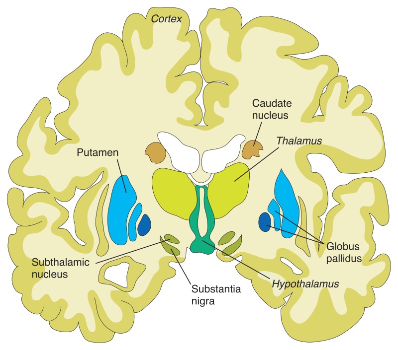
Figure 8.1. Structures of the basal ganglia.


The cells in the Substantial Nigra secrete a neurotransmitter, called Dopamine, which is responsible for the coordinated and smooth functioning of voluntary movements of the body and maintaining the tone of muscles. The enzyme AAAD  (aromatic L-amino acid decarboxylase) converts levodopa(L-DOPA) to dopamine. The final inactive degraded product of dopamine is 3-0 methyldopamine by the action of the Catechol- 0- methyltransferase (COMT) enzyme.

Nerve cells of the peripheral nervous system and adrenal medulla and endothelial cells of the GI tract also take up L-dopa. From amino acid Tyrosine and L-dopa, the adrenal glands make dopamine, norepinephrine and adrenaline. An enzyme Monoamine oxidase (MAO) degrades dopamine, norepinephrine and adrenaline to inactive compounds. The central nervous system has no MAO and MAO cannot cross into the brain from the blood (blood brain barrier).

In Parkinson's disease substantia nigra cells die prematurely, as a result, the movements become jerky, slow and disturbance of gait and balance, tremors and stiffness develop.

Parkinson's disease clinically may appear as 3 entities initially. In Type 1- the tremors of one or more limbs predominate. Muscle rigidity is absent and ambulation remains normal. In Type 2 disease the ataxic gait and mask like face and pill rolling movements are dominant symptoms. In Type 3 cases all the typical symptoms of PD are exhibited.

Progressive Supranuclear Palsy and Lowy Body Dementia are closely related to Parkinson's disease but are not a part of this discussion.

The standard medical treatment for Parkinson's disease is to supply Dopamine and reduce or stop MAO-B activities in the body so at the end more L-dopa will be available for the brain. A group of drugs is available for that purpose and are collectively called MAO inhibitors and are in regular use in PD.

In addition, direct interventions for tremors, depression, stiffness of the body and constipation, etc. are treated with well-known medications. Physical exercise, physiotherapy, occupation therapy, speech and languages therapy are also included in the management of Parkinson's disease.

New drugs and new methods of drug delivery to CNS, Stem cell transplantation, keeping dopamine secreting cells alive and surgical treatment are parts of this article.

In between doses of medication, rescue therapy.

Levodopa is a standard drug for Parkinson's disease. It is converted into Dopamine in the brain. Levodopa therapy replenishes decreased levels of dopamine in the brain. A drug called Carbidopa is an MAO inhibitor. Carbidopa and L-dopa are combined into one pill for use in PD. But in between doses, sudden deterioration of symptoms happens due to an unexpected fall of available L-dopa in the brain cells. To prevent this from happening, several drugs are now available.

  1. Istradefylline is marketed as Nouriazen. It is an antagonist to Adenosine A2A receptors. When this receptor is blocked the nerve cells release more gamma aminobutyric acid (GABA) which is another neurotransmitter. GABA helps to ease symptoms related to the "OFF episodes" from the sudden drop of dopamine. Istradefylline belongs to a small molecule drug and is given orally once a day dose.

  2. Entacapone is an inhibitor of the MAO-B enzyme. Available as Comptan. Other available MAO-B inhibitors are Rasagiline and Safinamide.

  3. New delivery system for Levodopa.

       New formulation allows Levodopa delivery via inhalation. It acts quickly.               It is marketed as Inbrija.

  1.  Apomorphine is a form of a thin sublingual tablet that acts quickly to counter the symptoms of off symptoms.

  2. Accordion pills. Layers of Levodopa are designed in such a way that they will release at a slow and steady level. Thus, decrease unusual dips of drug blood levels and decreases the incidence of “off” episodes."

     6. Pump and patch pumps are also used and have improved for steady drug         delivery

GDNF.

Glial cells produce a neurotrophic factor. It is a naturally occurring protein that protects many types of brain cells.

In Parkinson's disease, the GDNF is delivered directly into the substantia nigra by implanted tubes in the brain, through a port is surgically placed behind the ear. When GDNF is delivered every 4 weeks for 9 months, patients receiving this therapy show remarkable improvement and PET scans detect regeneration of dying dopamine producing cells.

Stem cell Transplantation.

In a study done at Massachusetts general hospital and Cornell Medical center, the dopamine producing stem cells were transplanted.

Pluripotent stem cells are harvested from the skin of a patient and the pluripotent cells are engineered to differentiate into dopamine generating stem cells. Then the stem cells are transfused. In this study, patients are followed for 2 years. During this follow-up period, the patients show symptomatic improvement and the transplant cells remained alive and functional.

Surgical treatment.

Deep brain stimulation by an implanted electrode in the ventrolateral nucleus of the Thalamus improves tremors in PD.

Pallidotomy.    Surgical removal of palladium improves dyskinesia. In bilateral lesions, the Subthalamic nucleus is removed instead of both pallidum to avoid the development of Hemiballismus.

Radiofrequency ablation has greatly replaced surgery of the basal ganglia and achieved the same results.

Deep brain stimulation in selected groups of nuclei of the basal ganglia for various uncontrollable symptoms has become an acceptable alternative to drug therapy.

Experimental therapy.

1. Dyskinesia results from the prolonged use of levodopa. An Insulin sensitizer – MSDC0160 therapy improves dyskinesia.

2 . Anti Alpha synuclein.

Alpha synuclein naturally occurring protein, which accumulates in the cells of substantia nigra of Parkinson's patients. Several drugs are now in development stages that show promise in removing the alpha synuclein.

Recently laboratory evidence shows similar alpha synuclein protein accumulation in the nerve cells in the GI tract and a new idea is developing that the gut bacterial products or certain bacteria initiate abnormal alpha synuclein production in the gut and that process is carried to the brain either via Vagus nerve or by circulation.


A small molecule, Anle 1386 is an example of such a drug. The initial results are encouraging and show a reduction of alpha synuclein accumulation in the brain cells.

Antibody against alpha synuclein. When PD patients are treated with alpha synuclein antibodies reduction of this protein in the brain cells and the clumping of protein molecules disappear.

Vaccine to stimulate antibodies to alpha synuclein is underway.

3. Repurposed drugs. Exenatide is a diabetic drug found to protect dying brain cells in Parkinson's disease patients. Inosine – is a nucleoside, when used in humans, it increases urate levels. Urates are antioxidantsand  and protect brain cells. Israpidine is a B.P drug, found to preserve brain cells. Nilotinib is a tyrosine kinase inhibitor and is used in Chronic myeloid leukemia, when used in Parkinson's patients, it helps clear the alpha synuclein by phagocytosis.

Treatment of the Gene mutation.

Mutation of the GBA gene causes cellular dysfunction from the accumulated of lipids in the cells. An experimental drug GZ/SAR 40267 reduces lipid accumulation in the cells.

An oral drug LTI-291 corrects Liposomal dysfunction which is the cause of lipid accumulation.

Hemanti's Neurotrophic factor. This neurotrophic factor protects cells from premature death.

Glutamate blockers.

In long term use of L-dopa in PD patients Glutamate is over expressed in neurons of basal ganglia due to phosphorylation of N-methyl D aspartate receptors and that is responsible for dyskinesia.

Foliglurax and Dipraglurant reduce Dyskinesia by blocking Glutamate in the brain.

Serotonin receptor blockers

In PD patients, in addition to the loss of dopamine producing cells in basal ganglia, serotonin producing neurons are also lost to a certain extent. As a result, serotonin terminals in basal ganglia become over abundant compared to dopamine terminals. Serotonin terminals convert L-dopa into dopamine in excess amounts and produce dyskinesia.

Serotonin receptor blockers mitigate this problem. Example: Eltoprazines.

Anti-Choline drugs.

Acetylcholine hyperactivity in dopamine depleted brain is implicated in gait disturbance and frequent falls. Varecline and Donepezil reduce brain acetylcholine levels and reduce these complications.

In recent years many private organizations came together and made funds available for research. The progress has been impressive. Improvement in stem cell transplants and gene therapy, perhaps, one day will make Parkinson's disease a mild form of disability in case an outright cure is not possible.

***********************************************************

Archive

  Archive. P.K.Ghatak, MD ghatak3@gmail.com 2025  March. Laser Endoscpoe 2025 February. Use of Ultrasound in Medicine MRI 2025 January. Bact...